

Diagnostic analysis of abnormal increase of PASP in fetus in middle- and late-stage pregnancy by color Doppler echocardiography
- PMID: 32160003
- PMCID: PMC10993218
- DOI: 10.1259/bjr.20191011
Diagnostic analysis of abnormal increase of PASP in fetus in middle- and late-stage pregnancy by color Doppler echocardiography
Abstract
Objective: Our study was conducted with an attempt to investigate the diagnostic analysis of abnormal increase of fetal pulmonary artery systolic pressure (PASP) in middle and late pregnancy by color Doppler echocardiography.
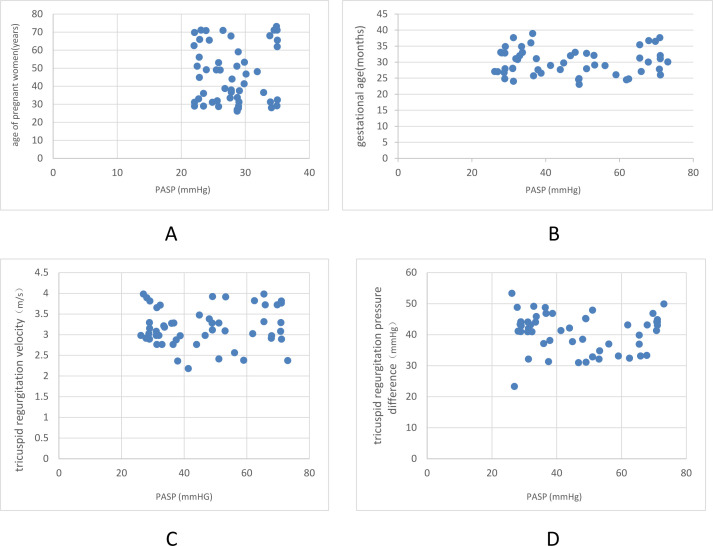
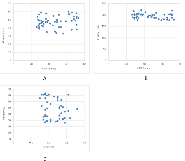
Methods: From August 2017 to January 2019, 52 fetuses with moderate or greater tricuspid high-speed regurgitation were retrospectively analyzed and selected as Group A. 88 fetuses with full-color blood flow of the two ventricles and symmetrical sizes of the cardiac cavities on both sides harboring tricuspid valve and mild regurgitation or a small amount of regurgitation were selected as Group B. The pulmonary artery blood flow acceleration time (AT) and right ventricular ejection time (ET) was measured, and the PASP was calculated.
Results: The tricuspid regurgitation velocity, tricuspid regurgitation pressure difference and PASP in Group A were higher than those in Group B (p < 0.05), and the AT and AT/ET values in Group A were lower than those in Group B (p < 0.05). Gestational age, tricuspid regurgitation velocity and tricuspid regurgitation pressure difference were positively correlated with PASP. However, AT/ET and AT value were negatively correlated with PASP.
Conclusion: The abnormal increase of pulmonary artery can be assessed by color Doppler echocardiography of fetal tricuspid regurgitation, which is worth popularizing and applying in clinic.
Advances in knowledge: It was suggested that the middle- and late-stage fetuses with moderate or greater tricuspid regurgitation and with >20 mmHg regurgitation pressure difference should be followed up in clinic. If PASP was ≥70 mmHg with symptoms of right heart failure, fetuses should be closely observed until 35-36 weeks old to ensure fetal safety and early delivery would be recommended.
Figures
References
-
- Kumar Vikraman S, Chandra V, Balakrishnan B, Jaiman S, Batra M, Kannoly G . Unguarded tricuspid orifice---a rare cause of fetal right atrial dilatation with characteristic color doppler sign: Case report with review of literature . J Clin Ultrasound 2017. ; 45: 370 – 4 . doi: 10.1002/jcu.22416 - DOI - PubMed
-
- Zhan H, Liu C, Yin H . Follow-Up value of ultrasound in fetal tricuspid regurgitation Chinese . Journal of Ultrasonography 2014. ; 23: 979 – 82 .
MeSH terms
LinkOut - more resources
Full Text Sources
Other Literature Sources
Medical
