
Severe Acute Respiratory Syndrome Coronavirus 2 from Patient with Coronavirus Disease, United States

Jennifer Harcourt et al. Emerg Infect Dis. 2020 Jun.

Abstract

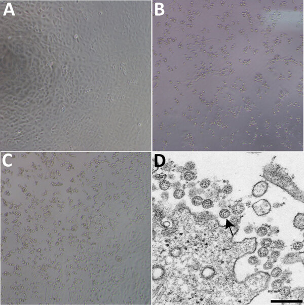
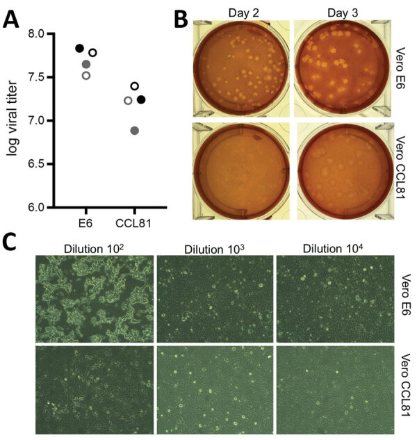
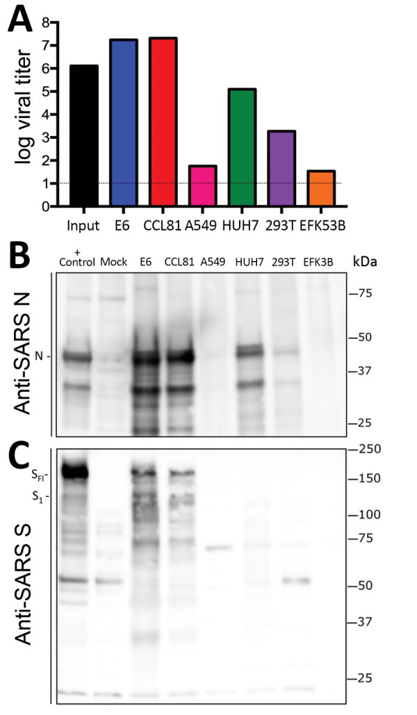
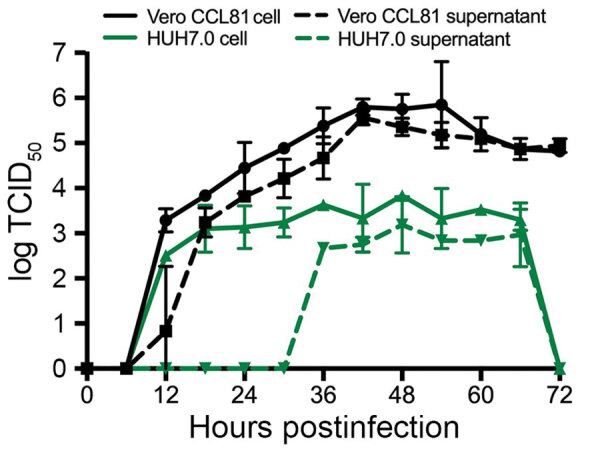
The etiologic agent of an outbreak of pneumonia in Wuhan, China, was identified as severe acute respiratory syndrome coronavirus 2 in January 2020. A patient in the United States was given a diagnosis of infection with this virus by the state of Washington and the US Centers for Disease Control and Prevention on January 20, 2020. We isolated virus from nasopharyngeal and oropharyngeal specimens from this patient and characterized the viral sequence, replication properties, and cell culture tropism. We found that the virus replicates to high titer in Vero-CCL81 cells and Vero E6 cells in the absence of trypsin. We also deposited the virus into 2 virus repositories, making it broadly available to the public health and research communities. We hope that open access to this reagent will expedite development of medical countermeasures.

Keywords: 2019 novel coronavirus disease; COVID-19; PCR; SARS-CoV-2; United States; characterization; coronavirus; isolation; respiratory infections; severe acute respiratory syndrome coronavirus 2; viruses; zoonoses.


Figures

Figure 1
Figure 1
Cytopathic effect caused by severe acute respiratory syndrome coronavirus 2 from patient with coronavirus disease, United States, 2020. A–C) Phase-contrast microscopy of Vero cell monolayers at 3 days postinoculation: A) Mock, B) nasopharyngeal specimen, C) oropharyngeal specimen. Original magnifications ×10). D) Electron microscopy of virus isolate showing extracellular spherical particles with cross-sections through the nucleocapsids (black dots). Arrow indicates a coronavirus virion budding from a cell. Scale bar indicates 200 nm.
Figure 2
Figure 2
Viral propagation and quantitation of severe acute respiratory syndrome coronavirus 2 from patient with coronavirus disease, United States, 2020. A) Two virus passage 4 stocks (black and gray circles) were quantified by using plaque assay at day 2 (solid circles) and day 3 (open circles) postinfection of Vero E6 and Vero CCL81 cells. B) Plaque morphology for virus on Vero E6 and Vero CCL81 at day 2 and day 3 postinoculation. C) Cell monolayers 2 days postinfection of Vero E6 (top) and Vero CCL81 (bottom) at 3 dilutions. Original magnifications ×40.
Figure 3
Figure 3
Cell lines from patient with coronavirus disease, United States, 2020, susceptible to SARS coronavirus 2 (SARS-CoV-2). Cell lines were infected with a high multiplicity of infection (>5), washed after adsorption, and subsequently harvested 24 h postinfection for viral titer and protein lysates. A) Viral titer for SARS-CoV-2 quantitated by plaque assay on Vero E6 cells 2 days postinoculation. Infected cell protein lysates were probed by using Western blotting with B) rabbit polyclonal anti-SARS N antibody or C) anti–SARS-CoV S protein antibody. Full-length spike protein (SFL) and spike protein S1 (S1) are indicated. N, nucleocapsid; S, spike protein; SARS, severe acute respiratory syndrome.
Figure 4
Figure 4
Multistep growth curve for severe acute respiratory syndrome coronavirus 2 from patient with coronavirus disease, United States, 2020. Vero CCL81 (black) and HUH7.0 cells (green) were infected at a multiplicity of infection of 0.1, and cells (solid line) and supernatants (dashed line) were harvested and assayed for viral replication by using TCID50. Circles, Vero CCL81 cells; squares, Vero CCL81 supernatants; triangles, HUH7.0 cells; inverted triangles, HUH7.0 supernatants. Error bars indicate SEM. TCID50, 50% tissue culture infectious dose.

Similar articles

Cited by

References

    1. Holshue ML, DeBolt C, Lindquist S, Lofy KH, Wiesman J, Bruce H, et al.; Washington State 2019-nCoV Case Investigation Team. Washington State 2019-nCoV Case Investigation Team. First case of 2019 novel coronavirus in the United States. N Engl J Med. 2020;382:929–36. 10.1056/NEJMoa2001191 - DOI - PMC - PubMed
    1. Zhu N, Zhang D, Wang W, Li X, Yang B, Song J, et al.; China Novel Coronavirus Investigating and Research Team. A novel coronavirus from patients with pneumonia in China, 2019. N Engl J Med. 2020;382:727–33. 10.1056/NEJMoa2001017 - DOI - PMC - PubMed
    1. Lu R, Zhao X, Li J, Niu P, Yang B, Wu H, et al. Genomic characterisation and epidemiology of 2019 novel coronavirus: implications for virus origins and receptor binding. Lancet. 2020;395:565–74. 10.1016/S0140-6736(20)30251-8 - DOI - PMC - PubMed
    1. Chan JF, Kok KH, Zhu Z, Chu H, To KK, Yuan S, et al. Genomic characterization of the 2019 novel human-pathogenic coronavirus isolated from a patient with atypical pneumonia after visiting Wuhan. Emerg Microbes Infect. 2020;9:221–36. 10.1080/22221751.2020.1719902 - DOI - PMC - PubMed
    1. Li Q, Guan X, Wu P, Wang X, Zhou L, Tong Y, et al. Early transmission dynamics in Wuhan, China, of novel coronavirus-infected pneumonia. N Engl J Med. 2020;NEJMoa2001316; Epub ahead of print. 10.1056/NEJMoa2001316 - DOI - PMC - PubMed

Publication types

LinkOut - more resources