. 2020 Mar 10;30(10):3368-3382.e7.
doi: 10.1016/j.celrep.2020.02.041.

MYC Dysregulates Mitosis, Revealing Cancer Vulnerabilities

Affiliations

MYC Dysregulates Mitosis, Revealing Cancer Vulnerabilities

Julia Rohrberg et al. Cell Rep. .

Abstract

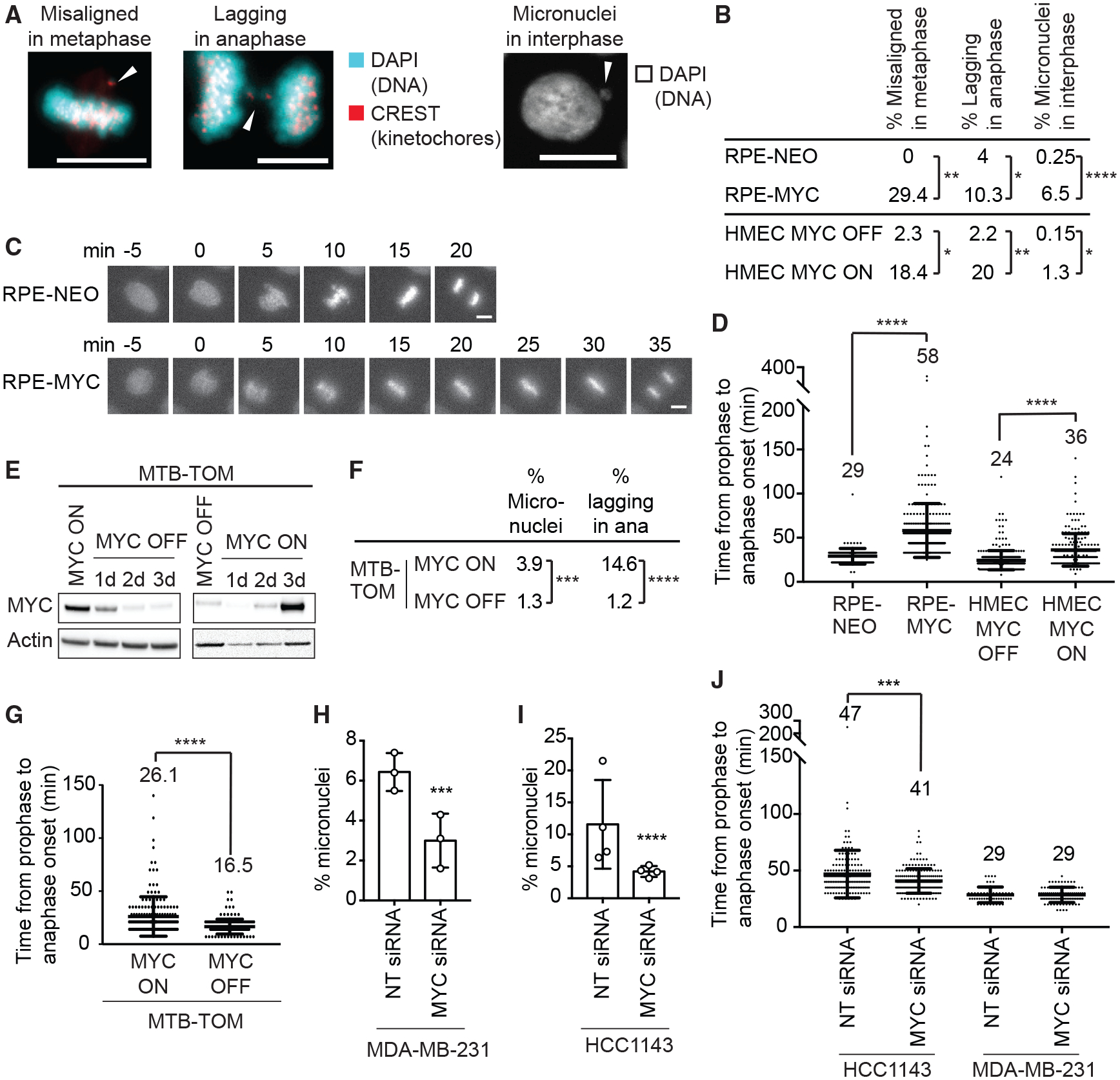
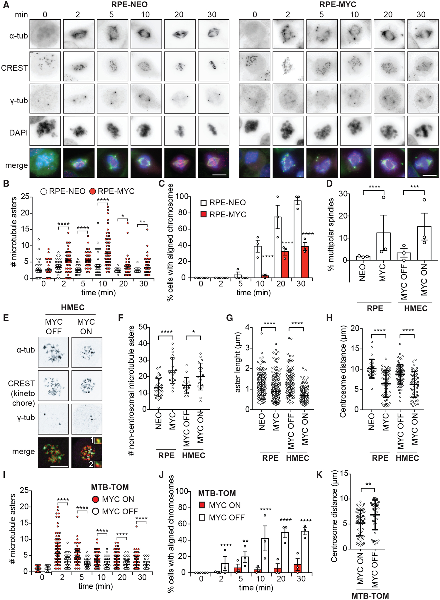
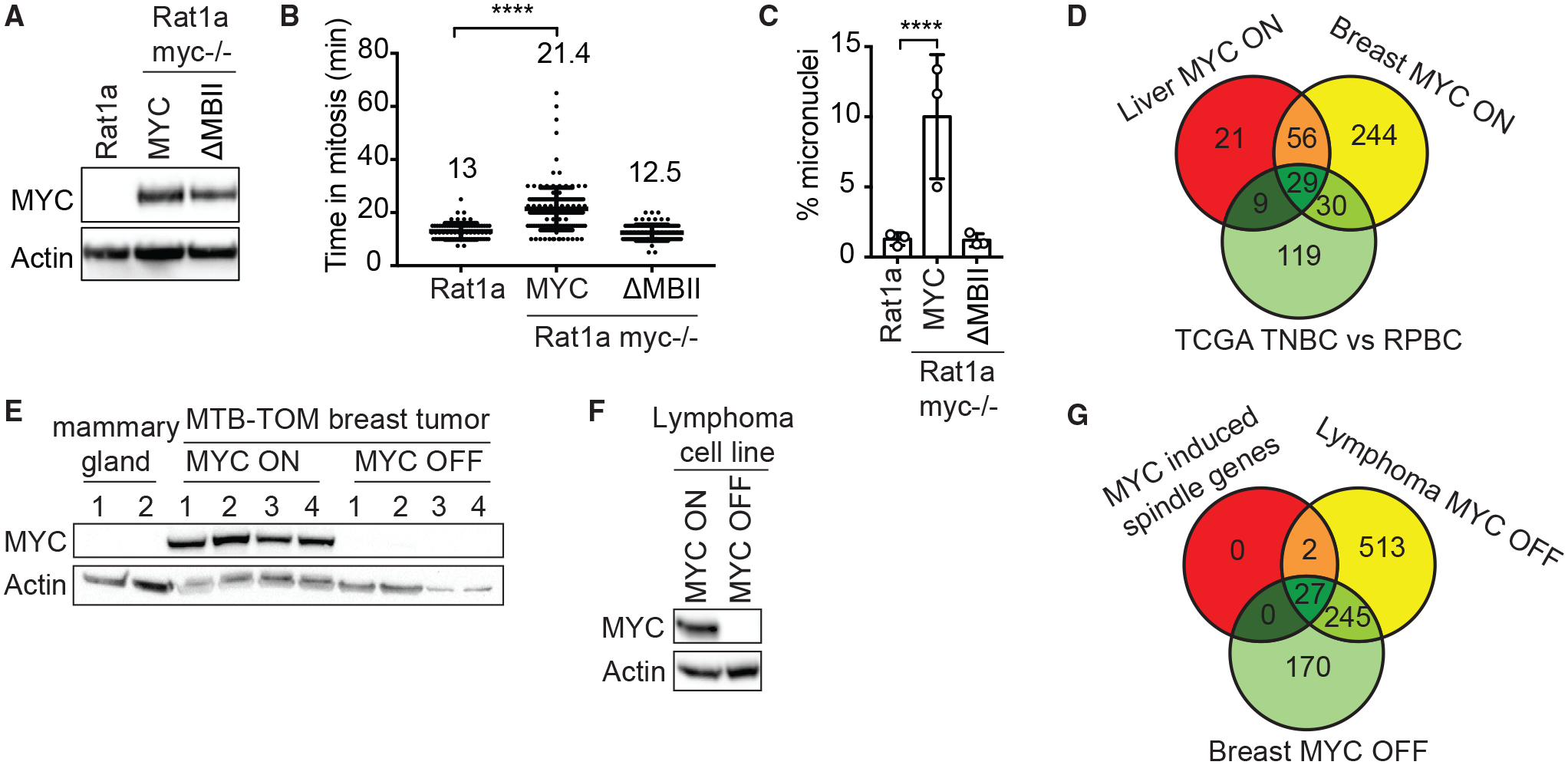
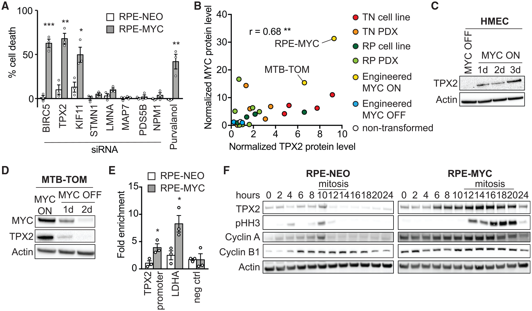
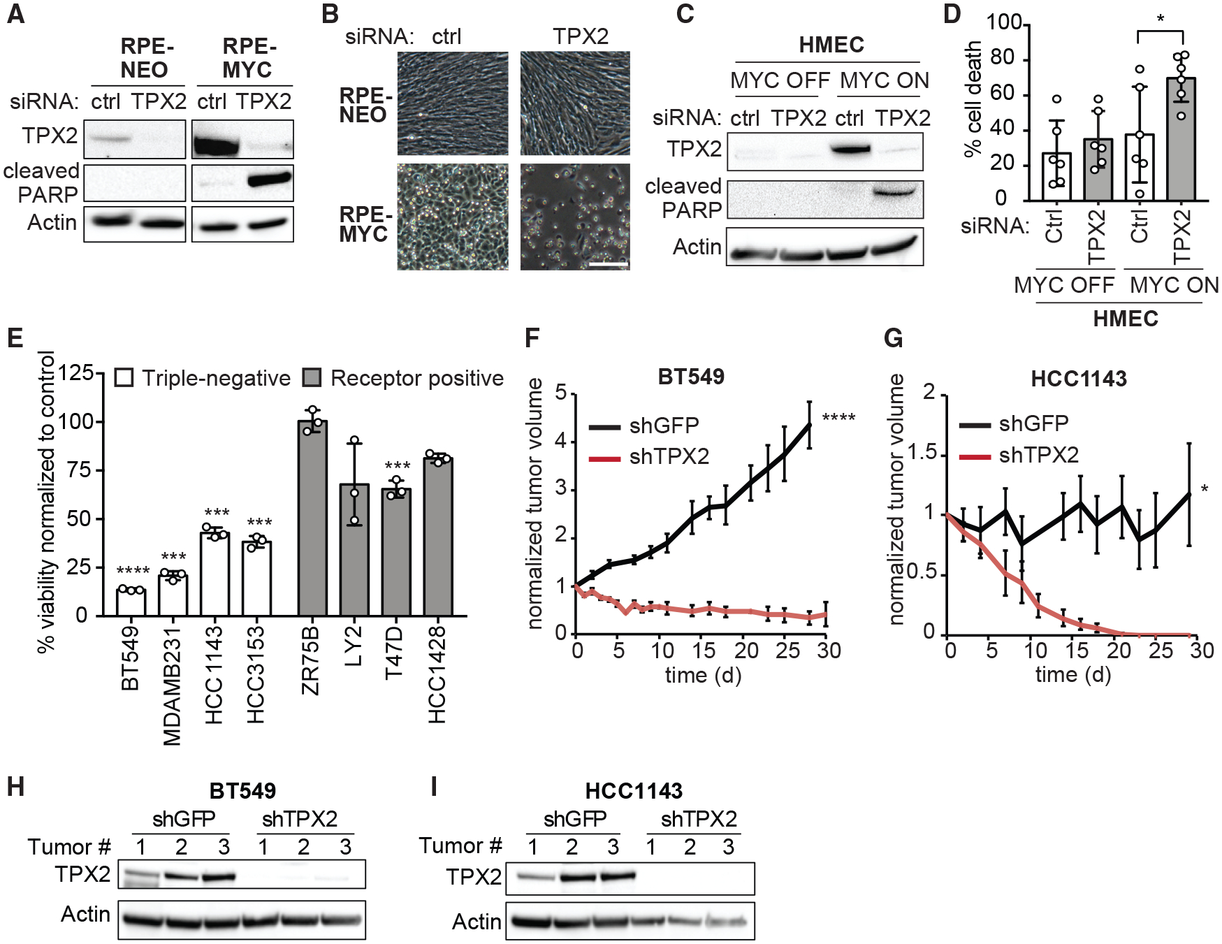
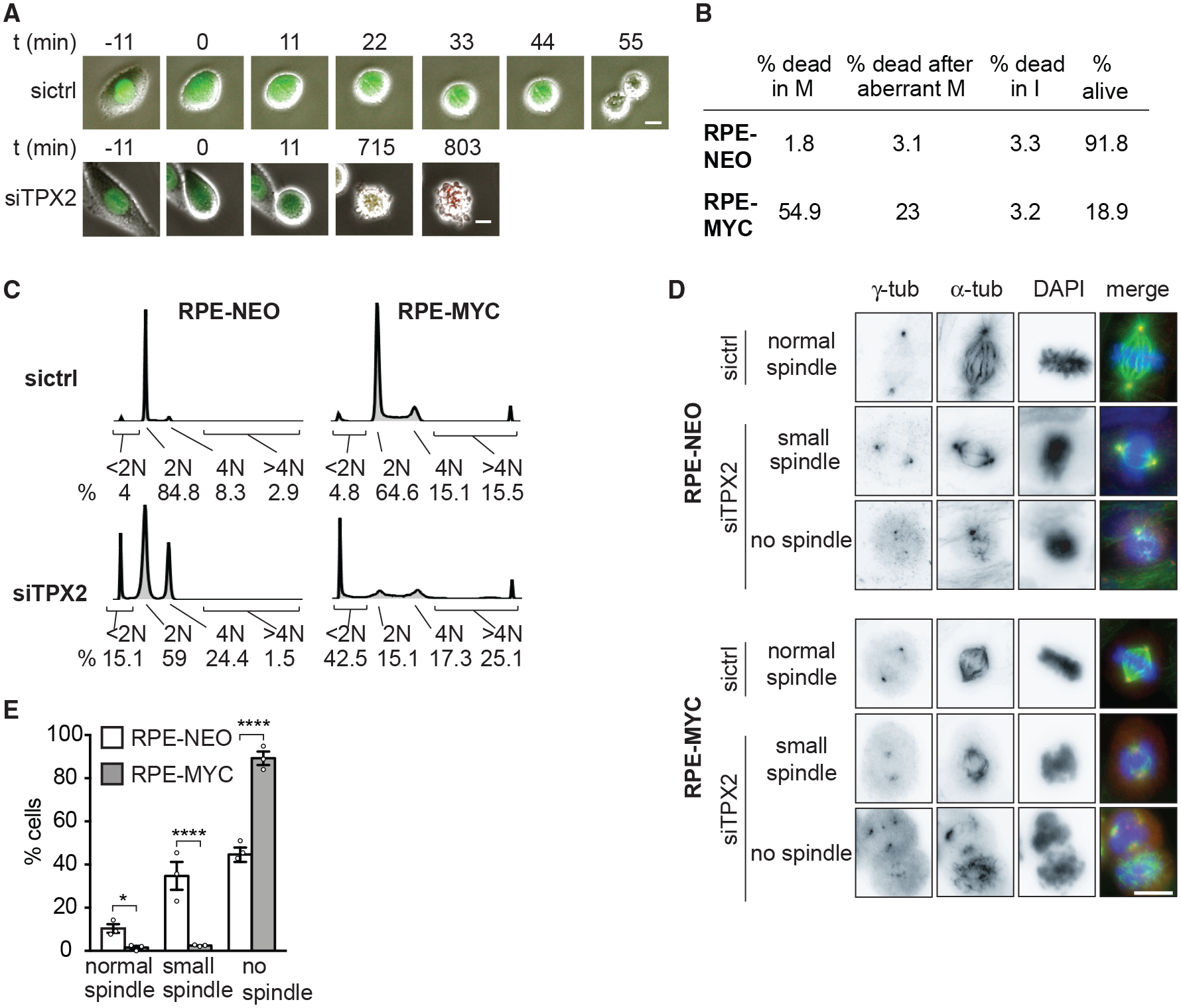
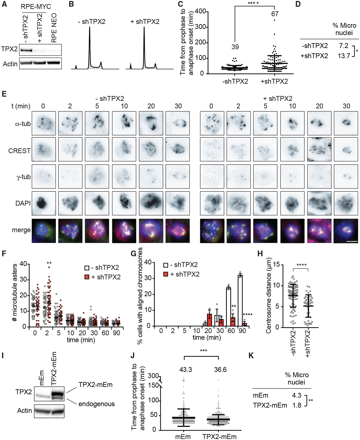
Tumors that overexpress the MYC oncogene are frequently aneuploid, a state associated with highly aggressive cancers and tumor evolution. However, how MYC causes aneuploidy is not well understood. Here, we show that MYC overexpression induces mitotic spindle assembly defects and chromosomal instability (CIN) through effects on microtubule nucleation and organization. Attenuating MYC expression reverses mitotic defects, even in established tumor cell lines, indicating an ongoing role for MYC in CIN. MYC reprograms mitotic gene expression, and we identify TPX2 to be permissive for spindle assembly in MYC-high cells. TPX2 depletion blocks mitotic progression, induces cell death, and prevents tumor growth. Further elevating TPX2 expression reduces mitotic defects in MYC-high cells. MYC and TPX2 expression may be useful biomarkers to stratify patients for anti-mitotic therapies. Our studies implicate MYC as a regulator of mitosis and suggest that blocking MYC activity can attenuate the emergence of CIN and tumor evolution.

Keywords: CIN; MYC; TNBC; TPX2; chromosomal instability; microtubules; mitosis; mitotic spindle assembly; receptor triple-negative breast cancer; synthetic-lethality.


Conflict of interest statement

Declaration of Interests The authors declare no competing interests.

Figures

Figure 1.
Figure 1.. MYC Reversibly Induces CIN
(A) Images of mitotic defects in RPE-MYC cells. Arrows indicate misaligned and missegregated chromosomes. Scale bars, 10 μm. (B) Percentage of mitotic defects. HMEC MYC ON, MYC activated for 3 days. Fisher’s exact test, n = 100–300 mitotic cells and n = 800–1,000 cells for micronuclei from 3 independent experiments. (C) Fluorescent time-lapse images of RPE-NEO and RPE-MYC cells expressing H2B-mCherry. Scale bars, 10 μm. (D) Time from chromosome condensation to anaphase onset (average time shown above each plot). Mean ± SD. Unpaired t test, n = 140–380 from 3 independent experiments. (E) Western blot analysis of MYC in MTB-TOM cells grown in the presence (MYC ON) and absence of doxycycline (MYC OFF). (F) Percentage of mitotic defects in MTB-TOM MYC ON and MYC OFF (3 days) cells. Fisher’s exact test, n = 1,628 and 520 for micronuclei and 178 and 164 for mitotic errors, 3 independent experiments. (G) Time from chromosome condensation to anaphase onset of MTB-TOM cells expressing H2B-mCherry (average time shown above each plot). Mean ± SD. t test, n = 178 and 164, 3 independent experiments. (H and I) Percentage of micronucleated MDA-MB-231 (H) and HCC1143 (I) cells 3 days after transfection with non-targeting (NT) or MYC siRNA. Mean ± SD. Fisher’s exact test, n = 628–1,056, 3 independent experiments. See also Figures S1H–S1L. (J) Time from chromosome condensation to anaphase onset 3 days after treatment with NT or MYC siRNA (average time shown above each plot). Mean ± SD. Unpaired t test, n = 171–179, 3 independent experiments. *p < 0.05, **p < 0.01, ***p < 0.001, ****p < 0.0001.
Figure 2.
Figure 2.. MYC Impairs Mitotic Spindle Formation
(A–C) Nocodazole washout assay in RPE-NEO and RPE-MYC cells. (A) Representative images. Scale bars, 10 μm. (B) Number of microtubule foci/asters. Mean ± SEM t test, n = 26–99, 3 independent experiments. See also Figure S2A. (C) Percentage of cells with aligned chromosomes. Mean ± SEM. Fisher’s exact test, n = 13–139, 3 independent experiments. (D) Percentage of multipolar spindles. HMEC MYC ON, MYC activated for 3 days. Mean ± SEM. Fisher’s exact test, n = 97–315, 3 independent experiments. See also Figures S2E and S2F. (E–G) Confocal microscopy after nocodazole washout. (E) Representative images of HMEC cells 2 min after washout. Scale bar, 10 μm. Insets show a 3-fold magnification of microtubule asters nucleating at non-centrosomal sites colocalizing with (1) or nucleating next to kinetochores (2). (F) Number of microtubule asters at non-centrosomal sites 2 min after washout. Mean ± SD. Unpaired t test, n = 16–35, 2 independent experiments. (G) Length of microtubule asters 5 min after washout. Mean ± SD. Fisher’s exact test, n = 139–286, 2 independent experiments. (H) Centrosome distance 30 min after nocodazole washout. Mean ± SD. Unpaired t test, n = 26–73, at least 3 independent experiments. (I–K) Nocodazole washout assay in MTB-TOM MYC ON and MYC OFF (3 days). (I) Number of microtubule asters. Mean ± SEM. t test, n = 47–208, 3 independent experiments. See also Figure S2G. (J) Percentage of cells with aligned chromosomes. Mean ± SEM. Fisher’s exact test, n = 81–209 cells, 3 independent experiments. (K) Centrosome distance 30 min after washout. Mean ± S.D. Unpaired t test, n = 41 and 63 cells, 3 independent experiments. *p < 0.05, **p < 0.01, ***p < 0.001, ****p < 0.0001.
Figure 3.
Figure 3.. MYC Regulates the Expression of Mitotic Spindle Genes
(A–C) Rat1a and Rat1a myc/ cells expressing mouse wild-type MYC or the transcriptionally inactive mutant MYCDMBII. (A) Western blot analysis of murine MYC; endogenous rat MYC expression was not detected. (B) Time from chromosome condensation to anaphase onset. Mean ± SD. Unpaired t test, n = 78–167, 3 independent experiments. (C) Percentage of micronucleated cells. Mean ± SD. Fisher’s exact test, n = 730–1,478, 3 independent experiments. (D) Deregulated mitotic spindle genes in murine MTB-TOM breast and liver tumors compared to the corresponding normal tissue and in human TNBC compared to RPBC (TCGA data). Log2 fold change >1, FDR < 0.05. (E) Western blot analysis of MYC in MTB-TOM tumors (MYC ON, n = 4), tumors off doxycycline for 3 days (MYC OFF, n = 4) and normal mammary gland (n = 2). (F) Western blot of MYC in Eμ-tTA/TRE-MYC lymphoma cells in the absence (MYC ON) and presence (MYC OFF) of doxycycline for 3 days. (G) Deregulated mitotic spindle genes when MYC expression is turned off in MTB-TOM breast tumors and Eμ-tTA/TRE-MYC lymphoma cells compared to the 29 MYC-induced spindle genes identified in (D). Log2 fold change > –1, FDR < 0.05. See also Table S2. ****p < 0.0001.
Figure 4.
Figure 4.. TPX2 Expression Correlates with MYC and Its Depletion Kills MYC-High Cells
(A) Percentage of cell death in RPE-NEO (white bars) and RPE-MYC cells (gray bars) 3 days after siRNA treatment normalized to control siRNA, and after treatment with 10 μM purvalanol A normalized to DMSO. Bars, mean ± SEM. t test, n = 3. (B) Correlation of MYC and TPX2 protein levels in RPE-MYC and MTB-TOM (engineered MYC ON, yellow), RPE-NEO, and MTB-TOM off doxycycline for 2 days (engineered MYC OFF, blue), 4 triple negative breast cancer (TNBC) cell lines (red), 4 receptor-positive breast cancer (RPBC) cell lines (dark green), 5 patient-derived xenograft (PDX) TNBC tumors (orange), 7 RPBC PDX tumors (light green), and non-transformed mouse mammary gland (white). See also Figures S3C–S3F. The correlation coefficient was computed using Pearson correlation. (C) Western blot of TPX2 in HMEC MYC ON and HMEC MYC OFF. (D) Western blot of TPX2 and MYC in MTB-TOM MYC ON and MYC OFF. (E) ChIP of MYC in RPE-NEO and RPE-MYC followed by qPCR against the promoter region of TPX2 and LDHA (positive control) and an upstream region of the LDHA promoter (negative control). Mean ± SEM. Unpaired t test, n = 3. See also Figure S3B. (F) Western blot of TPX2, pHH3, Cyclin A, and Cyclin B1 in RPE-NEO and RPE-MYC cells; time after release from a double thymidine block is indicated. Mitotic cell rounding observed by light microscopy is indicated with line. See also Figure S3G. *p < 0.05, **p < 0.01, ***p < 0.001.
Figure 5.
Figure 5.. TPX2 Is Required for the Survival of MYC-High Cells
(A and B) Western blot of TPX2 and cleaved PARP (A) and micrographs (B) of RPE-NEO and RPE-MYC cells 3 days after transfection with control (ctrl) or TPX2 siRNA. Scale bar, 100 μm. (C and D) Western blot of TPX2 and cleaved PARP (C) and percentage of cell death (D) of HMEC MYC OFF and HMEC MYC ON 3 days after transfection with control (ctrl) or TPX2 siRNA. Mean ± S.D., t test, n = 6. See also Figure S4C. (E) Percent viability of breast cancer cell lines three days after transfection with TPX2 siRNA normalized to control siRNA. Mean ± SEM, t test, n = 3. See also Figure S4D. (F and G) Relative volume of BT549 (F) and HCC1143 (G) xenograft tumors expressing doxycycline inducible shRNA against TPX2 (shTPX2) or GFP (shGFP). Mean ± SEM. BT549 shGFP (n = 7), shTPX2 (n = 7), HCC1143 shGFP (n = 6), and HCC1143 shTPX2 (n = 5). Unpaired t test. See also Figures S4E–S4F. (H and I) Western blot of TPX2 in BT549 (H) and HCC1143 (I) xenograft tumors at endpoint (n = 3). *p < 0.05, **p < 0.01, ***p < 0.001, ****p < 0.0001.
Figure 6.
Figure 6.. TPX2 Protects Mitotic Spindle Function in MYC-High Cells
(A) Time-lapse images of RPE-MYC cells expressing the FUCCI cell-cycle marker 12 h after transfection with control (sictrl) or TPX2 (siTPX2) siRNA. Fluorescence and phase-contrast images were overlaid. Scale bars, 20 μm. (B) Percentage of cells undergoing cell death in mitosis (M), after aberrant mitosis and in interphase (I) 12–24 h after TPX2 knockdown in RPE-NEO and RPE-MYC cells. n = 3. (C) Cell-cycle profiles 48 h after siRNA treatment with percentage of dead cells (<2N), cells in G1 (2N), and G2-M (4N) phases of the cell cycle and cells with >4N DNA content. (D) Images of RPE-NEO (top) and RPE-MYC (bottom) cells 24 h after transfection. Scale bar, 10 μm. (E) Percentage of mitotic cells with normal, small, or no spindles 24 h after transfection with siTPX2. Mean ± SEM. Fisher’s exact test, n = 36–126 mitotic cells, 3 independent experiments. See also Figure S5. *p < 0.05, **p < 0.01, ****p < 0.0001.
Figure 7.
Figure 7.. TPX2 Overexpression Is Necessary for MYC-High Cells to Progress through Mitosis
(A–H) RPE-MYC cells expressing doxycycline-inducible shRNA against TPX2 in the absence (−shTPX2) and presence (+shTPX2) of doxycycline for 4 days. (A) Western blot analysis of TPX2. (B) Cell-cycle profiles. (C) Time from nuclear envelope breakdown to anaphase onset quantified from time-lapse microscopy experiments. Mean ± SD. t test, n = 164 and 164, 3 independent experiments. (D) Percentage of micronucleated cells. Fisher’s exact test, n = 223 and 182, 3 independent experiments. (E–H) Nocodazole wash-out assay. (E) Representative images. Scale bar, 10 μm. (F) Number of microtubule asters. Mean ± SEM. Unpaired t test, n = 45–117, 3 independent experiments. (G) Percentage of cells with aligned chromosomes. Mean ± SEM. Fisher’s exact test, n = 45–117, 3 independent experiments. (H) Centrosome distance 90 min after washout. Mean ± SEM. Unpaired t test, n = 20–84, 3 independent experiments. (I–K) RPE-MYC cells expressing TPX2-mEmerald (TPX2mEm) or empty vector (mEm). (I) Western blot of TPX2. Exogenous TPX2-mEm is shifted upward. (J) Time from nuclear envelope breakdown to anaphase onset from time-lapse microscopy experiments. Mean ± SD. t test, n = 307 and 383, 3 independent experiments. (K) Percentage of micronucleated cells. Fisher’s exact test, n = 1,579 and 2,008, 3 independent experiments. *p < 0.05, **p < 0.01, ***p < 0.001, ****p < 0.0001.

References

    1. Aguirre-Portolés C, Bird AW, Hyman A, Cañamero M, Pérez de Castro I, and Malumbres M (2012). Tpx2 controls spindle integrity, genome stability, and tumor development. Cancer Res. 72, 1518–1528. - PubMed
    1. Amati B, Alevizopoulos K, and Vlach J (1998). Myc and the cell cycle. Front. Biosci 3, d250–d268. - PubMed
    1. Anderton B, Camarda R, Balakrishnan S, Balakrishnan A, Kohnz RA, Lim L, Evason KJ, Momcilovic O, Kruttwig K, Huang Q, et al. (2017). MYC-driven inhibition of the glutamate-cysteine ligase promotes glutathione depletion in liver cancer. EMBO Rep. 18, 569–585. - PMC - PubMed
    1. Bakhoum SF, Thompson SL, Manning AL, and Compton DA (2009). Genome stability is ensured by temporal control of kinetochore-microtubule dynamics. Nat. Cell Biol 11, 27–35. - PMC - PubMed
    1. Bakhoum SF, Ngo B, Laughney AM, Cavallo J-A, Murphy CJ, Ly P, Shah P, Sriram RK, Watkins TBK, Taunk NK, et al. (2018). Chromosomal instability drives metastasis through a cytosolic DNA response. Nature 553, 467–472. - PMC - PubMed

Publication types

Substances