. 2020 Mar 10;30(10):3552-3565.e6.
doi: 10.1016/j.celrep.2020.02.037.

Individual Limb Muscle Bundles Are Formed through Progressive Steps Orchestrated by Adjacent Connective Tissue Cells during Primary Myogenesis

Affiliations

Individual Limb Muscle Bundles Are Formed through Progressive Steps Orchestrated by Adjacent Connective Tissue Cells during Primary Myogenesis

Laurianne Besse et al. Cell Rep. .

Abstract

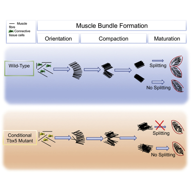
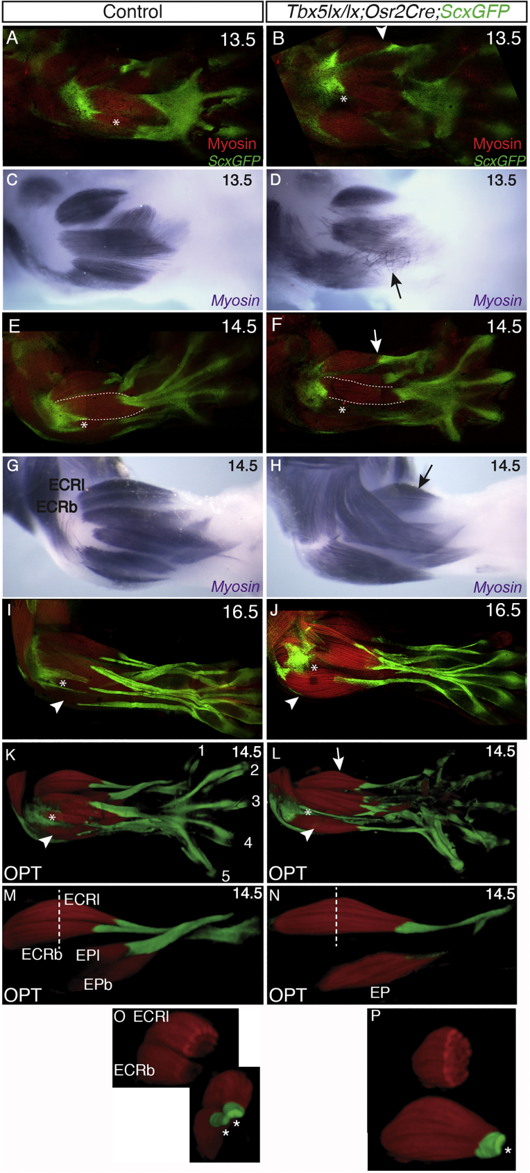
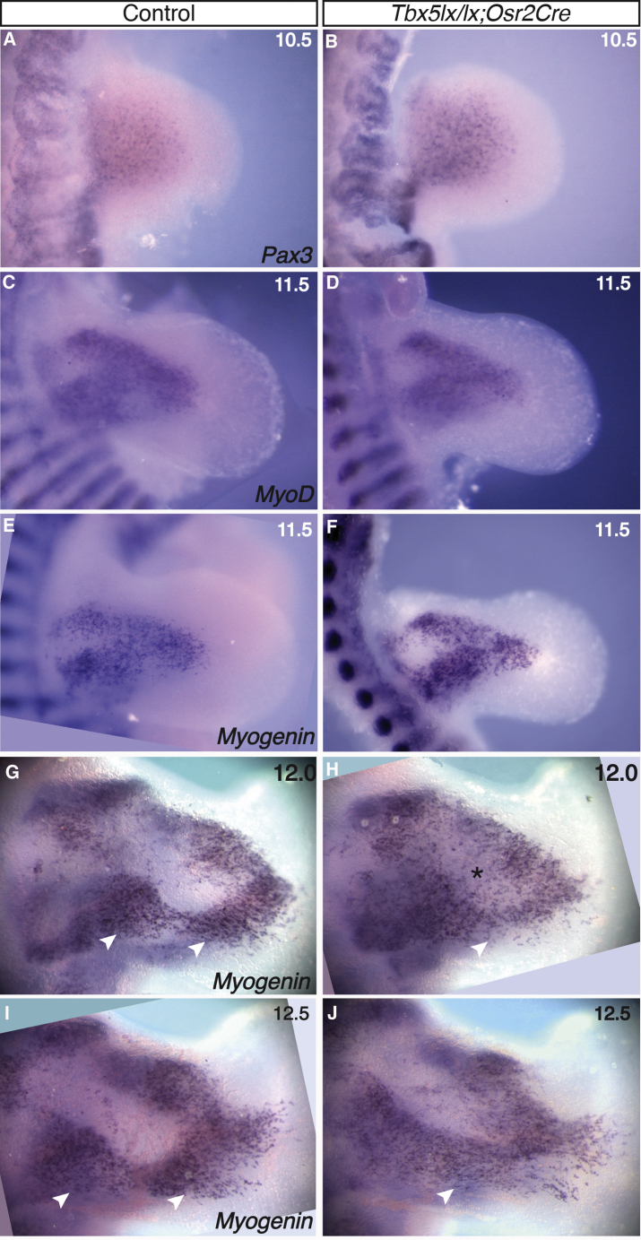
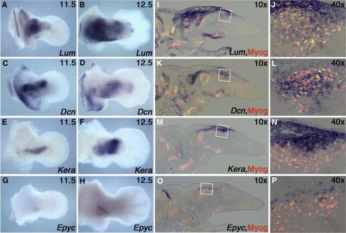
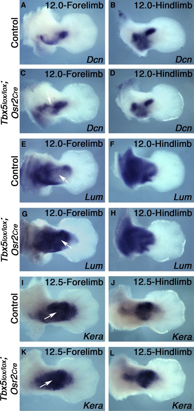
Although the factors regulating muscle cell differentiation are well described, we know very little about how differentiating muscle fibers are organized into individual muscle tissue bundles. Disruption of these processes leads to muscle hypoplasia or dysplasia, and replicating these events is vital in tissue engineering approaches. We describe the progressive cellular events that orchestrate the formation of individual limb muscle bundles and directly demonstrate the role of the connective tissue cells that surround muscle precursors in controlling these events. We show how disruption of gene activity within or genetic ablation of connective tissue cells impacts muscle precursors causing disruption of muscle bundle formation and subsequent muscle dysplasia and hypoplasia. We identify several markers of the populations of connective tissue cells that surround muscle precursors and provide a model for how matrix-modifying proteoglycans secreted by these cells may influence muscle bundle formation by effects on the local extracellular matrix (ECM) environment.

Keywords: HOS; Holt-Oram syndrome; Tbx5; irregular connective tissue; muscle; muscle connective tissue; tissue morphogenesis.


Conflict of interest statement

Declaration of Interests The authors declare no competing interests.

Figures

None
Graphical abstract
Figure 1
Figure 1
Individual Muscle Bundles Form through Progressive Series of Steps Dorsal view of control forelimbs from E10.5 (A) to E14.5 (M) embryos. Muscle progenitors are detected by in situ hybridization for MyoD (A) or Myogenin (B, C, G, H, and I) and immunohistochemistry to detect Myosin (E, F, J, K, L, and M). The limbs are oriented from proximal to distal as shown in the schematic E12.5 forelimb (D). Arrow in (C) shows an area devoid of myogenin positive cells. Arrow in (F) represents the beginning of myocyte fusion. Individual muscles are identified and listed in (M).
Figure 2
Figure 2
Orientation, Clustering, and Compaction of Nascent Muscle Fibers Prefigure Muscle Bundle Formation and Are Disrupted Following Conditional Deletion of Tbx5 (A–D) Dorsal view of E11.5 (A), E11.75 (B), E12.0 (C), and E12.5 (D) control forelimbs with muscle cells detected by Myogenin in situ hybridization to illustrate the region analyzed by immunohistochemistry in (E)–(K). Dotted line squares in (A)–(D) show the approximate area where cell vectors shown in (E)–(H) have been drawn. (E–K) Projection of cell vectors drawn from a Z series of confocal scans of limbs at the stages indicated, stained by whole mount immunohistochemistry for myogenin and myosin in control E11.5 (E), E11.75 (F), E12.0 (G), E12.5 (H), and Tbx5lx/lx;Osr2Cre mutant E11.5 (I), E12.0 (J), and E12.5 (K). The vector lines outline cell orientation. (E'–K') Projection of the color-coded cell vectors for control (E'–H') and mutant (I'–K') forelimbs. Each cell vector has been assigned a color value corresponding to a range of angle values from 0° to 180°, shown on the rainbow ruler.
Figure 3
Figure 3
Cleavage of Muscle Bundle Is a Step in the Morphogenesis of Only Some Muscles Consecutively staged dorsal views of four E13.5 forelimb zeugopods indicate the progressive cleavage of the single extensor carpi radialis (ECR) bundle into two distinct ECR longus and ECR brevis muscles. Muscles (blue/purple) are stained by whole-mount immunohistochemistry to detect Myosin. (A) In the most immature specimen, cleavage of the single extensor carpi radialis (ECR) bundle has started at the distal end (black arrow). (B) Cleavage extends from distal to proximal and is almost complete. (C) Separation of the single bundle into two discrete units is complete at the proximal end of the bundles (black arrows). (D) At the end of the cleavage event, two distinct ECR longus and ECR brevis bundles are formed. Scale bar, 200 μm.
Figure 4
Figure 4
Deletion of Tbx5 by Osr2Cre Produces Muscle and Tendon Patterning Defects Muscles and tendons labeled for myosin and ScxGFP, respectively. Dorsal view of control E13.5 (A and C), E14.5 (E, G, K, M, and O), and E16.5 (I) forelimbs and equivalent stages of mutant E13.5 (B and D), E14.5 (F, H, L, N, and P), and E16.5 (J) forelimbs. Black and white arrows indicate single ECR bundle (H) and a single tendon (B, F, and L) in mutant. Optical projection tomography (OPT) scans of control (K) and mutant (L) limbs at E14.5. Optical dissection of the ECRl, ECRb, EPl, and EPb of the sample shown in (K) (M). Equivalent optical dissection of the mutant shown in (L) showing single ECR and EP bundles (N). Dotted white lines in (M) and (N) show approximate position of optical slice through the 3D reconstruction made to generate the top images in (O) and (P), respectively. Bottom panels in (O) and (P) show rotated views of ECR to show the 2 tendons of ECRl and ECRb in the control and the single ECR tendon in the mutant (asterisks).
Figure 5
Figure 5
Muscle Patterning Defects in Tbx5lx/lx;Osr2Cre Mutants Are Detected at E12.0 Dorsal view of control (A, C, E, G, and I) and mutant (B, D, F, H, and J) forelimbs at E10.5 (A and B), E11.5 (C–F), E12.0 (G and H), and E12.5 (I and J). Muscle cells are detected by in situ hybridization against Pax3 (A and B), MyoD (C and D), or Myogenin (E, F, G, H, I, and J). Arrowheads and asterisks point to clustering defects in the mutant forelimbs (J) compared to the controls (I).
Figure 6
Figure 6
Expression Profiles of SLRP Genes in the Forelimbs Identify ICT Subdomains Dorsal view of wild-type forelimbs at E11.5 (A, C, E, and G) and E12.5 (B, D, F, and H) processed with probes for Lum (A and B), Dcn (C and D), Kera (E and F), and Epyc (G and H) by whole mount in situ hybridization. Co-localization of SLRP expression with muscle cells were detected by section in situ hybridization followed by immunofluorescence for Myogenin at E12.5 (I–P) and shown at 10× magnification (I, K, M, and O). The region boxed in 10× panels is shown at 40× magnification (J, L, N, and P).
Figure 7
Figure 7
SLRP Expression Domains Are Altered in Tbx5lx/lx;Osr2Cre Mutants Dorsal view of wild-type forelimbs (A, E, and I) and hindlimbs (B, F, and J), and mutant forelimbs (C, G, and K) and hindlimbs (D, H, and L) between E12 to E12.5 processed for Dcn (A–D), Lum (E–H), and Kera (I–L) whole mount in situ hybridization. Arrow and arrowhead indicates the ectopic domain of Dcn and absence of its expression in the posterior zeugopodal region in mutant, respectively (C). Arrows in (E) and (I) show the central zeugopodal domain where Lum and Kera are generally excluded or expressed at low levels and arrows in (G) and (K) show the ectopic expression of these SLRPs in the central domain.

References

    1. Abmayr S.M., Pavlath G.K. Myoblast fusion: lessons from flies and mice. Development. 2012;139:641–656. - PMC - PubMed
    1. Ameye L., Aria D., Jepsen K., Oldberg A., Xu T., Young M.F. Abnormal collagen fibrils in tendons of biglycan/fibromodulin-deficient mice lead to gait impairment, ectopic ossification, and osteoarthritis. FASEB J. 2002;16:673–680. - PubMed
    1. Baldock R., Bard J., Brune R., Hill B., Kaufman M., Opstad K., Smith D., Stark M., Waterhouse A., Yang Y., Davidson D. The Edinburgh Mouse Atlas: using the CD. Brief. Bioinform. 2001;2:159–169. - PubMed
    1. Basson C.T., Bachinsky D.R., Lin R.C., Levi T., Elkins J.A., Soults J., Grayzel D., Kroumpouzou E., Traill T.A., Leblanc-Straceski J. Mutations in human TBX5 [corrected] cause limb and cardiac malformation in Holt-Oram syndrome. Nat. Genet. 1997;15:30–35. - PubMed
    1. Brandan E., Gutierrez J. Role of skeletal muscle proteoglycans during myogenesis. Matrix Biol. 2013;32:289–297. - PubMed

Publication types

LinkOut - more resources