. 2020 May;40(5):1220-1230.
doi: 10.1161/ATVBAHA.120.314045. Epub 2020 Mar 12.

Sickle Cell Anemia Mediates Carotid Artery Expansive Remodeling That Can Be Prevented by Inhibition of JNK (c-Jun N-Terminal Kinase)

Affiliations

Sickle Cell Anemia Mediates Carotid Artery Expansive Remodeling That Can Be Prevented by Inhibition of JNK (c-Jun N-Terminal Kinase)

Hannah Song et al. Arterioscler Thromb Vasc Biol. 2020 May.

Abstract

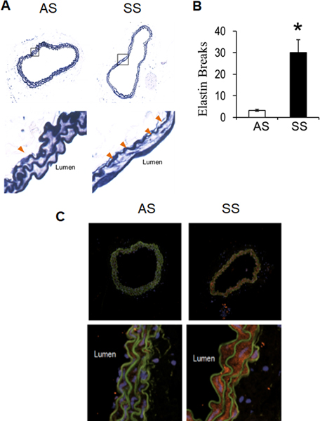
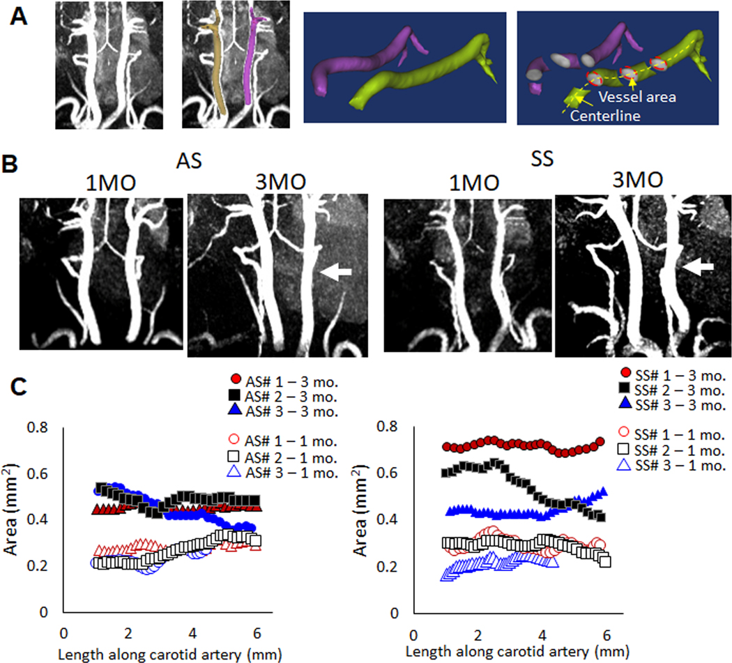
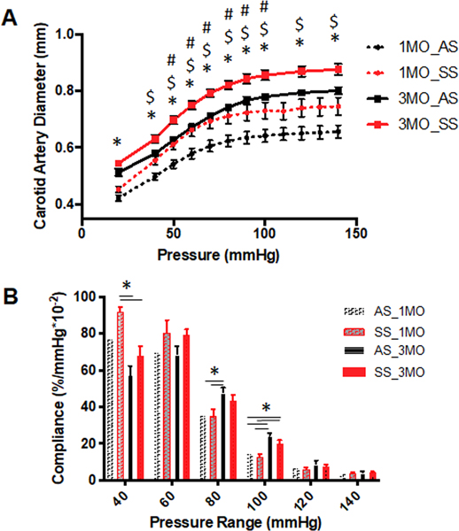
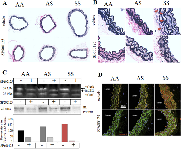
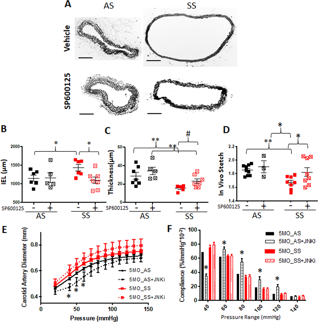
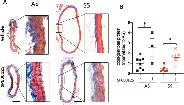
Objective: Sickle cell anemia (SCA) causes chronic inflammation and multiorgan damage. Less understood are the arterial complications, most evident by increased strokes among children. Proteolytic mechanisms, biomechanical consequences, and pharmaceutical inhibitory strategies were studied in a mouse model to provide a platform for mechanistic and intervention studies of large artery damage due to sickle cell disease. Approach and Results: Townes humanized transgenic mouse model of SCA was used to test the hypothesis that elastic lamina and structural damage in carotid arteries increased with age and was accelerated in mice homozygous for SCA (sickle cell anemia homozygous genotype [SS]) due to inflammatory signaling pathways activating proteolytic enzymes. Elastic lamina fragmentation observed by 1 month in SS mice compared with heterozygous littermate controls (sickle cell trait heterozygous genotype [AS]). Positive immunostaining for cathepsin K, a powerful collagenase and elastase, confirmed accelerated proteolytic activity in SS carotids. Larger cross-sectional areas were quantified by magnetic resonance angiography and increased arterial compliance in SS carotids were also measured. Inhibiting JNK (c-jun N-terminal kinase) signaling with SP600125 significantly reduced cathepsin K expression, elastin fragmentation, and carotid artery perimeters in SS mice. By 5 months of age, continued medial thinning and collagen degradation was mitigated by treatment of SS mice with JNK inhibitor.

Conclusions: Arterial remodeling due to SCA is mediated by JNK signaling, cathepsin proteolytic upregulation, and degradation of elastin and collagen. Demonstration in Townes mice establishes their utility for mechanistic studies of arterial vasculopathy, related complications, and therapeutic interventions for large artery damage due to SCA.

Keywords: carotid arteries; collagen; compliance; elastin; hematology.


Figures

Figure 1.
Figure 1.. Sickle cell disease promotes elastin fragmentation and cathepsin K expression in mice by 3 weeks of age.
(A) Perfusion fixed carotid arteries from 3-week AS and SS mice were isolated, sectioned, and stained for elastin morphology using modified Verhoeff elastic-van Gieson stain. (B) elastin fragmentation was quantified along with vessel area with significant increases in the SS mice compared to littermate AS controls. (* denotes p<0.05, n=4) (C) Cathepsin K expression was upregulated in carotid arteries from SS mice.
Figure 2.
Figure 2.. Longitudinal analysis of carotid arteries with magnetic resonance also suggests expansive remodeling in SS carotid arteries compared to AS carotids, but with variability.
(A) Reconstruction diagram. (B) Longitudinal magnetic resonance angiography was used on AS, or SS mice beginning at one month of age and those same mice were again imaged at three months of age to compare animal specific growth and remodeling. White arrows are shown to indicate diameter differences between AS and SS arteries in the representative images shown. C) Reconstructed images were used to measure luminal areas of the carotid arteries and to compare both age-related and genotype-related differences. While the one month carotid artery areas were similar between AS and SS, there was greater variability in the SS diameters by 3 months of age. Numbers indicate the mouse label, and the age to show specific change per animal. Area along the length of the artery are presented. Labeling: AS#1 – 1 month indicates sickle cell genotype (AS), animal replicate number of 3 (#1), and age at which MR imaging was conducted (1 mo = 1 month). The same mouse imaged at 1 month was again imaged at 3 months of age (i.e. AS#1 – 1 mo and AS#1 – 3 mo are images from the same mouse). Circle, square, and triangle represent each mouse, open indicates measurements made at 1 mo. and closed indicates measurements made of that mouse at 3 mo.
Figure 3.
Figure 3.. Carotid arteries from mice with sickle cell disease are more expanded than heterozygous littermate controls.
(A) Biomechanical testing was performed on 1 month and 3 months AS and SS mice to generate pressure-diameter curves from pressures 0 to 150 mmHg, (n=5 for 1 month old, n=9 for 3 months old) (* p<0.05, One-way ANOVA, # p<0.05 followed by Tukey’s multiple comparison test between AS and SS at 1 month of age, or $ p<0.05, at 3-month age) (B) Local compliance differences assessed at increments of 20 mmHg (* p<0.05, One-way ANOVA)
Figure 4.
Figure 4.. JNK inhibition preserves elastic lamina integrity in SS mice.
After an 8-week regimen of SP600125 intraperitoneal injections, 3 month old mice were sacrificed and perfusion fixed carotid arteries from AA, AS, and SS mice were isolated, sectioned, and (A) stained for elastin morphology using modified Verhoeff elastic-van Gieson stain. (B) Zoomed in images are shown as well. (C) Thoracic aortas were prepared for multiplex cathepsin zymography and for c-Jun phosphorylation by Western blot to demonstrate efficacy of SP600125 treatment, and confirmed that SS mice had higher baseline cathepsin activity, that was substantially reduced by JNKi, SP600125. (D) Carotid arteries from 3-month old AA, AS and SS mice were immunostained for cathepsin K. Only SS mice showed strong staining for catK (red); elastin fibers are autofluorescent (green).
Figure 5.
Figure 5.. Expansive remodeling of carotid arteries continues through 5 months of age for sickle cell disease mouse but can be reduced with JNK inhibition.
(A) JNK inhibitor (SP600125, 10mg/kg) was injected daily from age 3 months to 5 months. Then, mice were euthanized and perfusion fixed, carotid arteries excised, and processed for sectioning and stained for Van Giesen elastin stain (scale bar= 100 μm). (B) Internal elastin lamina was traced and length was measured using ImageJ. (C) For thickness measurement, medial thickness from four locations were measured and averaged (n of 4–6 animals per genotype and treatment, * p<0.05, ** p<0.01, Two-way ANOVA followed by Fisher’s LSD, # p<0.05, t-test). (D) Biomechanical testing was performed and in vivo stretch was determined and compared between treatments. (E) Pressure-diameter curves from mechanical testing. (F) Local compliance was calculated from P-D curves (One-way ANOVA followed by Tukey’s multiple comparison test. *p<0.05).
Figure 6.
Figure 6.. Carotid arteries from 5 month old sickle cell disease mouse (SS) lost medial collagen and JNKi treatment rescued it.
(A) The fixed and processed carotid arteries were stained with Mason’s Trichrome Stain (scale bar= 100 mm). (B) Carotid arteries post mechanical testing were homogenized and hydrolyzed using NaOH at 120’c, and measured total collagen using perchlorate free total collagen assay kit (abcam) was normalized to total protein amount. One way ANOVA was used for statistical analysis.

References

    1. Francis RB. Large-vessel occlusion in sickle cell disease: Pathogenesis, clinical consequences, and therapeutic implications. Med Hypotheses. 1991;35:88–95. - PubMed
    1. Adams R, McKie V, Nichols F, Carl E, Zhang DL, McKie K, Figueroa R, Litaker M, Thompson W, Hess D. The use of transcranial ultrasonography to predict stroke in sickle cell disease. N Engl J Med. 1992;326:605–610. - PubMed
    1. Powars D, Wilson B, Imbus C, Pegelow C, Allen J. The natural history of stroke in sickle cell disease. Am J Med. 1978;65:461–471. - PubMed
    1. Merkel KH, Ginsberg PL, Parker JC, Post MJ. Cerebrovascular disease in sickle cell anemia: A clinical, pathological and radiological correlation. Stroke. 1978;9:45–52. - PubMed
    1. Switzer JA, Hess DC, Nichols FT, Adams RJ. Pathophysiology and treatment of stroke in sickle-cell disease: Present and future. Lancet neurology. 2006;5:501–512. - PubMed

Publication types

MeSH terms