Review
. 2020 Mar 11;147(5):dev179325.
doi: 10.1242/dev.179325.

Skeletal stem cells: insights into maintaining and regenerating the skeleton

Affiliations
Review

Skeletal stem cells: insights into maintaining and regenerating the skeleton

Maxwell A Serowoky et al. Development. .

Abstract

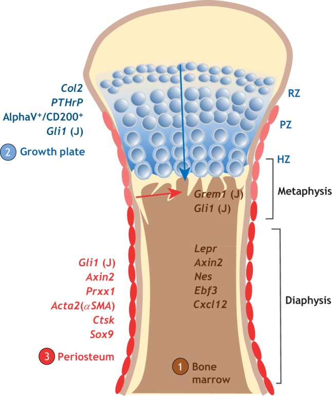
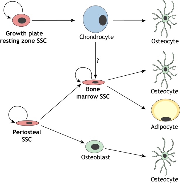
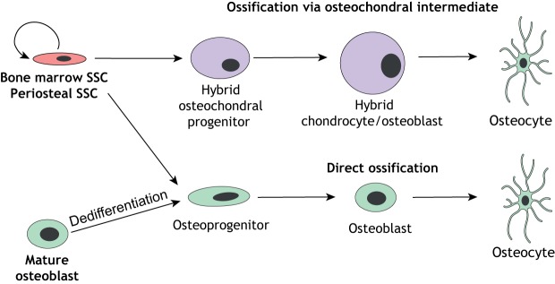
Skeletal stem cells (SSCs) generate the progenitors needed for growth, maintenance and repair of the skeleton. Historically, SSCs have been defined as bone marrow-derived cells with inconsistent characteristics. However, recent in vivo tracking experiments have revealed the presence of SSCs not only within the bone marrow but also within the periosteum and growth plate reserve zone. These studies show that SSCs are highly heterogeneous with regard to lineage potential. It has also been revealed that, during digit tip regeneration and in some non-mammalian vertebrates, the dedifferentiation of osteoblasts may contribute to skeletal regeneration. Here, we examine how these research findings have furthered our understanding of the diversity and plasticity of SSCs that mediate skeletal maintenance and repair.

Keywords: Bone marrow; Growth plate; Periosteum; Skeletal repair; Skeleton; Stem cells.


Conflict of interest statement

Competing interestsThe authors declare no competing or financial interests.

Figures

Fig. 1.
Fig. 1.
Skeletal stem cell populations and niches. Several populations of skeletal stem cells (SSCs) have been identified to date. (1) SSCs can be identified in the marrow cavity (brown) with some populations being enriched in the metaphysis region, particularly at early postnatal or juvenile stages. These populations can be identified using various Cre lines for the genes indicated (with ‘J’ indicating labeling at juvenile stages). (2) SSCs can also be found in the resting zone (RZ) region of the growth plate (blue), expressing the genes indicated. These cells contribute to more lineages than just cartilage. In a growing bone, the chondrocytes of the growth plate proliferate (in the proliferation zone, PZ) and become larger and hypertrophic (within the hypertrophic zone, HZ) near the juncture with the marrow cavity. Some of these cells do not undergo apoptosis but are ejected from the growth plate into the marrow cavity (represented by blue arrow) where they contribute to osteoblasts, adipocytes, other marrow cells, and potentially marrow SSCs. (3) The periosteum (indicated in dark red) is also known to contain SSCs (marked by expression of the genes indicated) involved in homeostasis and repair. During development, progenitor cells within the perichondrium (light red) translocate into the marrow (represented by red arrow) during initial vascularization of the bone.
Fig. 2.
Fig. 2.
Redundant pathways to make bone during development and homeostasis. An SSC (red) in the growth plate resting zone is proposed to self-renew and give rise to hypertrophic chondrocytes (blue) that can undergo transdifferentiation to give rise to osteocytes (green) (Park et al., 2015; Yang et al., 2014; Zhou et al., 2014b) and possibly (indicated by ‘?’) to bone marrow SSCs (Giovannone et al., 2019; Mizuhashi et al., 2019). Bone marrow SSCs can then give rise to osteocytes and adipocytes (yellow) in the bone marrow compartment (Zhou et al., 2014b). Better evidence for the origin of bone marrow SSCs comes from a study (Maes et al., 2010) showing that periosteal/perichondrial SSCs contribute to the marrow compartment during development. Whether this also happens postnatally is not clear. Osteocytes can also arise (via an osteoblast intermediate) from periosteal SSCs at the periosteal surface (Debnath et al., 2018).
Fig. 3.
Fig. 3.
Redundant pathways to make bone during repair. In response to injury, SSCs (red) from the periosteum and/or the bone marrow compartment generate bone through via an osteochondral intermediate (giving rise to cells with cartilage/bone properties, i.e. hybrid osteochondral progenitors, purple) or through direct ossification (giving rise to osteoprogenitors, green). In some contexts, such as the zebrafish fin and murine digit tip, osteoblasts can dedifferentiate and re-differentiate to produce new bone.

Similar articles

Cited by

References

    1. Akiyama H., Kim J.-E., Nakashima K., Balmes G., Iwai N., Deng J. M., Zhang Z., Martin J. F., Behringer R. R., Nakamura T. et al. (2005). Osteo-chondroprogenitor cells are derived from Sox9 expressing precursors. Proc. Natl. Acad. Sci. USA 102, 14665-14670. 10.1073/pnas.0504750102 - DOI - PMC - PubMed
    1. Allen M. R., Hock J. M. and Burr D. B. (2004). Periosteum: biology, regulation, and response to osteoporosis therapies. Bone 35, 1003-1012. 10.1016/j.bone.2004.07.014 - DOI - PubMed
    1. Alman B. A. (2015). The role of hedgehog signalling in skeletal health and disease. Nat Rev Rheumatol 11, 552-560. 10.1038/nrrheum.2015.84 - DOI - PubMed
    1. Ando K., Shibata E., Hans S., Brand M. and Kawakami A. (2017). Osteoblast production by reserved progenitor cells in zebrafish bone regeneration and maintenance. Dev. Cell 43, 643-650.e643. 10.1016/j.devcel.2017.10.015 - DOI - PubMed
    1. Baker M. (2005). Stem cell therapy or snake oil? Nat. Biotechnol. 23, 1467-1469. 10.1038/nbt1205-1467 - DOI - PubMed

Publication types