

Neurophysiological signatures in Alzheimer's disease are distinctly associated with TAU, amyloid-β accumulation, and cognitive decline
- PMID: 32161102
- PMCID: PMC7138514
- DOI: 10.1126/scitranslmed.aaz4069
Neurophysiological signatures in Alzheimer's disease are distinctly associated with TAU, amyloid-β accumulation, and cognitive decline
Abstract
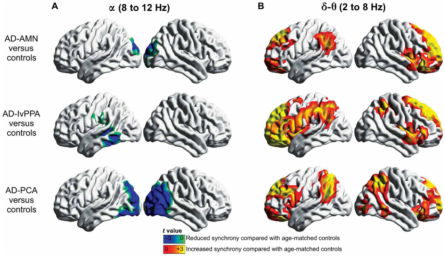
Neural synchrony is intricately balanced in the normal resting brain but becomes altered in Alzheimer's disease (AD). To determine the neurophysiological manifestations associated with molecular biomarkers of AD neuropathology, in patients with AD, we used magnetoencephalographic imaging (MEGI) and positron emission tomography with amyloid-beta (Aβ) and TAU tracers. We found that alpha oscillations (8 to 12 Hz) were hyposynchronous in occipital and posterior temporoparietal cortices, whereas delta-theta oscillations (2 to 8 Hz) were hypersynchronous in frontal and anterior temporoparietal cortices, in patients with AD compared to age-matched controls. Regional patterns of alpha hyposynchrony were unique in each neurobehavioral phenotype of AD, whereas the regional patterns of delta-theta hypersynchrony were similar across the phenotypes. Alpha hyposynchrony strongly colocalized with TAU deposition and was modulated by the degree of TAU tracer uptake. In contrast, delta-theta hypersynchrony colocalized with both TAU and Aβ depositions and was modulated by both TAU and Aβ tracer uptake. Furthermore, alpha hyposynchrony but not delta-theta hypersynchrony was correlated with the degree of global cognitive dysfunction in patients with AD. The current study demonstrates frequency-specific neurophysiological signatures of AD pathophysiology and suggests that neurophysiological measures from MEGI are sensitive indices of network disruptions mediated by TAU and Aβ and associated cognitive decline. These findings facilitate the pursuit of novel therapeutic approaches toward normalizing network synchrony in AD.
Copyright © 2020 The Authors, some rights reserved; exclusive licensee American Association for the Advancement of Science. No claim to original U.S. Government Works.
Conflict of interest statement
Figures


References
-
- Selkoe DJ, Alzheimer’s disease is a synaptic failure. Science 298, 789–791 (2002). - PubMed
-
- Terry RD, Masliah E, Salmon DP, Butters N, DeTeresa R, Hill R, Hansen LA, Katzman R, Physical basis of cognitive alterations in Alzheimer’s disease: Synapse loss is the major correlate of cognitive impairment. Ann. Neurol 30, 572–580 (1991). - PubMed
-
- Alzheimer’s Association, 2018 Alzheimer’s disease facts and figures. Alzheimers Dement. 14, 367–425 (2018).
-
- Busche MA, Eichhoff G, Adelsberger H, Abramowski D, Wiederhold K-H, Haass C, Staufenbiel M, Konnerth A, Garaschuk O, Clusters of hyperactive neurons near amyloid plaques in a mouse model of Alzheimer’s disease. Science 321, 1686–1689 (2008). - PubMed
-
- Ahnaou A, Moechars D, Raeymaekers L, Biermans R, Manyakov NV, Bottelbergs A, Wintmolders C, Van Kolen K, Van De Casteele T, Kemp JA, Drinkenburg WH, Emergence of early alterations in network oscillations and functional connectivity in a tau seeding mouse model of Alzheimer’s disease pathology. Sci. Rep 7, 14189 (2017). - PMC - PubMed
Publication types
MeSH terms
Substances
Grants and funding
- K08 AG058749/AG/NIA NIH HHS/United States
- U01 AG052943/AG/NIA NIH HHS/United States
- P30 AG062422/AG/NIA NIH HHS/United States
- RF1 AG062196/AG/NIA NIH HHS/United States
- R01 NS100440/NS/NINDS NIH HHS/United States
- R01 DC013979/DC/NIDCD NIH HHS/United States
- R01 AG034570/AG/NIA NIH HHS/United States
- P50 AG023501/AG/NIA NIH HHS/United States
- K23 AG038357/AG/NIA NIH HHS/United States
- R01 EB022717/EB/NIBIB NIH HHS/United States
- R21 NS076171/NS/NINDS NIH HHS/United States
- P01 AG019724/AG/NIA NIH HHS/United States
- R56 NS050915/NS/NINDS NIH HHS/United States
- R01 NS050915/NS/NINDS NIH HHS/United States
- R01 AG045611/AG/NIA NIH HHS/United States
- K24 DC015544/DC/NIDCD NIH HHS/United States
- F32 AG050434/AG/NIA NIH HHS/United States
- R01 NS066654/NS/NINDS NIH HHS/United States
LinkOut - more resources
Full Text Sources
Other Literature Sources
Medical
