. 2020 Apr 17;295(16):5496-5508.
doi: 10.1074/jbc.RA119.010467. Epub 2020 Mar 11.

Repression of sphingosine kinase (SK)-interacting protein (SKIP) in acute myeloid leukemia diminishes SK activity and its re-expression restores SK function

Affiliations

Repression of sphingosine kinase (SK)-interacting protein (SKIP) in acute myeloid leukemia diminishes SK activity and its re-expression restores SK function

Essam A Ghazaly et al. J Biol Chem. .

Abstract

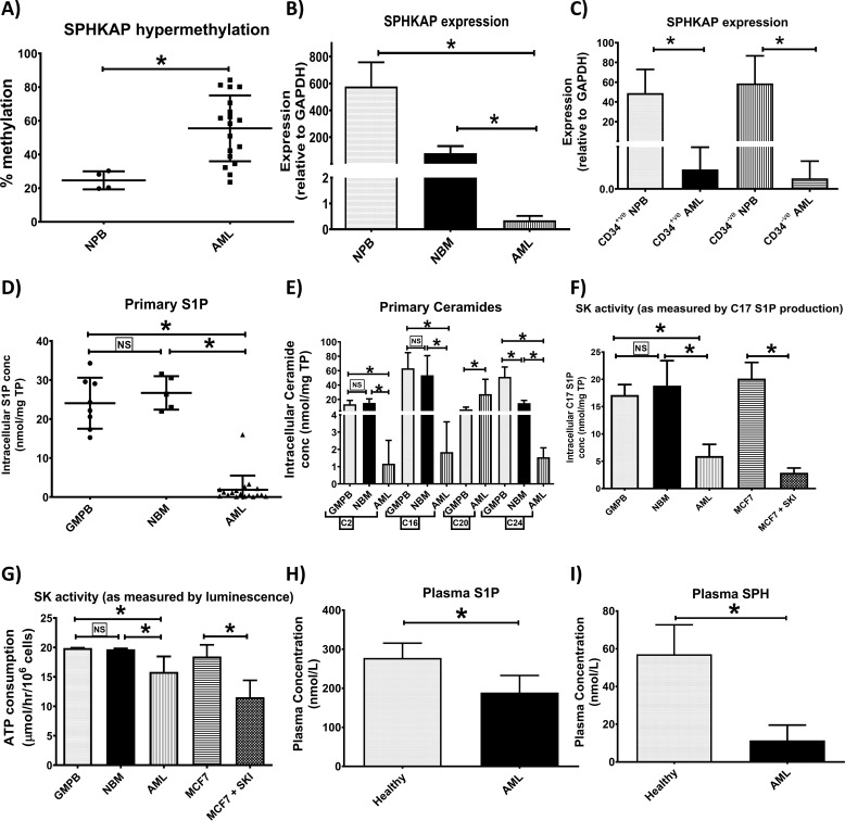
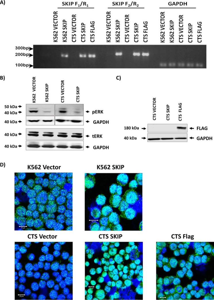
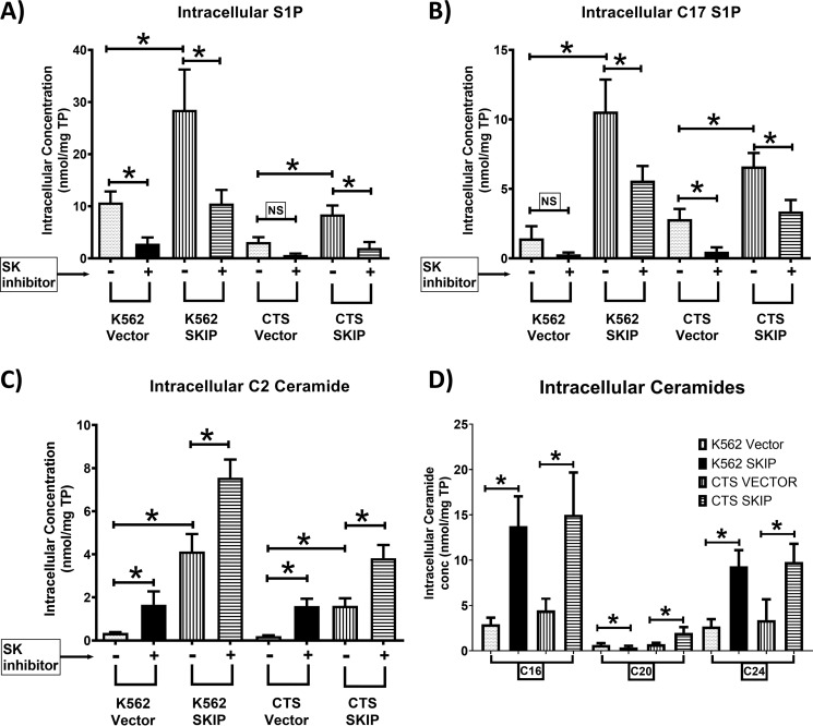
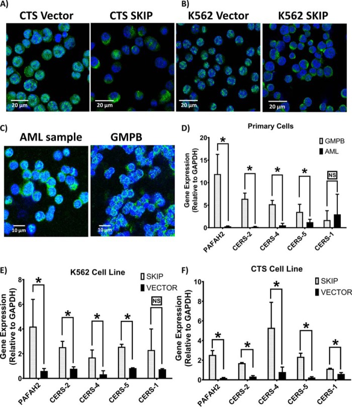
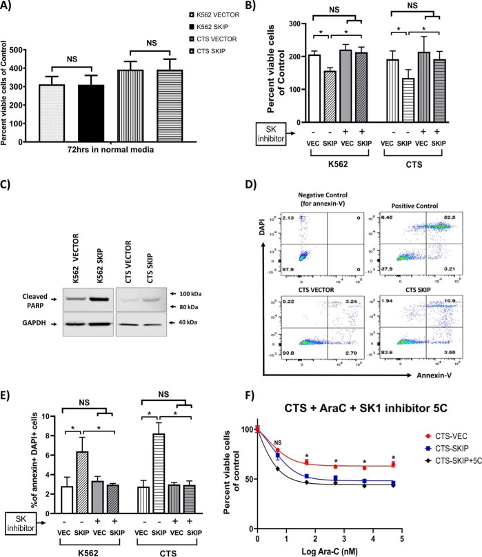
Previous studies have shown that sphingosine kinase interacting protein (SKIP) inhibits sphingosine kinase (SK) function in fibroblasts. SK phosphorylates sphingosine producing the potent signaling molecule sphingosine-1-phosphate (S1P). SKIP gene (SPHKAP) expression is silenced by hypermethylation of its promoter in acute myeloid leukemia (AML). However, why SKIP activity is silenced in primary AML cells is unclear. Here, we investigated the consequences of SKIP down-regulation in AML primary cells and the effects of SKIP re-expression in leukemic cell lines. Using targeted ultra-HPLC-tandem MS (UPLC-MS/MS), we measured sphingolipids (including S1P and ceramides) in AML and control cells. Primary AML cells had significantly lower SK activity and intracellular S1P concentrations than control cells, and SKIP-transfected leukemia cell lines exhibited increased SK activity. These findings show that SKIP re-expression enhances SK activity in leukemia cells. Furthermore, other bioactive sphingolipids such as ceramide were also down-regulated in primary AML cells. Of note, SKIP re-expression in leukemia cells increased ceramide levels 2-fold, inactivated the key signaling protein extracellular signal-regulated kinase, and increased apoptosis following serum deprivation or chemotherapy. These results indicate that SKIP down-regulation in AML reduces SK activity and ceramide levels, an effect that ultimately inhibits apoptosis in leukemia cells. The findings of our study contrast with previous results indicating that SKIP inhibits SK function in fibroblasts and therefore challenge the notion that SKIP always inhibits SK activity.

Keywords: Acute myeloid leukemia; Sphingosine kinase (SphK); cell signaling; ceramide; chemotherapy; cytarabine; hypermethylation; leukemia; lipid metabolism; sphingolipid; sphingosine kinase interacting protein; sphingosine-1-phosphate (S1P).


Conflict of interest statement

The authors declare that they have no conflicts of interest with the contents of this article

Figures

Figure 1.
Figure 1.
S1P and ceramides are down-regulated in AML due to SK hypofunction. Using bisulfite pyrosequencing, SPHKAP (the gene that produces SKIP) hypermethylation was confirmed in primary AML (n = 18) compared with NPB (n = 4) samples (A). SKIP underexpression was confirmed in blood samples from patients with AML (n = 18) compared with healthy volunteer NPB (n = 4) and normal bone marrow samples (NBM, n = 5) as studied by qPCR (B). Reduced SKIP expression involved both CD34+ and CD34 components of AML primary samples (n = 4) compared with NBP (n = 4) (C) as studied by qPCR. Scatter plots show lower S1P (D) and ceramide (E) concentrations in primary AML cells (n = 18) versus NBM (n = 5) and G-mobilized peripheral blood cells (GMPB) (n = 8). Bar charts show lower SK function as measured by UPLC-MS/MS detection of C17 S1P production (F) after 24 h incubation with 1 μm C17 sphingosine substrate in primary AML cells (n = 6) versus NBM (n = 5) and GMPB (n = 6) MCF7 cell line was used as positive control and 10 μm SKI 5C was used to inhibit SK activity. Lower SK function in primary AML cells (n = 18) versus NBM (n = 3) and GMPB (n = 3) was confirmed using another method for measuring SK activity depending on ELISA detection of ATP consumption due to SK enzymatic activity (G). Cell lysate from the MCF7 cell line was used as source for SK enzyme (positive control) and 10 μm SKI 5C was used to inhibit SK activity. Plasma S1P (H) and sphingosine (I) concentrations were lower in AML patients (n = 15) versus healthy volunteers (n = 5) as measured by UPLC-MS/MS. * = p < 0.05; NS, not significant (p > 0.05) as measured by t test.
Figure 2.
Figure 2.
Successful transfection and re-expression of SKIP protein in K562 and CTS cell lines. A, SKIP gene expression in SKIP-transfected cells was confirmed by RT-PCR using two different primers (F1/R1 and F2/R2), GAPDH expression was used as control for loading. B, Western blots show low pERK phenotype associated with SKIP re-expression in K562 and CTS cell lines and equal tERK expression in the two cell lines. C, Western blotting confirming the expression of FLAG-tagged SKIP protein in FLAG-tagged transfected CTS cell line. D, immunofluorescent detection of SKIP using anti-SKIP antibody and confocal microscopy in SKIP-transfected versus vector alone-transfected K562 and CTS cell lines. Fluorescence was detected using a Zeiss LSM 510 META, confocal microscope system. All immunofluorescent images were taken at a magnification of ×40, scale bar = 10 μm.
Figure 3.
Figure 3.
SKIP overexpression in leukemia cell lines increases S1P and ceramide levels and SK activity. A, higher SIP concentrations in SKIP-transfected leukemia cell lines (n = 4) compared with vector alone (n = 4) as detected by UPLC-MS/MS. The SK1 inhibitor 5C reduced S1P concentrations. B, higher SK activity in SKIP-transfected (n = 4) compared with vector-alone transfected (n = 4) leukemia cells as measured by the ability of the cells to produce C17 S1P production after 24 h incubation with C17 sphingosine substrate (n = 4). The SK1 inhibitor partially reversed the increased SK activity in SKIP-transfected cells. C, C2 ceramide was significantly higher in SKIP-transfected cells (n = 5) and cells cultured with the SK1 inhibitor 5C were compared with vector-alone transfected cells (n = 5). D, the intracellular long chain (C16, C20, and C24) ceramide levels are shown in SKIP-transfected cell lines (n = 4) compared with vector-transfected cells (n = 4) as detected by UPLC-MS/MS ceramide 14 and C18 were undetectable. * = p < 0.05; NS, not significant (p > 0.05) as measured by t test.
Figure 4.
Figure 4.
SKIP protein localizes SK to the cell cytoplasm and increases expression of ceramide synthases in SKIP-transfected cells. SK1 shows localized expression to cell cytoplasm in SKIP-transfected CTS (A) and K562 (B) versus nuclear expression in vector-alone transfected leukemia cell lines. C, SK1 expression shows nuclear localization in AML primary cells, whereas localized to the cell cytoplasm in GMPB cells. Fluorescence was detected using a Zeiss LSM 510 META confocal microscope system. All immunofluorescent images were taken at a magnification of ×40 with each scale bar = 10 m or 20 μm as indicated in each panel. D, lower expression of the genes encoding for ceramide synthases (CERSs) and platelet activating factor acetylhydrolase (PAFAH2) in primary AML samples (n = 11) compared with GMPB (n = 6) except for CERS-1 as determined by RT-qPCR. Lower expression of PAFAH2 and CERSs (except for CERS-1) genes in vector-alone transfected (n = 3) K562 (E) and CTS (F) compared with SKIP-transfected leukemia cell line (n = 3). * = p < 0.05; NS, not significant (p > 0.05) as measured by t test.
Figure 5.
Figure 5.
SKIP re-expression is associated with increased apoptotic signals. A, SKIP transfection did not affect growth of CTS and K562 cells under normal conditions. B, after 24 h serum starvation there were significantly less viable SKIP-transfected cells than vector-transfected cells for both K562 and CTS cells (n = 3). The SK1 inhibitor 5C reversed the pro-apoptotic effect of serum starvation in SKIP-transfected cells. C, Western blots showing higher expression of cleaved PARP (89-kDa fragment) in SKIP-transfected leukemia cell lines compared with vector-alone after 24 h of serum starvation. D, flow cytometry plots showing staining of cells with DAPI and Annexin-V (Annexin-V binds apoptotic cells). In the top left panel is the negative control for Annexin V. In the top right panel is the positive control (irradiated cells) with a high proportion of apoptotic cells (52%). In the bottom panels are vector- and SKIP-transfected CTS cells showing 3.2 and 10.9% apoptotic cells, respectively. E, a higher percentage of cells were apoptotic (positive for both Annexin V and DAPI stains) in SKIP-transfected leukemia cell lines exposed to 24 h of serum starvation compared with vector-alone cells (n = 3). The proapoptotic effect of SKIP transfection was reversed by the SK1 inhibitor 5C in the context of serum starvation. F, SKIP-transfected CTS cells were more sensitive to ara-c chemotherapy than vector-transfected cells (n = 3). The SK1 inhibitor 5C did not reverse the chemosensitivity to ara-c. * = p < 0.05 as measured by unpaired t test.

References

    1. Lacaná E., Maceyka M., Milstien S., and Spiegel S. (2002) Cloning and characterization of a protein kinase A anchoring protein (AKAP)-related protein that interacts with and regulates sphingosine kinase 1 activity. J. Biol. Chem. 277, 32947–32953 10.1074/jbc.M202841200 - DOI - PubMed
    1. Spiegel S., and Milstien S. (2003) Sphingosine-1-phosphate: an enigmatic signalling lipid. Nat. Rev. Mol. Cell Biol. 4, 397–407 10.1038/nrm1103 - DOI - PubMed
    1. Olivera A., and Spiegel S. (1993) Sphingosine-1-phosphate as second messenger in cell proliferation induced by PDGF and FCS mitogens. Nature 365, 557–560 10.1038/365557a0 - DOI - PubMed
    1. Xia P., Gamble J. R., Wang L., Pitson S. M., Moretti P. A., Wattenberg B. W., D'Andrea R. J., and Vadas M. A. (2000) An oncogenic role of sphingosine kinase. Curr. Biol. 10, 1527–1530 10.1016/S0960-9822(00)00834-4 - DOI - PubMed
    1. Morita Y., Perez G. I., Paris F., Miranda S. R., Ehleiter D., Haimovitz-Friedman A., Fuks Z., Xie Z., Reed J. C., Schuchman E. H., Kolesnick R. N., and Tilly J. L. (2000) Oocyte apoptosis is suppressed by disruption of the acid sphingomyelinase gene or by sphingosine-1-phosphate therapy. Nat. Med. 6, 1109–1114 10.1038/80442 - DOI - PubMed

Publication types

MeSH terms

Substances