Review
. 2020 Apr;21(4):213-229.
doi: 10.1038/s41583-020-0278-2. Epub 2020 Mar 11.

Presynaptic calcium channels: specialized control of synaptic neurotransmitter release

Affiliations
Review

Presynaptic calcium channels: specialized control of synaptic neurotransmitter release

Annette C Dolphin et al. Nat Rev Neurosci. 2020 Apr.

Abstract

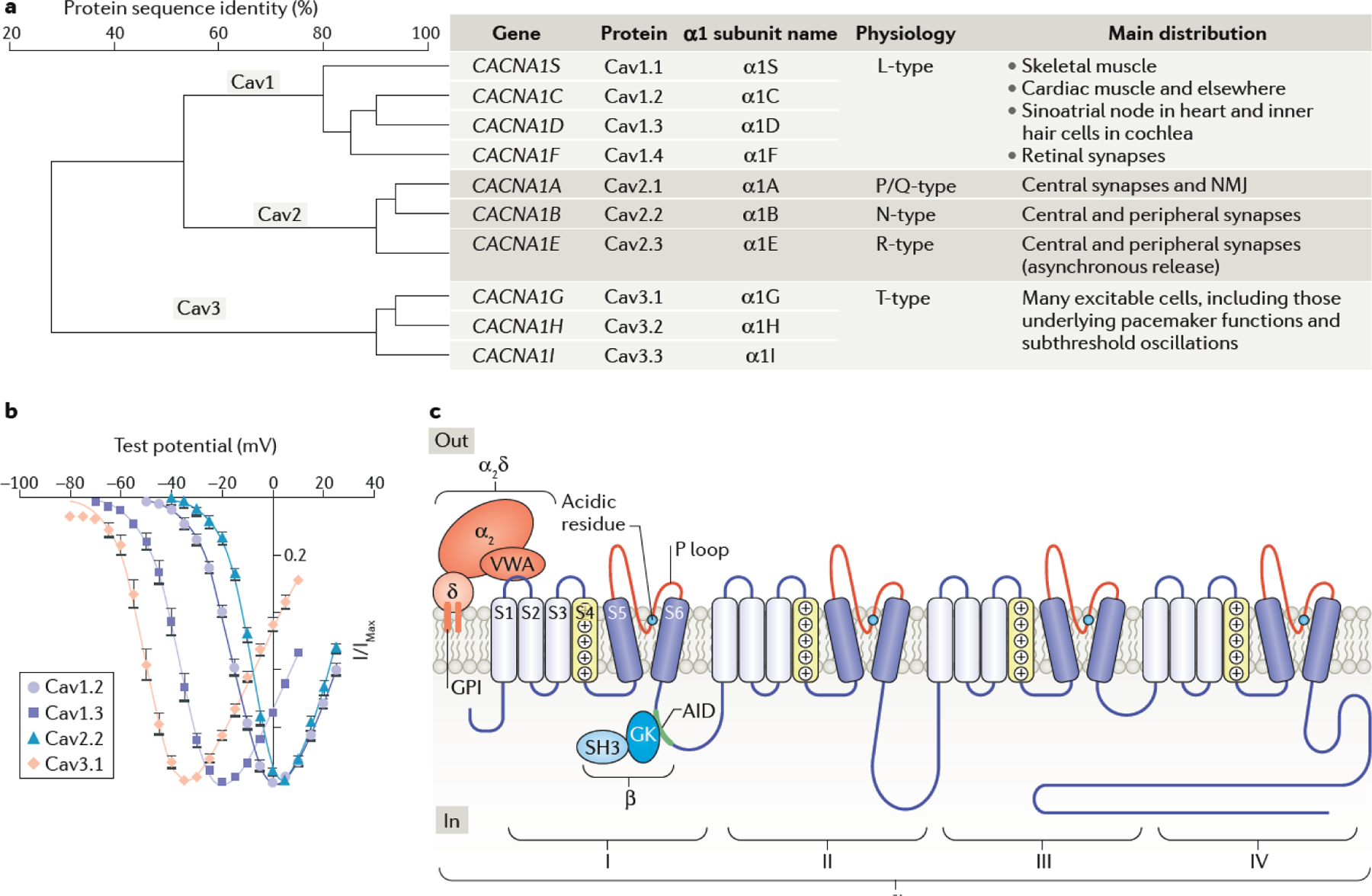
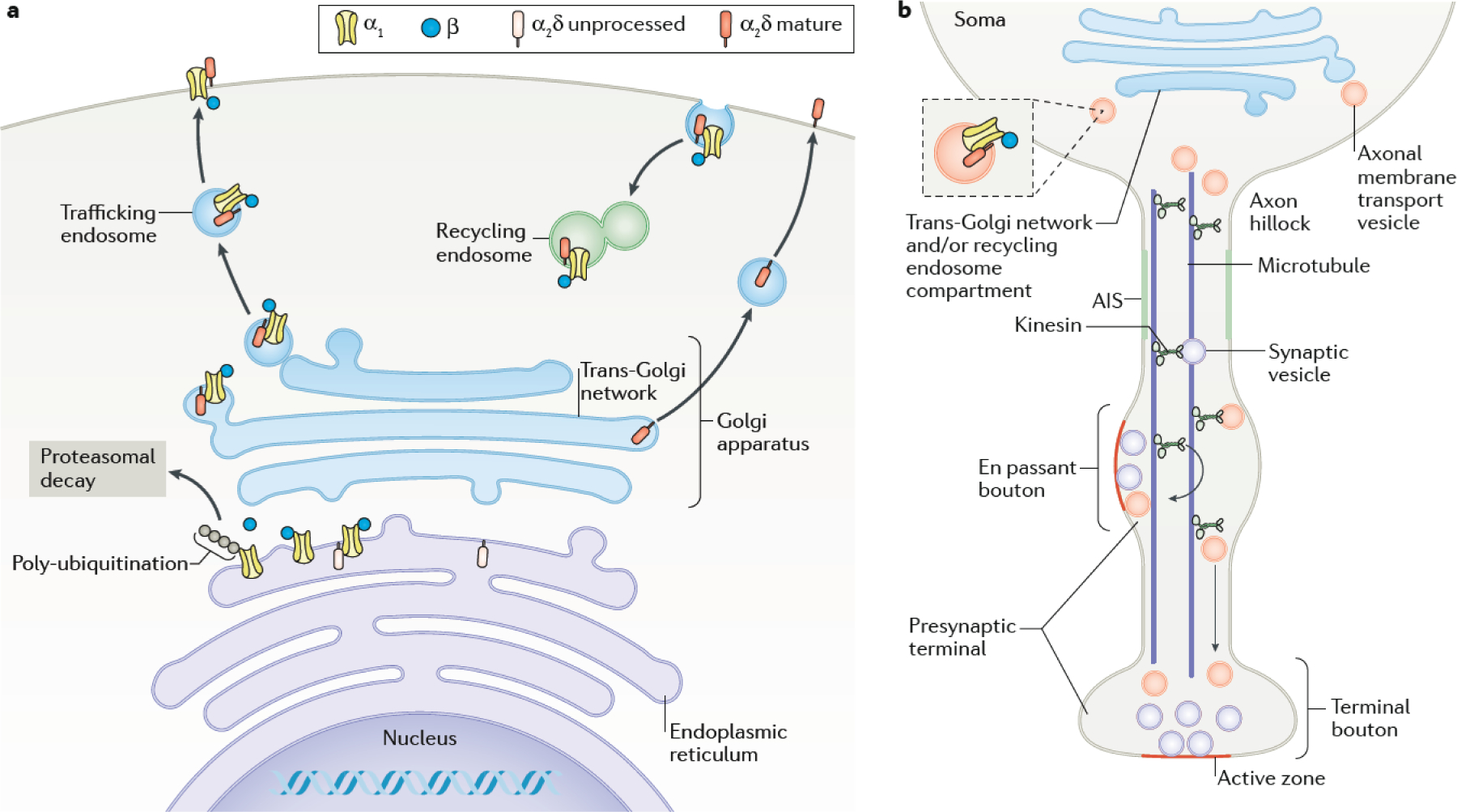
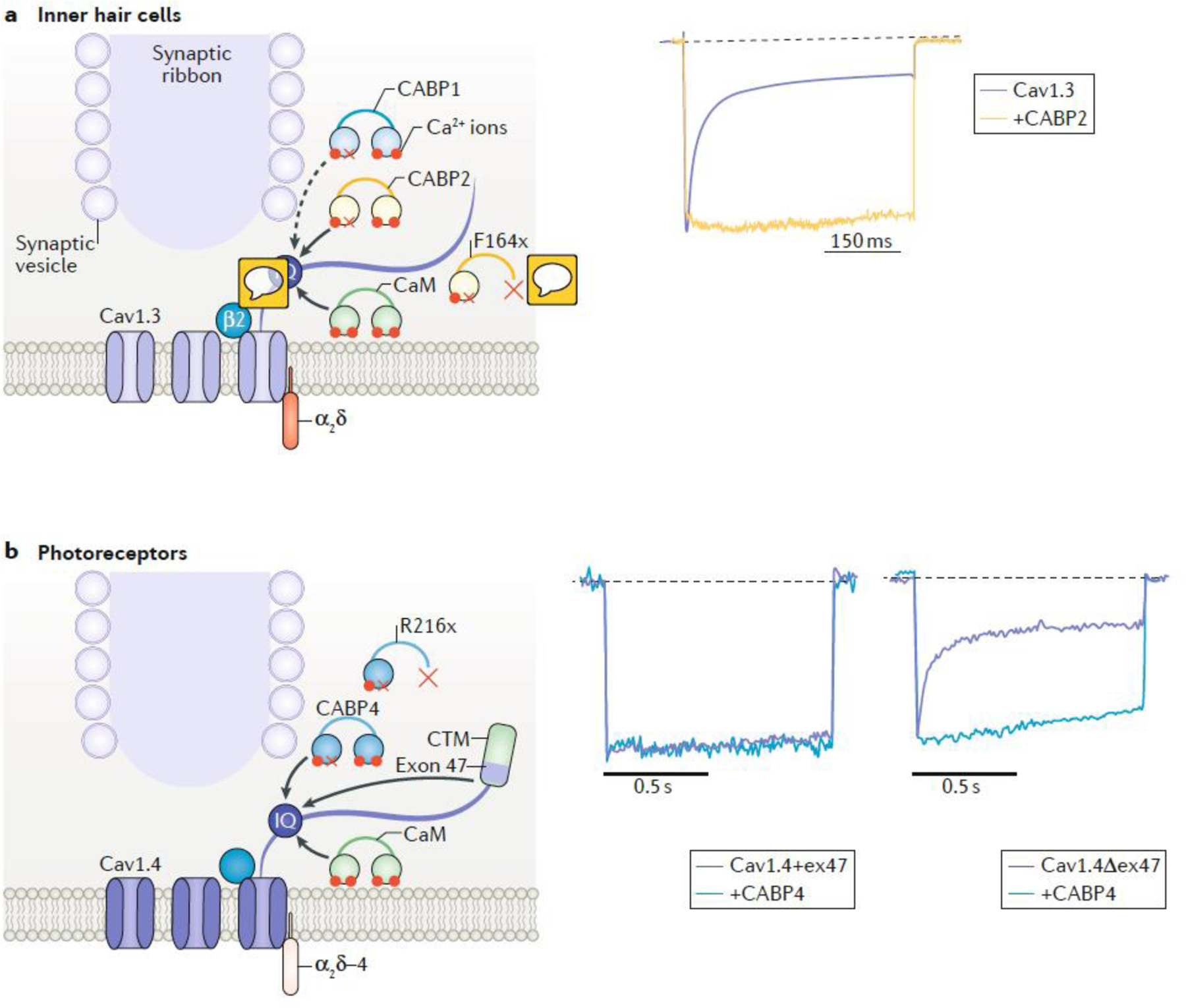
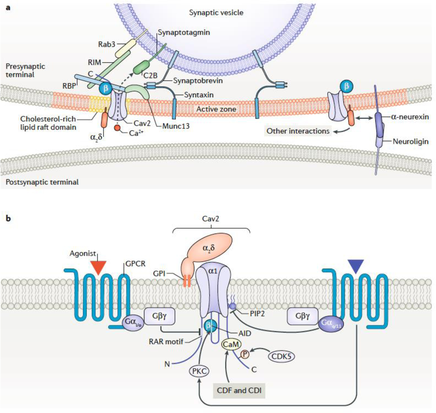
Chemical synapses are heterogeneous junctions formed between neurons that are specialized for the conversion of electrical impulses into the exocytotic release of neurotransmitters. Voltage-gated Ca2+ channels play a pivotal role in this process as they are the major conduits for the Ca2+ ions that trigger the fusion of neurotransmitter-containing vesicles with the presynaptic membrane. Alterations in the intrinsic function of these channels and their positioning within the active zone can profoundly alter the timing and strength of synaptic output. Advances in optical and electron microscopic imaging, structural biology and molecular techniques have facilitated recent breakthroughs in our understanding of the properties of voltage-gated Ca2+ channels that support their presynaptic functions. Here we examine the nature of these channels, how they are trafficked to and anchored within presynaptic boutons, and the mechanisms that allow them to function optimally in shaping the flow of information through neural circuits.


Conflict of interest statement

Competing interests

The authors declare no competing interests.

Figures

Fig. 1|
Fig. 1|. Cav channel nomenclature and properties
a| Schematic showing Cav channel homology (the % identity between protein sequence of the difference Cav channel isoforms), human genetic nomenclature and protein classification. The channels are divided into 3 main groups, CaV1, CaV2 and CaV3, based on homology, and then subdivided according to the individual gene products. The original name for each of the cloned α1 subunits, as well as the names derived from electrophysiological experiments are also shown. The tissue distribution and main functions of each isoform are listed. b| Normalized current-voltage relationships for Cav currents recorded from tsA201 cells. A comparison of the activation voltage ranges of Cav3.1, Cav1.3, Cav1.2 and Cav2.2 channels shows that there is a continuum of activation voltages for the different channels, rather than a clear division into low voltage activated (LVA) and high voltage activated (HVA) channels–,–. c| Schematic illustrating Cav channel subunit interactions,,. The α1 subunit, contains four homologous domains (I-IV) each with 6 transmembrane segments (S1-S6), an S4 voltage sensor (yellow), containing a motif of positively charged amino acid residues, and a P-loop between S5 and S6 (red segments), which comprise the pore domain, containing key acidic residues (generally glutamate, cyan circles) involved in the selectivity filter. The α2δ subunit is shown with its GPI anchor linking it into the membrane and von Willebrand factor A (VWA) domain binding to the first extracellular loop of the α1 subunit. The two domains of the β subunit (Src homology domain (SH3) and guanylate kinase-like domain (GK)) are also shown, with the GK interacting with the α1-interaction domain (AID) motif on the I-II linker intracellular loop. Part b is reproduced, with permission from ref. NMJ, neuromuscular junction.
Fig. 2|
Fig. 2|. Synthesis and trafficking of Cav channels
a| The synthesis of the α1 and α2δ subunits of Cav2 channels occurs on endoplasmic reticulum-associated ribosomes. The unprocessed form of α2δ is therefore synthesised entirely within the endoplasmic reticulum and attached to the endoplasmic reticulum membrane by a GPI anchor. In the endoplasmic reticulum, the α1 subunit associates with the ß subunit, which is a cytoplasmic protein. This interaction protects the α1 subunit from polyubiquitination and ER-associated degradation via the proteasome. The α2δ subunits are heavily glycosylated in the endoplasmic reticulum (there are up to 18 N-glycosylation sites to which glycans are attached) and the glycosyl moieties are further processed in the Golgi apparatus, which is also probably the main site of its proteolytic cleavage into α2 and δ to form the mature protein. α2δ proteins may associate with the α1 and ß subunit complex in the endoplasmic reticulum or in the Golgi apparatus. The complex is subsequently transported in trafficking endosomes and incorporated into the plasma membrane by fusion. The calcium channel complexes are also subject to endocytosis and are recycled to the plasma membrane via recycling endosomes,. The α2δ proteins are also able to reach the cell surface alone. b| The transport of axonal membrane transport vesicles containing Cav channels destined for active zone membranes involves binding of axonal cargo-containing transport vesicles (axonal endosomes) from the trans-Golgi network / recycling endosome compartment to microtubules in the pre-axonal exclusion zone at the axon hillock,. The axon initial segment (AIS) represents a specialized region in which particular Na+ and K+ channels are concentrated, and action potentials are initiated, which may also restrict axonal trafficking. The cargo destined for en passant or terminal boutons is attached by axonal kinesins to axonal microtubules, and is released from the microtubules at presynaptic sites, as are synaptic vesicles .
Fig. 3|
Fig. 3|. Regulation of Cav1 channel inactivation at ribbon synapses.
The schematics show the location of Cav1 channels at the active zones of inner hair cells (IHCs) and photoreceptors near the synaptic ribbon. The traces on the right show the normalized Ca2+ current recorded from HEK293T cells expressing Cav1 channels alone or Cav1 channels together with a calcium-binding protein (CABP). a| In many cell-types, Cav1 channels are thought to be constitutively associated with calmodulin (CaM) which is bound to an IQ-domain (IQ) in the C-terminal domain of the channel. The binding of 4 Ca2+ ions to the N- and C-terminal lobes of CaM triggers a conformational change in the channel that favors Ca2+-dependent inactivation (CDI) (reviewed in). In IHCs, Cav1.3 channels exhibit limited inactivation in comparison to that present in other cell types,. This is due in part to the co-assembly of Cav1.3 with β2 subunits, which cause slow voltage-dependent inactivation. In addition, CABP1 and/or CABP2 are Ca2+ binding proteins with a dysfunctional Ca2+ binding site in the N-terminal lobe (red x). These CABPs are thought to compete with CaM for binding to the channel thus suppressing the effects of CaM on inactivation. Right panel shows that Ca2+ currents inactivate more slowly in cells cotransfected with Cav1.3 and CABP2 than in those transfected with Cav1.3 alone,. CABP1 has a similar effect (not shown) ,. A mutation (F164X) that causes premature truncation of CABP2 inhibits its ability to suppress inactivation of Cav1.3 and causes autosomal recessive hearing loss. b|In photoreceptor terminals, Cav1.4 channels, which are associated with β2 and extracellular α2δ−4 subunits, show little CDI. Channels containing exon 47 (Cav1.4+ex47) possess a C-terminal modulatory domain (CTM) that is thought to compete with CaM for binding to the channel (left current trace). Splice variants lacking exon 47 (Cav1.4Δex47) show stronger CDI, which may be due to a reduced ability of the CTM to compete with CaM for binding to the channel (right purple current trace). CABP4 slows inactivation of Cav1.4Δex47 but not of Cav1.4+ex47, possibly because the loss of exon 47 enables CaBP4 to compete effectively with CaM for binding to the channel. A mutation (R216X) that causes premature truncation of CABP4 inhibits its ability to suppress CDI of Cav1.4 and causes vision impairment in humans. Trace in part a is adapted, with permission from. Traces in part b are adapted, with permission from ref .
Fig. 4|
Fig. 4|. Organization and modulation of Cav2 channels in synapses
a| Schematic depicts some of the proteins known to be involved in anchoring the Cav2 channels sufficiently near to synaptic vesicles to form a nanodomain within the presynaptic active zone (for reviews see ,). These include Rab3, synaptotagmin (the major Ca2+ sensor, with one of its C2 domains shown) and synaptobrevin, all of which are associated with the vesicular membrane. Rab3-interacting molecules (RIM) and RIM binding proteins (RBP) are cytosolic, whereas Munc13 and syntaxin are associated with the plasma membrane. The association of the Cav ß subunit with the channel is shown. CaVß interacts with RBP and also with another scaffolding protein, CAST/ELKS (not shown). The α2δ subunit is extracellular, and may (via its GPI anchor) preferentially associate with cholesterol rich lipid raft membrane domains. It also mediates effects on Cav channels of α-neurexins, which also interact with neuroligin (postsynaptic except in Caenorhabditis elegans). Other interactions of α2δ subunits are also likely to occur at the synapse but have not been depicted for clarity. b| Schematic showing some of the pathways modulating CaV2 calcium channel function. Some G protein-coupled receptors (GPCRs) inhibit CaV2 channel activity via their Gßγ subunits. This requires the presence of a conserved RAR motif in the N-terminal sequence of the channel, and also involves Cav ß binding to the I-II linker,. GPCRs coupled to Gq/11 inhibit Cav2 channels by reducing levels of PIP2 (and activating PKC, which phosphorylates the AID and elsewhere). Modulation involving the C-terminal domain of Cav2 channels includes CDK5-mediated phosphorylation of a conserved serine of CaV2.2, which increases channel open probability. Ca2+-dependent inactivation (CDI) (in Cav2.2 and Cav2.1) and Ca2+-dependent facilitation (CDF (in CaV2.1) are mediated by calmodulin (CaM) binding to sites in the proximal C-terminal domain,.
Fig. 5|
Fig. 5|. Differential synaptic recruitment of Cav channel regulatory mechanisms
a| Cav2 channel–protein interactions that promote or inhibit presynaptic Cav channel function (depicted along the color-coded gradient to indicate strength of stimulatory (red) and inhibitory (blue) modulation) are major determinants of the amount of neurotransmitter release at the synapse,,,,,,,,,–,,,–,,,,,. Arrows between proteins indicate that they exhibit opposing forms of regulation. Distinct forms of alternative splicing, different auxiliary subunits, and bidirectional regulators of trafficking/mobility are also expected to have either stimulatory or inhibitory effects on presynaptic Cav channel function.b| These diverse modulatory mechanisms may add to observed variations in the nanoscale topographies of synapses, resulting in heterogeneity in synapse strength and plasticity that is needed for the computational robustness of neural circuits. c| In cell-types that utilize multiple active zones such as inner hair cells (IHCs), inter-synaptic differences in Cav channel modulation and therefore neurotransmission could increase the complexity of information that can be encoded and integrated postynaptically. The individual active zones of a single IHC provide input to functionally distinct spiral ganglion afferent neurons. Such presynaptic diversity of Cav channel properties likely contributes to the ability of spiral ganglion neurons to represent the richness of sound information through their variable firing rates. Colored arcs represent presynaptic inputs to postsynaptic sites of a neuron (B) and IHC (C) and are coded to represent intensity of presynaptic Cav Ca2+ signals based on modulatory mechanisms described in part a.
Box 2
Box 2. Models of Cav channel arragment in the active zone

References

    1. Katz B & Miledi R Ionic requirements of synaptic transmitter release. Nature 215, 651, (1967). - PubMed
    1. Wu J, Yan Z, Li Z, Qian X, Lu S, Dong M, Zhou Q & Yan N Structure of the voltage-gated calcium channel Cav1.1 at 3.6 A resolution. Nature 537, 191–196, (2016). - PubMed
    1. Zhao Y, Huang G, Wu Q, Wu K, Li R, Lei J, Pan X & Yan N Cryo-EM structures of apo and antagonist-bound human Cav3.1. Nature 576, 492–497, (2019). - PubMed
    1. Hagiwara S, Ozawa S & Sand O Voltage clamp analysis of two inward current mechanisms in the egg cell membrane of a starfish. J. Gen. Physiol 65, 617–644, (1975). - PMC - PubMed
    1. Carbone E & Lux HD A low voltage-activated fully inactivating Ca channel in vertebrate sensory neurones. Nature 310, 501–502, (1984). - PubMed

Publication types

Substances