. 2020 Apr;17(4):442-449.
doi: 10.1038/s41592-020-0792-1. Epub 2020 Mar 11.

Machine learning analysis of whole mouse brain vasculature

Affiliations

Machine learning analysis of whole mouse brain vasculature

Mihail Ivilinov Todorov et al. Nat Methods. 2020 Apr.

Abstract

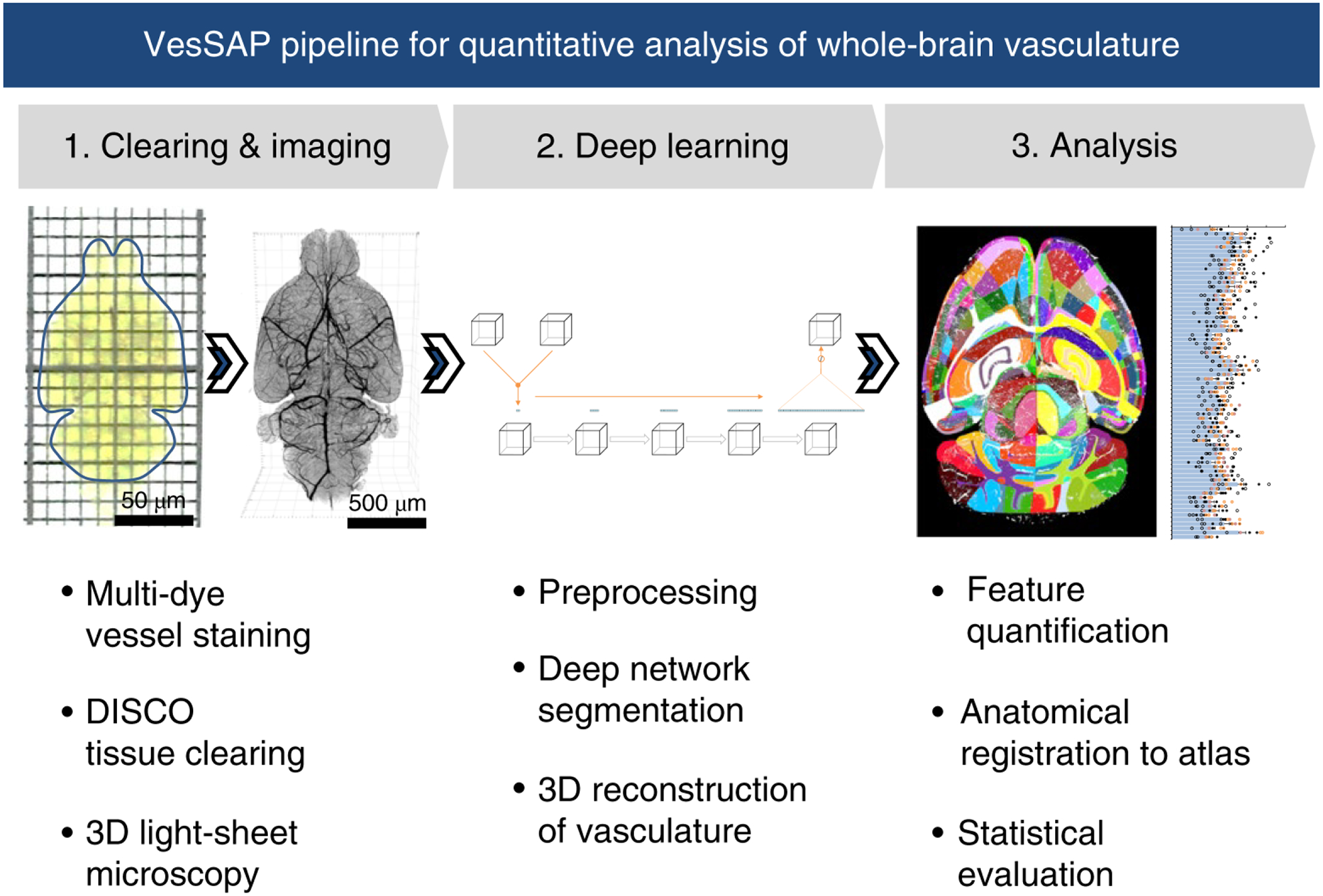
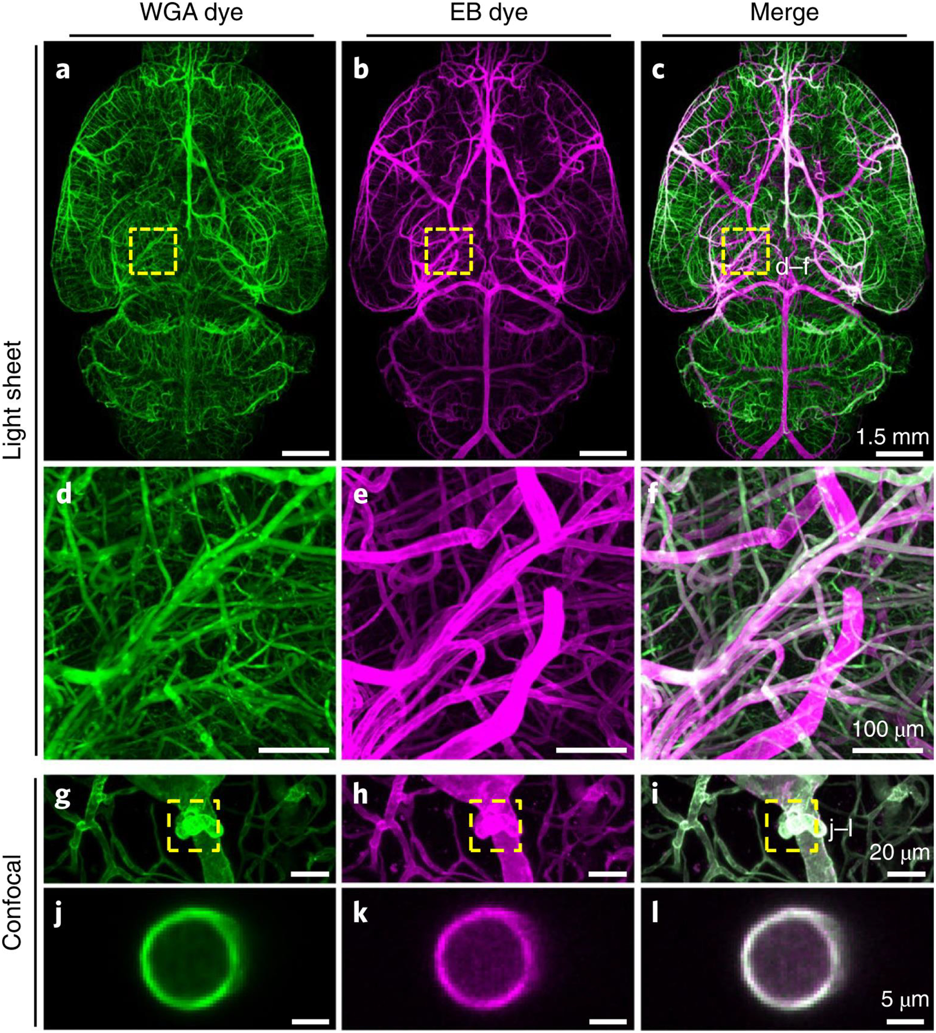
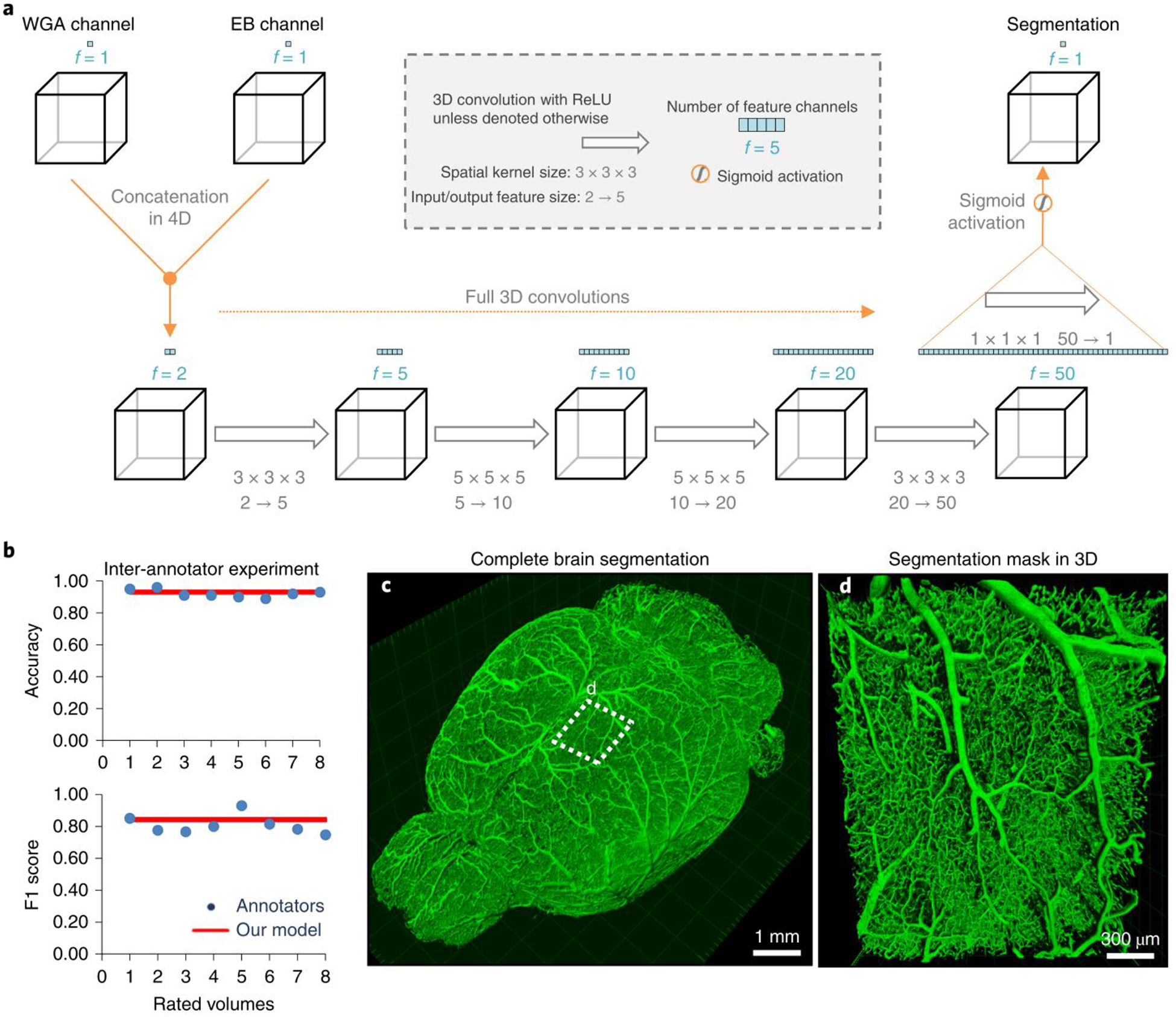
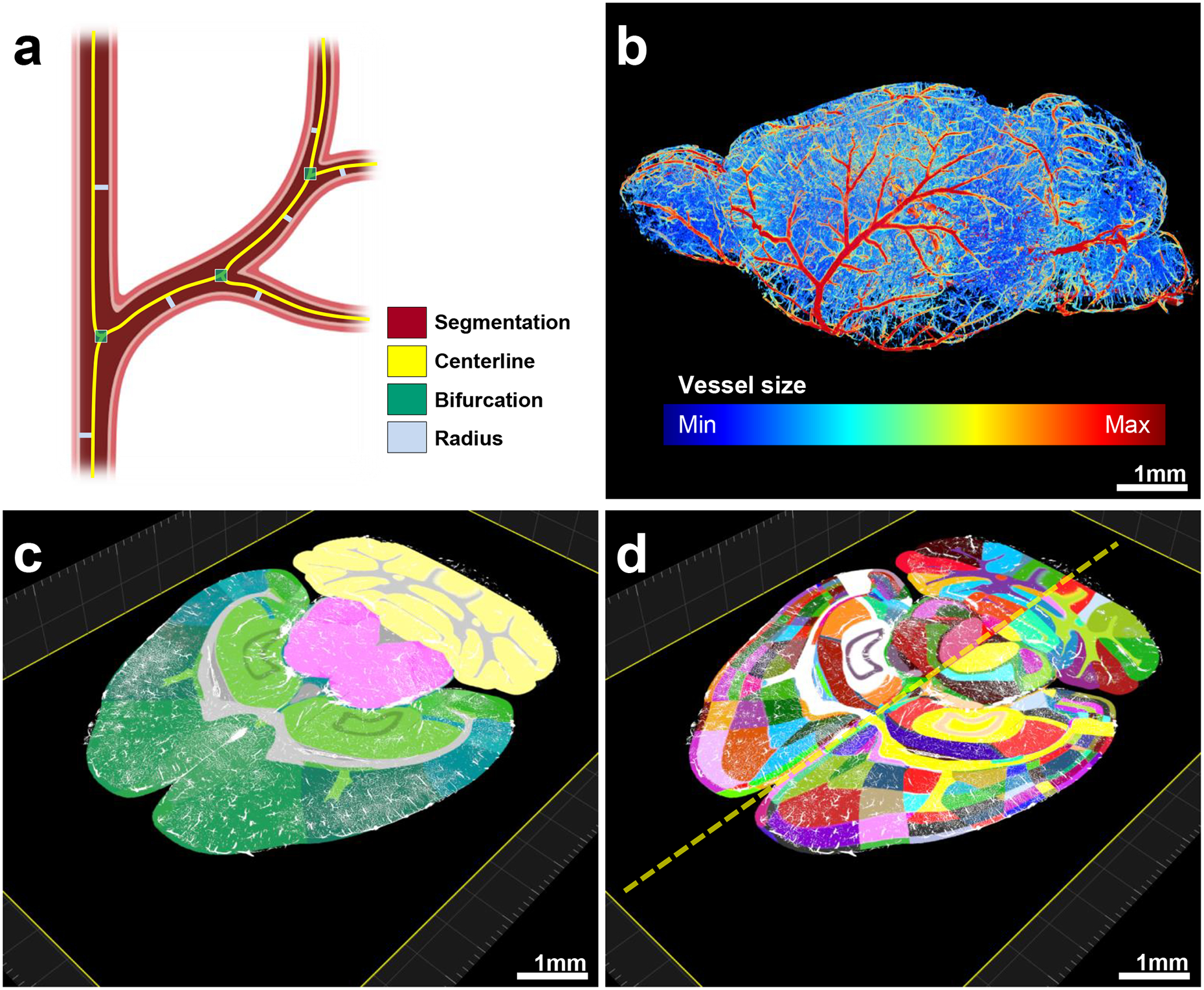
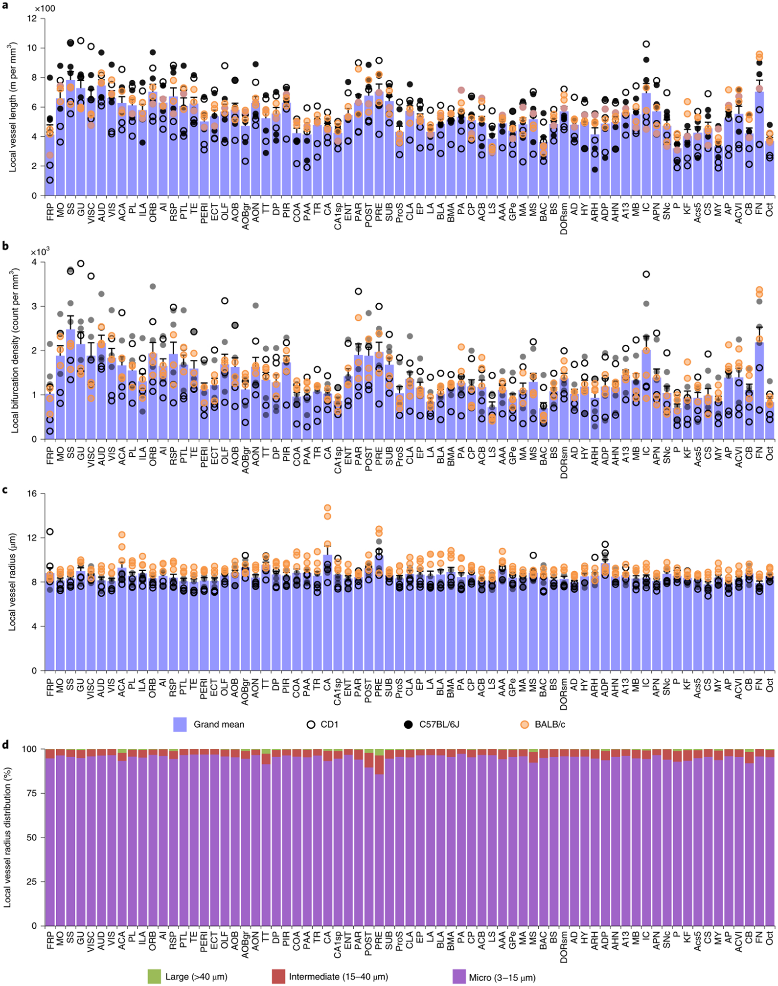
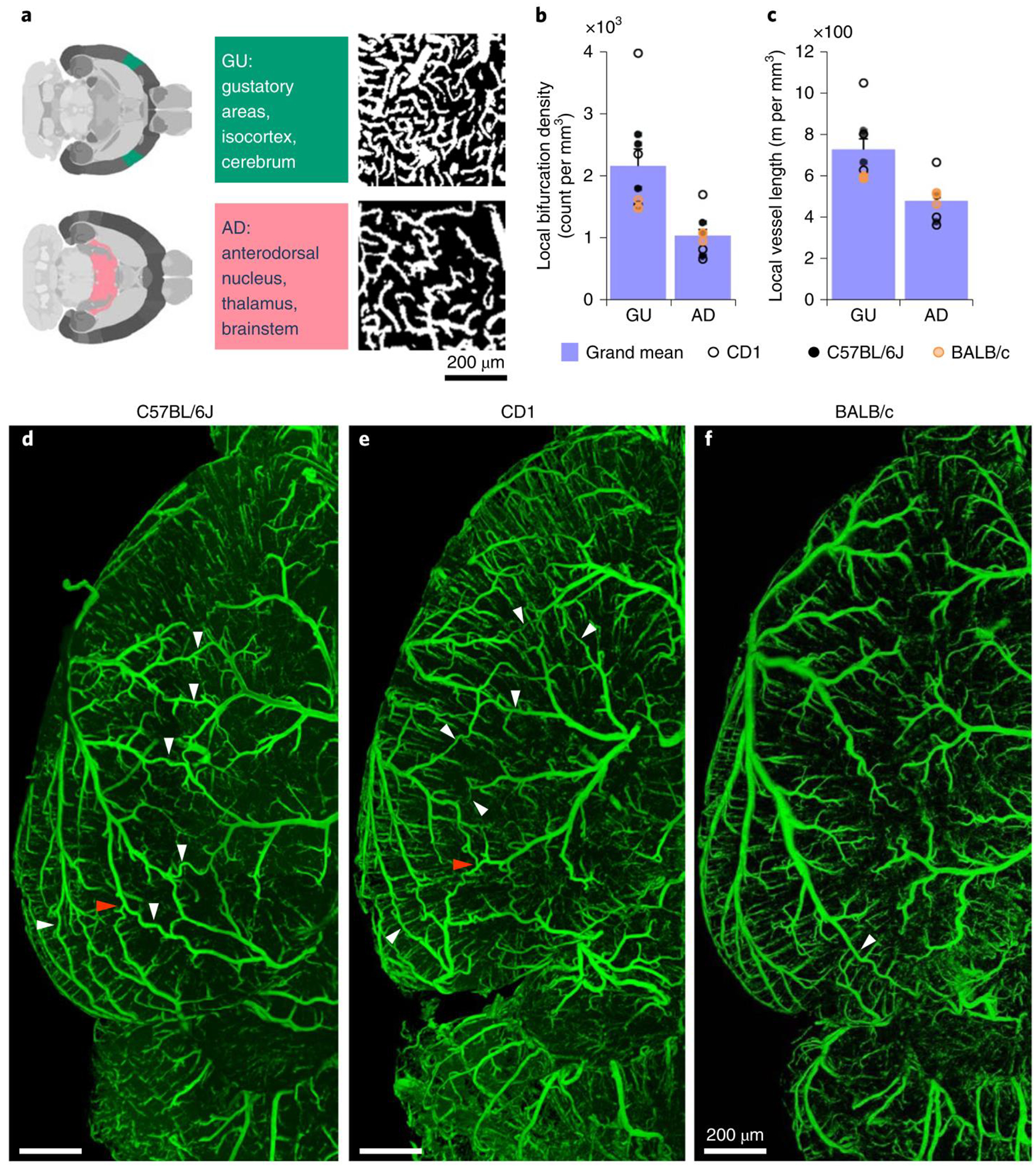
Tissue clearing methods enable the imaging of biological specimens without sectioning. However, reliable and scalable analysis of large imaging datasets in three dimensions remains a challenge. Here we developed a deep learning-based framework to quantify and analyze brain vasculature, named Vessel Segmentation & Analysis Pipeline (VesSAP). Our pipeline uses a convolutional neural network (CNN) with a transfer learning approach for segmentation and achieves human-level accuracy. By using VesSAP, we analyzed the vascular features of whole C57BL/6J, CD1 and BALB/c mouse brains at the micrometer scale after registering them to the Allen mouse brain atlas. We report evidence of secondary intracranial collateral vascularization in CD1 mice and find reduced vascularization of the brainstem in comparison to the cerebrum. Thus, VesSAP enables unbiased and scalable quantifications of the angioarchitecture of cleared mouse brains and yields biological insights into the vascular function of the brain.


Conflict of interest statement

CONFLICT OF INTEREST STATEMENT

The authors declare that the research was conducted in the absence of any commercial or financial relationships that could be construed as a potential conflict of interest.

Figures

Figure 1 |
Figure 1 |. Summary of the VesSAP pipeline
The method consists of three modular steps: 1, multi dye vessel staining and DISCO tissue clearing for high imaging quality using 3D light-sheet microscopy; 2, dep-learning based segmentation of blood vessels with 3D reconstruction and 3, anatomical feature extraction and mapping of the entire vasculature to the Allen adult mouse brain atlas for statistical analysis.
Figure 2 |
Figure 2 |. Enhancement of vascular staining using two complementary dyes
a-c, Maximum intensity projections of the automatically reconstructed tiling scans of WGA (a) and Evans blue (b) signals in the same sample and the merged view (c). d-f: Close-up of marked region in (c). g–l, Confocal images of WGA- and EB-stained vessels and vascular wall (g–i, maximum intensity projections of 112 μm and j–l, single slice of 1 μm). The experiment was performed on 9 different mice with similar results.
Figure 3 |
Figure 3 |. Deep learning architecture of VesSAP and performance on vessel segmentation
a, The 3D VesSAP network architecture consisting of five convolutional layers and a sigmoid activation for the last layer including the kernel sizes and input/output of the feature size. b, The F1-score for inter-annotator experiments (blue) compared to VeSAP (red). c, 3D rendering of full brain segmentation from a CD1-E mouse. d, 3D rendering of a small volume marked in (c). The experiment was performed on 9 different mice with similar results.
Figure 4 |
Figure 4 |. Pipeline showing feature extraction and registration process
a, Representation of the features extracted from vessels. b, Radius illustration of the vasculature in a CD1 mouse brain. C–d, Vascular segmentation results overlaid on the hierarchically (c) and randomly color-coded atlas to reveal all annotated regions (d) available including hemispheric difference (dashed-line in d). The experiment was performed on 9 different mice with similar results.
Figure 5 |
Figure 5 |. Anatomical properties of the neurovasculature in the adult mouse brain mapped to the Allen brain atlas clusters
a–c, Representation of the local vessel length (a), the density of bifurcations (b) and the average radius (c) in each of the 71 main anatomical clusters of the Allen brain atlas. Open, black and orange circles denote measurements in CD1, C57BL/6J and BALB/c strains respectively; each circle represents a single mouse (a–c). d, Local distribution of the large, middle and micro vessels in the same anatomical clusters. All abbreviations are listed in the Supplementary table 1. All data values are given as mean ± SEM and n=3 mice per strain (a–c).
Figure 6 |
Figure 6 |. Exemplary quantitative analysis enabled by VesSAP
a, The respective location of AD and GU areas in the mouse brain (left panels), maximum intensity projections of representative volumes from the gustatory areas (GU) and anterodorsal nucleus (AD) segmentation (600 × 600 × 33 μm) (right panels). b, c, Quantification of the bifurcation density and local vessel length for the AD and GU clusters. CD1 mice shown by open circles, BALB/C by orange circles, and C57BL/6J by black circles. Values are mean ± SEM, n=3 mice per strain. D–f, Images of the vasculature in a representative C57BL/6J (d), CD1 (e) and BALB/c mouse (f) where the white arrowheads indicate anastomoses between the major arteries. Direct vascular connections between the medial cerebral artery (MCA), the anterior cerebral artery (ACA) and the posterior cerebral artery (PCER) are indicated by red arrowheads. The experiment was performed 3 times with similar results.

References

    1. Bennett RE et al. Tau induces blood vessel abnormalities and angiogenesis-related gene expression in P301L transgenic mice and human Alzheimer’s disease. Proc Natl Acad Sci U S A 115, E1289–E1298, doi:10.1073/pnas.1710329115 (2018). - DOI - PMC - PubMed
    1. Obenaus A et al. Traumatic brain injury results in acute rarefication of the vascular network. Sci Rep 7, 239, doi:10.1038/s41598-017-00161-4 (2017). - DOI - PMC - PubMed
    1. Völgyi K et al. Chronic Cerebral Hypoperfusion Induced Synaptic Proteome Changes in the rat Cerebral Cortex. Molecular Neurobiology 55, 4253–4266, doi:10.1007/s12035-017-0641-0 (2018). - DOI - PubMed
    1. Klohs J et al. Contrast-enhanced magnetic resonance microangiography reveals remodeling of the cerebral microvasculature in transgenic ArcAbeta mice. J Neurosci 32, 1705–1713, doi:10.1523/JNEUROSCI.5626-11.2012 (2012). - DOI - PMC - PubMed
    1. Edwards-Richards A et al. Capillary rarefaction: an early marker of microvascular disease in young hemodialysis patients. Clin Kidney J 7, 569–574, doi:10.1093/ckj/sfu106 (2014). - DOI - PMC - PubMed

Publication types