. 2020 Feb 25:14:60.
doi: 10.3389/fnhum.2020.00060. eCollection 2020.

A Guide to Literature Informed Decisions in the Design of Real Time fMRI Neurofeedback Studies: A Systematic Review

Affiliations

A Guide to Literature Informed Decisions in the Design of Real Time fMRI Neurofeedback Studies: A Systematic Review

Samantha J Fede et al. Front Hum Neurosci. .

Abstract

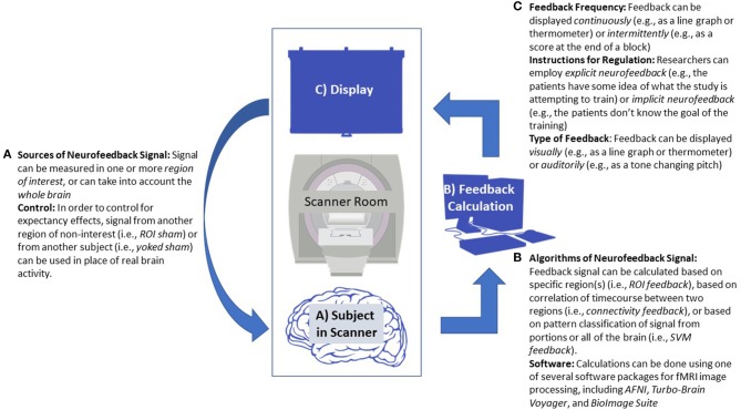
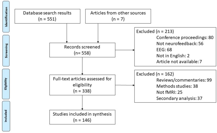
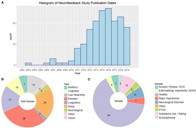
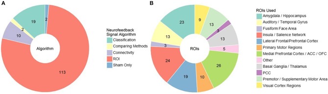
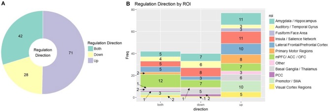
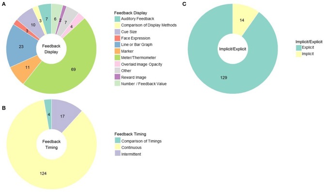
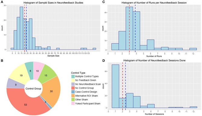
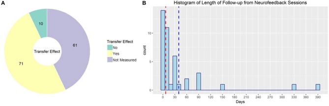
Background: Although biofeedback using electrophysiology has been explored extensively, the approach of using neurofeedback corresponding to hemodynamic response is a relatively young field. Real time functional magnetic resonance imaging-based neurofeedback (rt-fMRI-NF) uses sensory feedback to operantly reinforce patterns of neural response. It can be used, for example, to alter visual perception, increase brain connectivity, and reduce depression symptoms. Within recent years, interest in rt-fMRI-NF in both research and clinical contexts has expanded considerably. As such, building a consensus regarding best practices is of great value. Objective: This systematic review is designed to describe and evaluate the variations in methodology used in previous rt-fMRI-NF studies to provide recommendations for rt-fMRI-NF study designs that are mostly likely to elicit reproducible and consistent effects of neurofeedback. Methods: We conducted a database search for fMRI neurofeedback papers published prior to September 26th, 2019. Of 558 studies identified, 146 met criteria for inclusion. The following information was collected from each study: sample size and type, task used, neurofeedback calculation, regulation procedure, feedback, whether feedback was explicitly related to changing brain activity, feedback timing, control group for active neurofeedback, how many runs and sessions of neurofeedback, if a follow-up was conducted, and the results of neurofeedback training. Results: rt-fMRI-NF is typically upregulation practice based on hemodynamic response from a specific region of the brain presented using a continually updating thermometer display. Most rt-fMRI-NF studies are conducted in healthy samples and half evaluate its effect on immediate changes in behavior or affect. The most popular control group method is to provide sham signal from another region; however, many studies do not compare use a comparison group. Conclusions: We make several suggestions for designs of future rt-fMRI-NF studies. Researchers should use feedback calculation methods that consider neural response across regions (i.e., SVM or connectivity), which should be conveyed as intermittent, auditory feedback. Participants should be given explicit instructions and should be assessed on individual differences. Future rt-fMRI-NF studies should use clinical samples; effectiveness of rt-fMRI-NF should be evaluated on clinical/behavioral outcomes at follow-up time points in comparison to both a sham and no feedback control group.

Keywords: fMRI; intervention; methods; neurofeedback; rt-fMRI.


Figures

Figure 1
Figure 1
Diagram of a generic neurofeedback experiment with study design elements highlighted. (A) Functional MRI data is collected from a patient, focusing either on a single region of interest (ROI), multiple ROIs, or the whole brain. (B) Data from the subject is processed in real-time, usually employing standard neuroimaging software packages. Feedback is calculated, sometimes based on ROI signal change, correlation between several ROIs, or using a classification algorithm. This may require an offline fMRI scan prior to neurofeedback training to establish baseline activity or to train the classifier. (C) The feedback value is transformed and given to the patient in the scanner, often via a continuous visual graph displayed on a screen. (A) This allows the participant to react and change their neural response, starting the feedback cycle over.
Figure 2
Figure 2
Diagram of literature search procedures across the primary and update literature search dates based on PRISMA 2009 Flow Diagram (Moher et al., 2009). fMRI, functional magnetic resonance imaging; rt-fMRI-NF, realtime functional magnetic resonance imaging neurofeedback training.
Figure 3
Figure 3
(A) Histogram of publication dates for all empirical rt-fMRI neurofeedback studies. (B) rt-fMRI neurofeedback studies categorized by task type. NA studies had no stimuli or instructions that fell into a particular task domain. Numbers indicate the number of studies in each category. (C) rt-fMRI neurofeedback studies by type of sample used in the study. Numbers indicate the number of studies in each category.
Figure 4
Figure 4
(A) rt-fMRI neurofeedback studies categorized by signal/algorithm type. Classification includes SVM, DecNef, MVPA etc. Numbers indicate the number of studies in each category. (B) rt-fMRI neurofeedback studies that employed an ROI signal approach by region used for that ROI. Numbers indicate the number of studies in each category. ROI, region of interest; ACC/PCC, anterior/posterior cingulate cortex; OFC, orbitofrontal cortex.
Figure 5
Figure 5
(A) rt-fMRI neurofeedback studies categorized by direction subjects were instructed to regulate Numbers indicate the number of studies in each category. (B) Stacked bar char of rt-fMRI neurofeedback studies using the ROI approach broken down by direction of regulation and ROI. Numbers indicate the number of studies in each category. ROI, region of interest; mPFC, medial prefrontal cortex; ACC/PCC, anterior/posterior cingulate cortex; OFC, orbitofrontal cortex; SMA, supplementary motor area.
Figure 6
Figure 6
(A) rt-fMRI neurofeedback studies categorized by the way feedback was displayed to subjects. Numbers indicate the number of studies in each category. (B) rt-fMRI neurofeedback studies by feedback timing. Numbers indicate the number of studies in each category. (C) rt-fMRI neurofeedback studies by nature of the instructions for regulating brain activity. Numbers indicate the number of studies in each category.
Figure 7
Figure 7
(A) Histogram of sample sizes used in rt-fMRI neurofeedback studies. Red line indicates median, blue line indicates mean. (B) rt-fMRI neurofeedback studies by nature of the control group used to compared the active neurofeedback condition. Numbers indicate the number of studies in each category. ROI, Region of interest. (C) Histogram of number of runs used in rt-fMRI neurofeedback training sessions. Red line indicates median, blue line indicates mean. (D) Histogram of number of sessions used in rt-fMRI neurofeedback training protocols. Red line indicates median, blue line indicates mean.
Figure 8
Figure 8
(A) rt-fMRI neurofeedback studies by nature of the outcome measure used to evaluate neurofeedback effectiveness. Numbers indicate the number of studies in each category. (B) Histogram of follow-up times used in rt-fMRI neurofeedback studies with follow-up sessions. Red line indicates median, blue line indicates mean.

References

    1. Alegria A. A., Wulff M., Brinson H., Barker G. J., Norman L. J., Brandeis D., et al. . (2017). Real-time fMRI neurofeedback in adolescents with attention deficit hyperactivity disorder. Hum. Brain Mapp. 38, 3190–3209. 10.1002/hbm.23584 - DOI - PMC - PubMed
    1. Amano K., Shibata K., Kawato M., Sasaki Y., Watanabe T. (2016). Learning to associate orientation with color in early visual areas by associative decoded fMRI neurofeedback. Curr. biol. 26, 1861–1866. 10.1016/j.cub.2016.05.014 - DOI - PMC - PubMed
    1. Arns M., Batail J. M., Bioulac S., Congedo M., Daudet C., Drapier D., et al. . (2017). Neurofeedback: one of today's techniques in psychiatry? Encephale 43, 135–145. 10.1016/j.encep.2016.11.003 - DOI - PubMed
    1. Baecke S., Lutzkendorf R., Mallow J., Luchtmann M., Tempelmann C., Stadler J., et al. . (2015). A proof-of-principle study of multi-site real-time functional imaging at 3T and 7T: implementation and validation. Sci Rep. 5:8413. 10.1038/srep08413 - DOI - PMC - PubMed
    1. Banca P., Sousa T., Duarte I. C., Castelo-Branco M. (2015). Visual motion imagery neurofeedback based on the hMT+/V5 complex: evidence for a feedback-specific neural circuit involving neocortical and cerebellar regions. J. Neural Eng. 12:066003. 10.1088/1741-2560/12/6/066003 - DOI - PubMed

Publication types