

Manganese-Enhanced Magnetic Resonance Imaging: Application in Central Nervous System Diseases
- PMID: 32161572
- PMCID: PMC7052353
- DOI: 10.3389/fneur.2020.00143
Manganese-Enhanced Magnetic Resonance Imaging: Application in Central Nervous System Diseases
Abstract
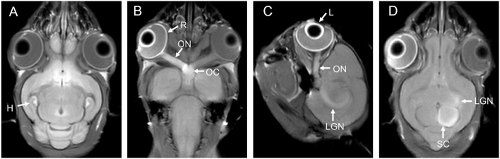
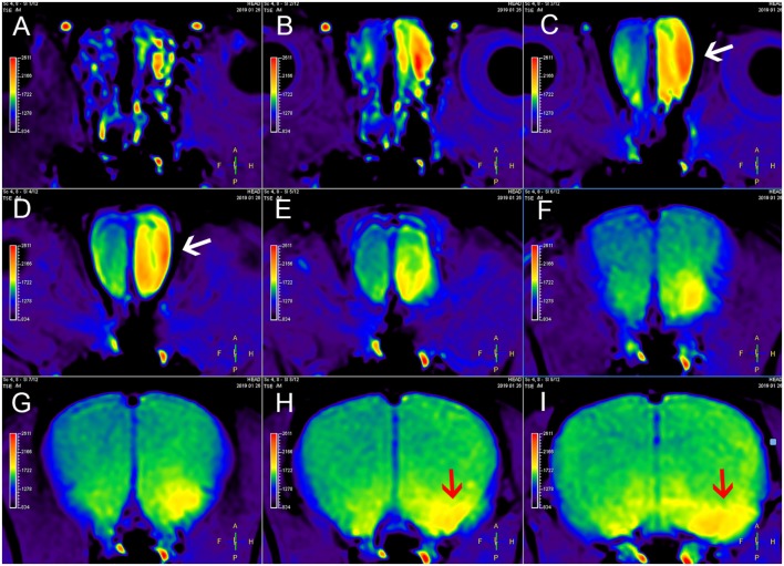
Manganese-enhanced magnetic resonance imaging (MEMRI) relies on the strong paramagnetism of Mn2+. Mn2+ is a calcium ion analog and can enter excitable cells through voltage-gated calcium channels. Mn2+ can be transported along the axons of neurons via microtubule-based fast axonal transport. Based on these properties, MEMRI is used to describe neuroanatomical structures, monitor neural activity, and evaluate axonal transport rates. The application of MEMRI in preclinical animal models of central nervous system (CNS) diseases can provide more information for the study of disease mechanisms. In this article, we provide a brief review of MEMRI use in CNS diseases ranging from neurodegenerative diseases to brain injury and spinal cord injury.
Keywords: MEMRI; brain injury; central nervous system; manganese; neurodegeneration.
Copyright © 2020 Yang and Li.
Figures
Similar articles
-
Manganese-enhanced magnetic resonance imaging (MEMRI).Methods Mol Biol. 2011;711:145-74. doi: 10.1007/978-1-61737-992-5_7. Methods Mol Biol. 2011. PMID: 21279601 Free PMC article.
-
Manganese Enhanced MRI for Use in Studying Neurodegenerative Diseases.Front Neural Circuits. 2019 Jan 7;12:114. doi: 10.3389/fncir.2018.00114. eCollection 2018. Front Neural Circuits. 2019. PMID: 30666190 Free PMC article. Review.
-
Longitudinal manganese-enhanced magnetic resonance imaging of neural projections and activity.NMR Biomed. 2022 Jun;35(6):e4675. doi: 10.1002/nbm.4675. Epub 2022 Mar 6. NMR Biomed. 2022. PMID: 35253280 Free PMC article. Review.
-
Studying Axonal Transport in the Brain by Manganese-Enhanced Magnetic Resonance Imaging (MEMRI).Methods Mol Biol. 2022;2431:111-142. doi: 10.1007/978-1-0716-1990-2_6. Methods Mol Biol. 2022. PMID: 35412274
-
Manganese enhanced MRI (MEMRI): neurophysiological applications.Rev Neurosci. 2011;22(6):675-94. doi: 10.1515/RNS.2011.048. Epub 2011 Nov 18. Rev Neurosci. 2011. PMID: 22098448 Free PMC article. Review.
Cited by
-
Development of Resorbable Phosphate-Based Glass Microspheres as MRI Contrast Media Agents.Molecules. 2024 Sep 10;29(18):4296. doi: 10.3390/molecules29184296. Molecules. 2024. PMID: 39339291 Free PMC article.
-
Magnetic resonance imaging to assess the brain response to fasting in glioblastoma-bearing rats as a model of cancer anorexia.Cancer Imaging. 2023 Apr 10;23(1):36. doi: 10.1186/s40644-023-00553-y. Cancer Imaging. 2023. PMID: 37038232 Free PMC article.
-
Synthetic and Nanotechnological Approaches for a Diagnostic Use of Manganese.Molecules. 2022 May 13;27(10):3124. doi: 10.3390/molecules27103124. Molecules. 2022. PMID: 35630601 Free PMC article.
-
In vivo MRI evaluation of anterograde manganese transport along the visual pathway following whole eye transplantation.J Neurosci Methods. 2022 Apr 15;372:109534. doi: 10.1016/j.jneumeth.2022.109534. Epub 2022 Feb 22. J Neurosci Methods. 2022. PMID: 35202613 Free PMC article.
-
Brain Network Allostasis after Chronic Alcohol Drinking Is Characterized by Functional Dedifferentiation and Narrowing.J Neurosci. 2022 May 25;42(21):4401-4413. doi: 10.1523/JNEUROSCI.0389-21.2022. Epub 2022 Apr 18. J Neurosci. 2022. PMID: 35437279 Free PMC article.
References
Publication types
LinkOut - more resources
Full Text Sources