Review
. 2020 Feb 26:8:43.
doi: 10.3389/fcell.2020.00043. eCollection 2020.

Hec1/Ndc80 Tail Domain Function at the Kinetochore-Microtubule Interface

Affiliations
Review

Hec1/Ndc80 Tail Domain Function at the Kinetochore-Microtubule Interface

Robert T Wimbish et al. Front Cell Dev Biol. .

Abstract

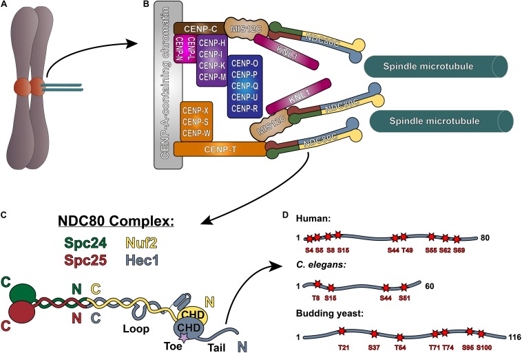
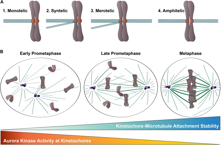
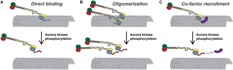
Successful mitotic cell division is critically dependent on the formation of correct attachments between chromosomes and spindle microtubules. Microtubule attachments are mediated by kinetochores, which are large proteinaceous structures assembled on centromeric chromatin of mitotic chromosomes. These attachments must be sufficiently stable to transduce force; however, the strength of these attachments are also tightly regulated to ensure timely, error-free progression through mitosis. The highly conserved, kinetochore-associated NDC80 complex is a core component of the kinetochore-microtubule attachment machinery in eukaryotic cells. A small, disordered region within the Hec1 subunit of the NDC80 complex - the N-terminal "tail" domain - has been actively investigated during the last decade due to its roles in generating and regulating kinetochore-microtubule attachments. In this review, we discuss the role of the NDC80 complex, and specifically the Hec1 tail domain, at the kinetochore-microtubule interface, and how recent studies provide a more unified view of Hec1 tail domain function.

Keywords: Hec1; NDC80; kinetochore; microtubule; mitosis.


Figures

FIGURE 1
FIGURE 1
The NDC80 complex at the kinetochore-microtubule interface. (A) Mitotic chromosome. (B) Organization of the kinetochore-microtubule interface in vertebrate cells. The foundation of the kinetochore is the CCAN, or the Constitutive Centromere Associated Network, which binds to CENP-A-containing centromeric chromatin. The CCAN is composed of 16 subunits, organized in multiple subcomplexes including: CENP-L/N; CENP-O/P/Q/U/R; CENP-H/I/K/M; CENP-T/W/S/X; and CENP-C. CENP-C recruits the KMN “network” (composed of KNL1, the MIS12 complex, and the NDC80 complex) through its direct association with the MIS12 complex. CENP-T also recruits the NDC80 complex alone, as well as the KMN network through binding the MIS12 complex. (C) Architecture of the NDC80 complex. The C-termini of Spc24 (green) and Spc25 (red) form the kinetochore-targeting domain which binds either the MIS12 complex or CENP-T. The N-terminal regions of Spc24 and Spc25 form a coiled-coil domain that tetramerizes with the C-termini of Nuf2 (yellow) and Hec1 (blue). The N-terminus of Hec1 is comprised of a well-ordered CH domain, which contains the high affinity microtubule-binding “toe” region, and the tail domain which is also implicated in microtubule binding. The ∼40 amino acid loop domain of Hec1 is also indicated on the schematic. (D) Representation of the Hec1/Ndc80 tail domains from human, Caenorhabditis elegans, and the budding yeast Saccharomyces cerevisiae. Shown are the mapped and putative Aurora kinase phosphorylation sites. The human sites shown are Ser4, Ser5, Ser8, Ser15, Ser44, Thr49, Ser55, Ser62, and Ser69. The C. elegans sites shown are Thr8, Ser18, Ser44, and Ser51. The budding yeast sites shown are Thr21, Ser37, Thr54, Thr71, Thr74, Ser95, and Ser100 (see text for references).
FIGURE 2
FIGURE 2
Kinetochore-microtubule attachments during mitosis. (A) Types of kinetochore-microtubule attachments. (1) Monotelic attachment: one sister kinetochore is attached to microtubules from one spindle pole and one sister is unattached; (2) Syntelic attachment: both sister kinetochores are attached to microtubules emanating from the same spindle pole; (3) Merotelic attachment: one sister kinetochore is attached to microtubules from both spindle poles; and (4) Amphitelic attachment (correct): one sister kinetochore is attached to microtubules from one pole and one sister kinetochore is attached to microtubules from the opposite pole. (B) Chromosome congression during mitotic progression. In early prometaphase, kinetochore-microtubule attachments errors are common, kinetochore-microtubule attachments are short-lived and labile, and Aurora B kinase activity at kinetochores is high. As mitosis progresses, erroneous kinetochore-microtubule attachments are corrected, kinetochore-microtubule attachments become long-lived and stable, and Aurora B kinase activity at kinetochores decreases.
FIGURE 3
FIGURE 3
Models for Hec1 tail domain function. (A) Direct microtubule binding. In this model, the tail domain directly interacts with the microtubule lattice to increase CH-domain-mediated NDC80 complex-microtubule interactions. Phosphorylation of the Hec1 tail reduces the positive charge of the tail domain and as a result, reduces the affinity of NDC80 complexes for the negatively charged microtubule lattice. (B) Oligomerization. In this model, a dephosphorylated tail domain functions to oligomerize adjacent NDC80 complexes, which promotes high affinity NDC80-complex-microtubule binding. Upon phosphorylation of the tail domain, complex oligomerization is no longer favored, possibly due to a decrease in affinity of a phosphorylated tail domain for a negatively charged region within the CH domain of Hec1. (C) Co-factor recruitment. In this model, a dephosphorylated Hec1 tail domain recruits kinetochore-associated microtubule binding proteins or protein complexes to promote high affinity NDC80 complex-microtubule binding. In contrast, a phosphorylated tail domain restricts co-factor recruitment. As discussed in the text, these models are not mutually exclusive.

References

    1. Akiyoshi B., Nelson C. R., Ranish J. A., Biggins S. (2009). Analysis of Ipl1-mediated phosphorylation of the Ndc80 kinetochore protein in Saccharomyces cerevisiae. Genetics 183 1591–1595. 10.1534/genetics.109.109041 - DOI - PMC - PubMed
    1. Alushin G. M., Musinipally V., Matson D., Tooley J., Stukenberg P. T., Nogales E. (2012). Multimodal microtubule binding by the Ndc80 kinetochore complex. Nat. Struct. Mol. Biol. 19 1161–1167. 10.1038/nsmb.2411 - DOI - PMC - PubMed
    1. Alushin G. M., Ramey V. H., Pasqualato S., Ball D. A., Grigorieff N., Musacchio A., et al. (2010). The Ndc80 kinetochore complex forms oligomeric arrays along microtubules. Nature 467 805–810. 10.1038/nature09423 - DOI - PMC - PubMed
    1. Auckland P., Clarke N. I., Royle S. J., McAinsh A. D. (2017). Congressing kinetochores progressively load Ska complexes to prevent force-dependent detachment. J. Cell Biol. 216 1623–1639. 10.1083/jcb.201607096 - DOI - PMC - PubMed
    1. Bakhoum S. F., Thompson S. L., Manning A. L., Compton D. A. (2009). Genome stability is ensured by temporal control of kinetochore-microtubule dynamics. Nat. Cell Biol. 11 27–35. 10.1038/ncb1809 - DOI - PMC - PubMed

LinkOut - more resources