. 2018 Oct 4;3(1):bpy010.
doi: 10.1093/biomethods/bpy010. eCollection 2018.

A cost-effective technique for generating preservable biomass smoke extract and measuring its effect on cell receptor expression in human bronchial epithelial cells

Affiliations

A cost-effective technique for generating preservable biomass smoke extract and measuring its effect on cell receptor expression in human bronchial epithelial cells

K C Rajendra et al. Biol Methods Protoc. .

Abstract

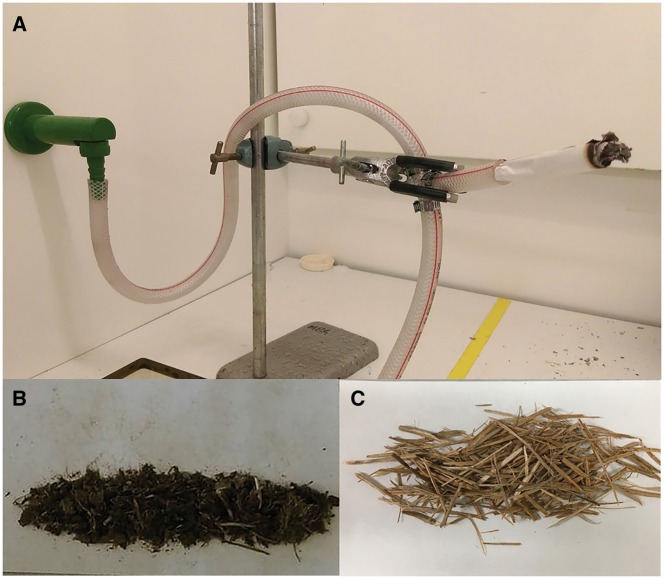
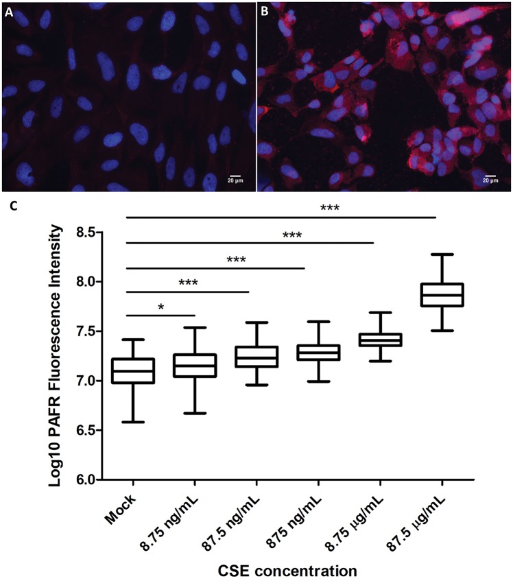
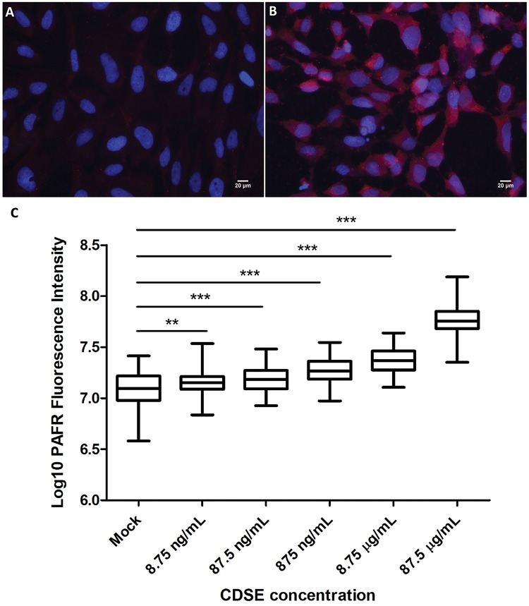
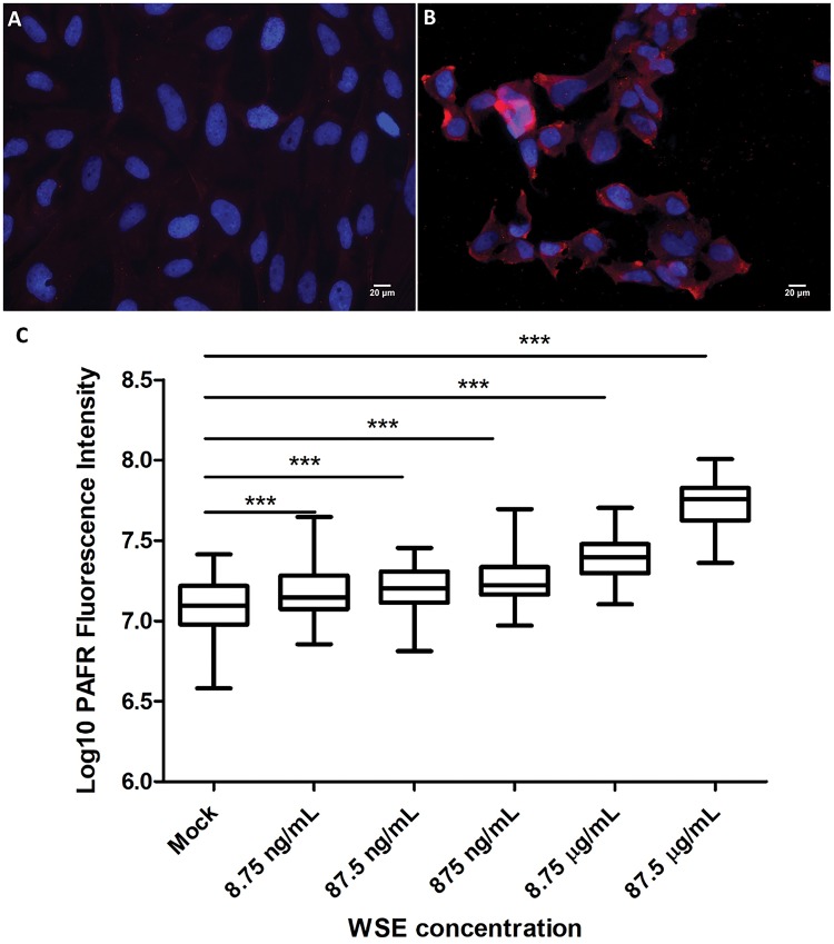
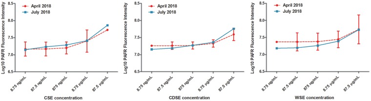
Nearly half of the world's population uses biomass fuel for the purposes of cooking and heating. Smoke derived from biomass increases the risk of the development of lung diseases, including pneumonia, chronic obstructive pulmonary disease, airway tract infections, and lung cancer. Despite the evidence linking biomass smoke exposure to pulmonary disease, only a small number of experimental studies have been conducted on the impact of biomass smoke on airway epithelial cells. This is in part due to the lack of a standard and easily accessible procedure for the preparation of biomass smoke. Here, we describe a cost-effective and reproducible method for the generation of different smoke extracts, in particular, cow dung smoke extract (CDSE) and wood smoke extract (WSE) for use in a range of biological applications. We examined the effect of the biomass smoke extracts on human bronchial epithelial cell expression of a known responder to cigarette smoke exposure (CSE), the platelet-activating factor receptor (PAFR). Similar to the treatment with CSE, we observed a dose-dependent increase in PAFR expression on human airway epithelial cells that were exposed to CDSE and WSE. This method provides biomass smoke in a re-usable form for cell and molecular bioscience studies on the pathogenesis of chronic lung disease.

Keywords: biomass smoke; chronic obstructive pulmonary disease; cigarette smoke extract; platelet-activating factor receptor.


Figures

Figure 1:
Figure 1:
Generation of CDSE and WSE. (A) A water aspirator was set up to draw smoke from a burning cow dung or wood shaving roll using the vacuum created by the flow of water. (B) Cow dung was sun-dried, crushed into fine particles, and rolled in paper. (C) Wood was cut into small chips and rolled in paper.
Figure 2:
Figure 2:
CSE exposure and PAFR expression on bronchial epithelial cells. (A) Mock treatment of BEAS-2B cells with 1% DMSO as a control. (B) BEAS-2B cells exposed to 87.5 µg/ml CSE. All immunofluorescence micrographs show BEAS-2B cells with PAFR expression (anti-PAFR monoclonal antibody; 2.5 μg/ml, red) and nuclei stained with 4′, 6-diamidino-2-phenylindole (1 μg/ml, blue). Magnification = 400×. (C) Response to different concentrations of CSE. PAFR expression corresponds to log10 of fluorescence intensity following labelling with Alexa Fluor 594 conjugate antibody targeting anti-PAFR mAb. PAFR expression was significantly increased in 8.75 ng/ml, 87.5 ng/ml, 875 ng/ml, 8.75 µg/ml and 87.5 µg/ml CSE exposed BEAS-2B cells. Data are representative of two independent experiments (*P < 0.05, ***P <0.0001, One-way ANOVA with Dunnett’s multiple comparison test).
Figure 3:
Figure 3:
CDSE exposure and PAFR expression on bronchial epithelial cells. (A) Mock treatment of BEAS-2B cells with 1% DMSO as a control. (B) BEAS-2B cells exposed to 87.5 µg/ml CDSE. All immunofluorescence micrographs show BEAS-2B cells with PAFR expression (anti-PAFR monoclonal antibody; 2.5 μg/ml, red) and nuclei stained with 4′, 6-diamidino-2-phenylindole (1 μg/ml, blue). Magnification = 400×. (C) Response to different concentrations of CDSE. PAFR expression corresponds to log10 of fluorescence intensity following labelling with Alexa Fluor 594 conjugate antibody targeting anti-PAFR mAb. The PAFR expression was significantly increased in 8.75 ng/ml, 87.5 ng/ml, 875 ng/ml, 8.75 µg/ml and 87.5 µg/ml CDSE treated BEAS-2B cells. Data are representative of two independent experiments (**P < 0.001, ***P < 0.0001, one-way ANOVA with Dunnett’s multiple comparison test).
Figure 4:
Figure 4:
Wood smoke extract (WSE) exposure and PAFR expression on bronchial epithelial cells. (A) Mock treatment of BEAS-2B cells with 1% DMSO as a control. (B) BEAS-2B cells exposed to 87.5 µg/ml WSE. All immunofluorescence micrographs show BEAS-2B cells with PAFR expression (anti-PAFR monoclonal antibody; 2.5 μg/ml, red) and nuclei stained with 4′, 6-diamidino-2-phenylindole (1 μg/ml, blue). Magnification = 400×. (C) Response to different concentrations of WSE. PAFR expression corresponds to log10 of fluorescence intensity following labelling with Alexa Fluor 594 conjugate antibody targeting anti-PAFR mAb. The PAFR expression was significantly increased in 8.75 ng/ml, 87.5 ng/ml, 875 ng/ml, 8.75 µg/ml and 87.5 µg/ml WSE exposed BEAS-2B cells. Data are representative of two independent experiments (***P < 0.0001, one-way ANOVA with Dunnett’s multiple comparison test).
Figure 5:
Figure 5:
Comparison of effects of smoke extracts on PAFR expression in experiments conducted 3 months apart. All of the smoke extracts, CSE, CDSE, and WSE, were run in the concentration range from 8.75 ng/ml to 87.5 µg/ml in the July 2018 experiment. The CSE extract was run in the same concentration range from 8.75 ng/ml to 87.5 µg/ml in the April 2018 experiment. For the April 2018 experiments, the CDSE and WSE data were interpolated to the 8.75 ng/ml to 87.5 µg/ml concentration range using non-linear least squares regression. The levels of PAFR expression in the July 2018 experiments with the CSE, CDSE, and WSE samples were within 95% CI of the levels obtained for the April 2018 experiments. Therefore, no significant decay in the PAFR inducing activity of the smoke extracts was detected following storage at −20°C over a 3-month period.

Similar articles

Cited by

References

    1. World Health Organisation. Household Air Pollution and Health Fact Sheet Number 292 Updated in February 2016: World Health Organisation 2016http://www.who.int/mediacentre/factsheets/fs292/en/.
    1. Lim SS, Vos T, Flaxman AD. et al. A comparative risk assessment of burden of disease and injury attributable to 67 risk factors and risk factor clusters in 21 regions, 1990-2010: a systematic analysis for the Global Burden of Disease Study 2010. Lancet 2012;380:2224–60. - PMC - PubMed
    1. Assad NA, Balmes J, Mehta S. et al. Chronic obstructive pulmonary disease secondary to household air pollution. Semin Respir Crit Care Med 2015;36:408–21. - PubMed
    1. Assad NA, Kapoor V, Sood A.. Biomass smoke exposure and chronic lung disease. Curr Opin Pulm Med 2016;22:150–7. - PubMed
    1. Gordon SB, Bruce NG, Grigg J. et al. Respiratory risks from household air pollution in low and middle income countries. Lancet Respir Med 2014;2:823–60. - PMC - PubMed