. 2020 Jan 17;5(1):bpz020.
doi: 10.1093/biomethods/bpz020. eCollection 2020.

A new method for measuring the size of nematodes using image processing

Affiliations

A new method for measuring the size of nematodes using image processing

David G H Andrews. Biol Methods Protoc. .

Abstract

Many studies have been made on nematodes, especially Caenorhabditis Elegans, which are used as a model organism. In many studies, the size of the nematode is important. This article describes a method of measuring the length, volume and surface area of nematodes from photographs. The method uses the imaging software ImageJ, which is in the public domain. Two macros are described. The first converts the images into binary form, and the second uses several built-in functions to measure the length of the worm and its diameter along its length. If it is assumed that the worm has a circular cross-section, then the volume and surface area of the nematode can be calculated. This is a cheap and easy technique.

Keywords: nematode; image processing; size.


Figures

Figure 1:
Figure 1:
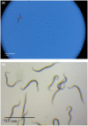
(a) Caenorhabditis elegans on agar photographed with a Nikon camera through a Leica microscope. (b) Nematodes photographed in water using a Leica microscope with a built-in camera.
Figure 2:
Figure 2:
Binary images of nematodes are shown in Figure 1.
Figure 3:
Figure 3:
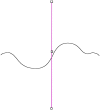
Line through the centre of ‘spine’ line.
Figure 4:
Figure 4:
‘Spine’ line of the worm, with the line to find its end.
Figure 5:
Figure 5:
Extending ‘spine’ line to the end of the worm.
Figure 6:
Figure 6:
Find the width of the worm.
Figure 7:
Figure 7:
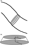
(a) Worm divided into truncated cones. (b) Truncated cone.
Figure 8:
Figure 8:
Worms outline are shown in Excel.
Figure 9:
Figure 9:
Straightened silhouettes of the worms in Figure 1.
Figure 10:
Figure 10:
Segment line through the worm.

Similar articles

Cited by

References

    1. Stroustrup N, Anthony WE, Nash ZM. et al. The temporal scaling of Caenorhabditis elegans ageing. Nature 2016;530:103–7. - PMC - PubMed
    1. Thutupalli S, Uppaluri S, Constable GWA. et al. Farming and public goods production in Caenorhabditis elegans population. Proc Natl Acad Sci USA 2017;114:2289–94. - PMC - PubMed
    1. Wang YA, Kammenga JE, Harvey SC.. Genetic variation in neurodegenerative diseases and its accessibility in the model organism Caenorhabditis elegans. Hum Genomics 2017;11:12. - PMC - PubMed
    1. Seth M, Shirayama M, Tang W. et al. The coding regions of germline mRNAs confer sensitivity to Argonaute regulation in C. elegans. Cell Reports 2018;22:2254–64. - PMC - PubMed
    1. Schneider CA, Rasband WS, Eliceiri KW.. NIH Image to ImageJ: 25 years of image analysis. Nat Methods 2012;9:671–5. - PMC - PubMed