Review
. 2020 Feb 14;4(3):10.1002/jbm4.10345.
doi: 10.1002/jbm4.10345. eCollection 2020 Mar.

Ex Vivo Organ Cultures as Models to Study Bone Biology

Affiliations
Review

Ex Vivo Organ Cultures as Models to Study Bone Biology

Teresita Bellido et al. JBMR Plus. .

Abstract

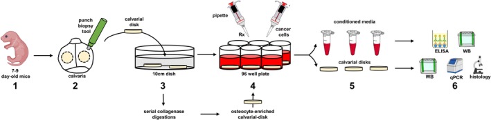
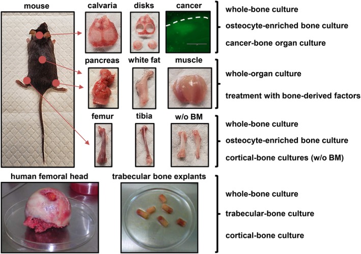
The integrity of the skeleton is maintained by the coordinated and balanced activities of the bone cells. Osteoclasts resorb bone, osteoblasts form bone, and osteocytes orchestrate the activities of osteoclasts and osteoblasts. A variety of in vitro approaches has been used in an attempt to reproduce the complex in vivo interactions among bone cells under physiological as well as pathological conditions and to test new therapies. Most cell culture systems lack the proper extracellular matrix, cellular diversity, and native spatial distribution of the components of the bone microenvironment. In contrast, ex vivo cultures of fragments of intact bone preserve key cell-cell and cell-matrix interactions and allow the study of bone cells in their natural 3D environment. Further, bone organ cultures predict the in vivo responses to genetic and pharmacologic interventions saving precious time and resources. Moreover, organ cultures using human bone reproduce human conditions and are a useful tool to test patient responses to therapeutic agents. Thus, these ex vivo approaches provide a platform to perform research in bone physiology and pathophysiology. In this review, we describe protocols optimized in our laboratories to establish ex vivo bone organ cultures and provide technical hints and suggestions. In addition, we present examples on how this technical approach can be employed to study osteocyte biology, drug responses in bone, cancer-induced bone disease, and cross-talk between bone and other organs © 2020 The Authors. JBMR Plus published by Wiley Periodicals, Inc. on behalf of American Society for Bone and Mineral Research.

Keywords: BONE; CANCER; EX VIVO; FAT; MUSCLE; OSTEOCYTES.


Figures

Figure 1
Figure 1
Step‐by‐step graphical depiction of the procedures described in the Practical Considerations in the Establishment of ex vivo Bone Organ Cultures section. 1. Obtain 7‐ to 9‐day‐old mice. 2. Isolate calvarial disks using a punch biopsy tool. 3. Wash the calvarial cells with PBS. If using osteocyte‐enriched bone organ cultures, perform serial collagenase digestions, wash with PBS, and incubate with complete media overnight. 4. Transfer calvarial disks to independent wells and start treatments (Rx) or add cancer cells to establish cancer–bone organ cultures. 7. At the end of the study, (a) collect conditioned media in Eppendorf tubes and either freeze or perform protein analysis by ELISA and/or Western blot (WB); and (b) collect calvarial disks and either freeze or process for protein (WB), RNA expression (qPCR), or histology.
Figure 2
Figure 2
Ex vivo organ cultures with mouse and human tissues. Ex vivo organ cultures preserve key cell–cell and cell–matrix interactions and allow the study of cells in their natural niche and thus reproduce and predict in vivo responses in a time‐ and cost‐efficient manner. Multiple organ cultures can be established with tissues from the mouse. Calvarias and long bones can be used to establish different types of ex vivo bone organ cultures, including whole‐bone organ cultures, osteocyte‐enriched organ cultures, and cancer–bone organ cultures. The effects of bone‐derived factors on other organ such as muscle, fat, and pancreas can also be examined using whole‐organ cultures treated with bone‐derived factors. Lastly, ex vivo bone organ cultures established with bone explants obtained from femoral heads collected after arthroplasty can be convenient to examine responses in human bone.

Similar articles

Cited by

References

    1. Parfitt AM. A new approach to iliac bone histomorphometry: implications for biomechanics and cell biology. J Clin Invest. 2014;124(1):70–1. - PMC - PubMed
    1. Frost HM. In vivo osteocyte death. J Bone Joint Surg Am. 1960;42‐A:138–43. - PubMed
    1. Marotti G. The structure of bone tissues and the cellular control of their deposition. Ital J Anat Embryol. 1996;101(4):25–79. - PubMed
    1. Plotkin LI, Bellido T. Osteocytic signalling pathways as therapeutic targets for bone fragility. Nat Rev Endocrinol. 2016;12(10):593–605. - PMC - PubMed
    1. Delgado‐Calle J, Bellido T. Osteocytes and skeletal pathophysiology. Curr Mol Biol Rep. 2015;1(4):157–67. - PMC - PubMed

LinkOut - more resources