

Evaluation of Root and Root Canal Morphology of Mandibular First and Second Molars in a Greek Population: A CBCT Study
- PMID: 32161889
- PMCID: PMC7006552
- DOI: 10.14744/eej.2019.19480
Evaluation of Root and Root Canal Morphology of Mandibular First and Second Molars in a Greek Population: A CBCT Study
Abstract
Objective: Τo study the number of roots, canal configurations, and frequency of morphological variations in mandibular first and second molars in a Greek population.
Methods: This study examined 478 mandibular first molars and 524 mandibular second molars using a high-resolution cone-beam computed tomography (CBCT). The number of roots was recorded and the root canal configuration was categorized based on the classification by Vertucci. The presence and configuration of C-shaped root canals were recorded and they were classified according to the Fan classification. The symmetry between the right and the left side was also evaluated.
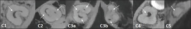
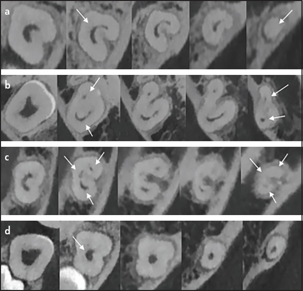
Results: Among the mandibular first molars, 0.2% teeth were single-rooted, 96.4% were two-rooted, and 3.3% were three-rooted. In the mandibular second molars, 12.2%, 82.8%, and 4.9% were single-rooted, two-rooted, and three-rooted, respectively. In two-rooted mandibular first and second molars, the most frequent root canal pattern observed was Vertucci's type II in the mesial root (69.8% and 64.1%, respectively) and Vertucci's type I in the distal root (81.7% and 97.7%, respectively). Three-rooted molars showed one oval-shaped mesial root and two distal roots (56.2% in first molars, 65.4% in second molars), where each distal root contained a single root canal (type I), and the mesial root presented either type II (53.3%), IV (26.6%), I (13.3%), or V (6.6%) canal configurations. C-shaped canals were only detected in mandibular second molars (5.3% of teeth, 10.8% of individuals), and bilateral occurrence was observed in 24.5% patients. The most frequent root canal pattern was Fan's C1 type at the orifice, followed by C3a and C3b in the coronal and middle third, which joined into a single canal (C4) apically.
Conclusion: The characteristics of the root and root canal anatomy of the mandibular first and second molars of Greek individuals were similar to those observed in Caucasians. However, the higher incidence of third roots in mandibular molars in Greek individuals compared to Caucasians requires absolute clinical awareness.
Keywords: Cone-beam computed tomography; greek population; mandibular molars; root canal morphology; root morphology.
Copyright: © 2019 European Endodontic Journal.
Conflict of interest statement
Disclosures Conflict of interest: The authors deny any conflicts of interest related to this study.
Figures


References
-
- Vertucci FJ. Root canal morphology and its relationship to endodontic procedures. Endod Topics. 2005;10(1):3–29.
-
- Vertucci FJ. Root canal anatomy of the human permanent teeth. Oral Surg Oral Med Oral Pathol. 1984;58(5):589–99. - PubMed
-
- Martins JNR, Marques D, Mata A, Caramês J. Root and root canal morphology of the permanent dentition in a Caucasian population:a cone-beam computed tomography study. Int Endod J. 2017;11(50):1013–26. - PubMed
-
- Plotino G, Tocci L, Grande NM, Testarelli L, Messineo D, Ciotti M, et al. Symmetry of root and root canal morphology of maxillary and mandibular molars in a white population:a cone-beam computed tomography study in vivo. J Endod. 2013;39(12):1545–8. - PubMed
LinkOut - more resources
Full Text Sources
Miscellaneous
