. 2020 Jul;30(7):3650-3659.
doi: 10.1007/s00330-020-06776-y. Epub 2020 Mar 11.

Radiomics for lung adenocarcinoma manifesting as pure ground-glass nodules: invasive prediction

Affiliations

Radiomics for lung adenocarcinoma manifesting as pure ground-glass nodules: invasive prediction

Yingli Sun et al. Eur Radiol. 2020 Jul.

Abstract

Objectives: To investigate the value of radiomics based on CT imaging in predicting invasive adenocarcinoma manifesting as pure ground-glass nodules (pGGNs).

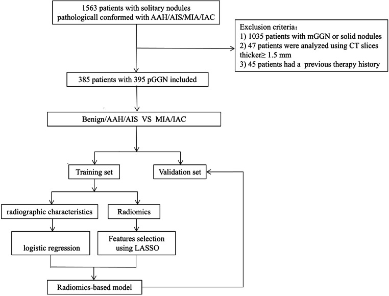
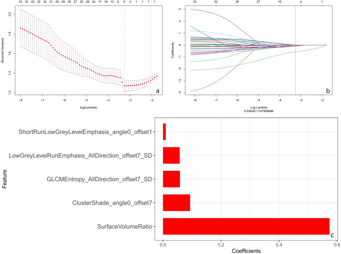
Methods: This study enrolled 395 pGGNs with histopathology-confirmed benign nodules or adenocarcinoma. A total of 396 radiomic features were extracted from each labeled nodule. A Rad-score was constructed with the least absolute shrinkage and selection operator (LASSO) in the training set. Multivariate logistic regression analysis was conducted to establish the radiographic model and the combined radiographic-radiomics model. The predictive performance was validated by receiver operating characteristic (ROC) curve. Based on the multivariate logistic regression analysis, an individual prediction nomogram was developed and the clinical utility was assessed.

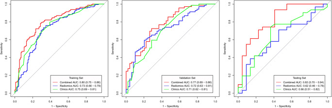
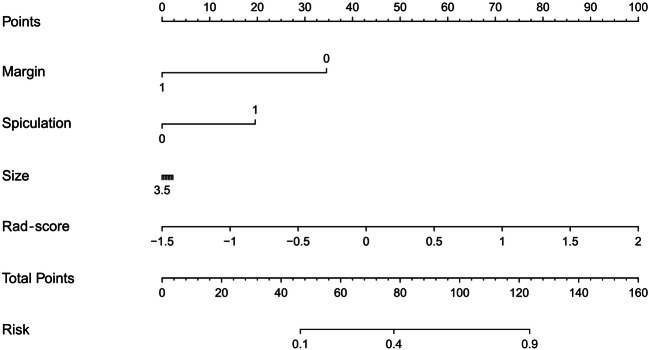
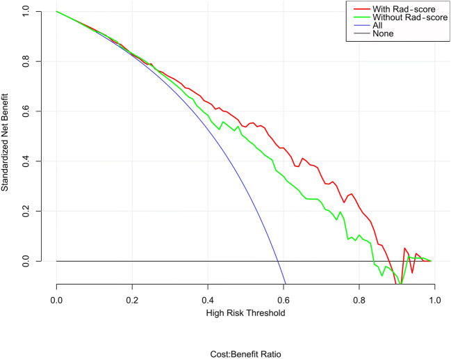
Results: Five radiomic features and four radiographic features were selected for predicting the invasive lesions. The combined radiographic-radiomics model (AUC 0.77; 95% CI, 0.69-0.86) performed better than the radiographic model (AUC 0.71; 95% CI, 0.62-0.81) and Rad-score (AUC 0.72; 95% CI, 0.63-0.81) in the validation set. The clinical utility of the individualized prediction nomogram developed using the Rad-score, margin, spiculation, and size was confirmed in the validation set. The decision curve analysis (DCA) indicated that using a model with Rad-score to predict the invasive lesion would be more beneficial than that without Rad-score and the clinical model.

Conclusions: The proposed radiomics-based nomogram that incorporated the Rad-score, margin, spiculation, and size may be utilized as a noninvasive biomarker for the assessment of invasive prediction in patients with pGGNs.

Key points: • CT-based radiomics analysis helps invasive prediction manifested as pGGNs. • The combined radiographic-radiomics model may be utilized as a noninvasive biomarker for predicting invasive lesion for pGGNs. • Radiomics-based individual nomogram may serve as a vital decision support tool to identify invasive pGGNs, obviating further workup and blind follow-up.

Keywords: Adenocarcinoma; Lung; Nomograms; Solitary pulmonary nodule; X-ray computed tomography.


Conflict of interest statement

One of the authors of this manuscript (Shaofeng Duan) is an employee of GE Healthcare. The remaining authors declare no relationships with any companies whose products or services may be related to the subject matter of the article.

Figures

Fig. 1
Fig. 1
The workflow of the study
Fig. 2
Fig. 2
Noninvasive lesion and invasive lesion appearing as pure GGNs. ad Transverse, coronal, sagittal, and pathology imaging (hematoxylin and eosin, × 100) of an 11-mm pure GGN in the right middle lobe. This nodule was confirmed as non-neoplastic lesion (fibrosis, with alveolar epithelial hyperplasia and dysplasia, vascular malformations). eh Transverse, coronal, sagittal, and pathology imaging (hematoxylin and eosin, × 100) of an 18-mm well-defined pure GGN in the right upper lobe of a 72-year-old woman. This nodule was confirmed as IPA at lobectomy
Fig. 3
Fig. 3
The AUC of Rad-score, radiographic model, and combined model in the training set, validation set, and testing set. The predictive performance of the combined model for an invasive lesion of pGGNs was better than that of the radiographic model and Rad-score in the training, validation, and testing sets
Fig. 4
Fig. 4
Texture feature selection using the least absolute shrinkage and the histogram of the Rad-score based on the selected features. a Selection of the tuning parameter (λ) in the LASSO model via 10-fold cross-validation based on minimum criteria. Binomial deviances from the LASSO regression cross-validation procedure were plotted as a function of log (λ). The optimal λ value of 0.038 was selected. b The black vertical line was drawn at the value selected using 10-fold cross-validation in a. The 5 resulting features with nonzero coefficients were indicated in the plot. c The y-axis indicates the selected five radiomics, and the x-axis represents the coefficient of radiomics
Fig. 5
Fig. 5
Radiomics-based nomogram was developed in the training set, and the Rad-score, margin, spiculation, and size were incorporated
Fig. 6
Fig. 6
Decision curve analysis for the model with and without Rad-score. The decision curve showed that if the threshold probability of a patient or a doctor is > 10%, using a model with the Rad-score to predict the invasive lesion would be more beneficial than that without the Rad-score

Similar articles

Cited by

References

    1. Austin JH, Muller NL, Friedman PJ, et al. Glossary of terms for CT of the lungs: recommendations of the Nomenclature Committee of the Fleischner Society. Radiology. 1996;200:327–331. doi: 10.1148/radiology.200.2.8685321. - DOI - PubMed
    1. Henschke CI, Yankelevitz DF, Mirtcheva R, et al. CT screening for lung cancer: frequency and significance of part-solid and nonsolid nodules. AJR Am J Roentgenol. 2002;178:1053–1057. doi: 10.2214/ajr.178.5.1781053. - DOI - PubMed
    1. Heidinger BH, Anderson KR, Nemec U, et al. Lung adenocarcinoma manifesting as pure ground-glass nodules: correlating CT size, volume, density, and roundness with histopathologic invasion and size. J Thorac Oncol. 2017;12:1288–1298. doi: 10.1016/j.jtho.2017.05.017. - DOI - PubMed
    1. Chang B, Hwang JH, Choi YH, et al. Natural history of pure ground-glass opacity lung nodules detected by low-dose CT scan. Chest. 2013;143:172–178. doi: 10.1378/chest.11-2501. - DOI - PubMed
    1. Kakinuma R, Muramatsu Y, Kusumoto M, et al. Solitary pure ground-glass nodules 5 mm or smaller: frequency of growth. Radiology. 2015;276:873–882. doi: 10.1148/radiol.2015141071. - DOI - PubMed