. 2020 May 6;15(9):738-743.
doi: 10.1002/cmdc.202000069. Epub 2020 Mar 19.

Resolving Binding Events on the Multifunctional Human Serum Albumin

Affiliations

Resolving Binding Events on the Multifunctional Human Serum Albumin

Lea Wenskowsky et al. ChemMedChem. .

Abstract

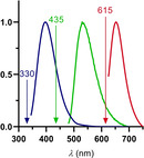
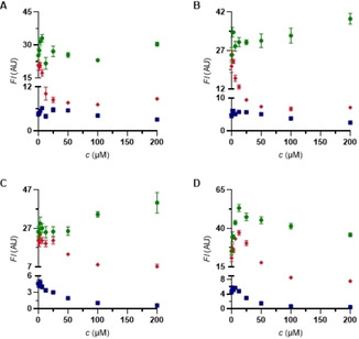
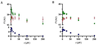
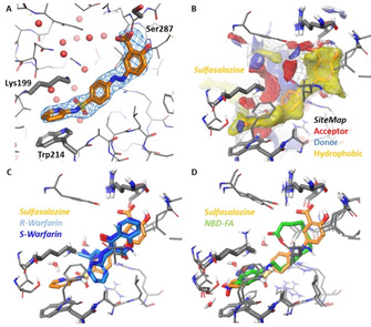
Physiological processes rely on initial recognition events between cellular components and other molecules or modalities. Biomolecules can have multiple sites or mode of interaction with other molecular entities, so that a resolution of the individual binding events in terms of spatial localization as well as association and dissociation kinetics is required for a meaningful description. Here we describe a trichromatic fluorescent binding- and displacement assay for simultaneous monitoring of three individual binding sites in the important transporter and binding protein human serum albumin. Independent investigations of binding events by X-ray crystallography and time-resolved dynamics measurements (switchSENSE technology) confirm the validity of the assay, the localization of binding sites and furthermore reveal conformational changes associated with ligand binding. The described assay system allows for the detailed characterization of albumin-binding drugs and is therefore well-suited for prediction of drug-drug and drug-food interactions. Moreover, conformational changes, usually associated with binding events, can also be analyzed.

Keywords: albumin binding; drug interactions; kinetics investigations; multicolor assays; switchSENSE technology.


Conflict of interest statement

The authors declare no conflict of interest.

Figures

Scheme 1
Scheme 1
Synthesis of BODIPY derivatives 5 ac.
Figure 1
Figure 1
A) Absorption and emission spectra (both normalized) of dimethylamino‐BODIPY 5 a (40 μM ethanol). λ exc=625 nm. B) Titration of dimethylamino‐BODIPY 5 a (0–50 μM) to HSA (12.5 μM) (K D=4.6±0.8 μM. R2=0.9176). λ exc=615 nm. λ em=690 nm. C) Determination of the binding stoichiometry of dimethylamino‐BODIPY 5 a to HSA (12.5 μM, Job plot). λ exc=615 nm. λ em=690 nm. D) Competition experiment of ibuprofen (1) against dimethylamino‐BODIPY 5 a on HSA (12.5 μM each). λ exc=615 nm. λ em=690 nm.
Scheme 2
Scheme 2
Synthesis of warfarin derivatives 8 ac.
Figure 2
Figure 2
A) Absorption and emission spectra (both normalized) of warfarin derivative 8 a (40 μM ethanol). λ exc=340 nm. B) Titration of warfarin derivative 8 a (0–400 μM) to HSA (25 μM) (K D=26.5±3.0 μM. R2=0.9607). λ exc=330 nm. λ em=384 nm. C) Determination of the binding stoichiometry of warfarin derivative 8 a to HSA (25 μM, Job plot). λ exc=330 nm. λ em=384 nm. D) Competition experiment of iophenoxic acid (3) against warfarin derivative 8 a on HSA (25 μM each). λ exc=330 nm. λ em=384 nm.
Figure 3
Figure 3
Absorption spectra (normalized) of warfarin derivative 8a (blue), NBD‐FA (green) and dimethylamino‐BODIPY 5a (red). Excitation wavelengths for each dye were drawn.
Figure 4
Figure 4
Competition experiments against NBD‐FA/BODIPY 5 a/coumarin 8 a (8 μM each) on HSA. NBD‐FA. BODIPY 5 a. warfarin derivative 8 a. A) ibuprofen (1). B) ketoprofen (4). C) indomethacin (5). D) iophenoxic acid (3).
Figure 5
Figure 5
Competition experiments against NBD‐FA/BODIPY 5 a/coumarin 8 a (8 μM each) on HSA. NBD‐FA. BODIPY 5 a. warfarin derivative 8 a. A) sulfasalazine (6). B) balsalazide (7).
Figure 6
Figure 6
X‐ray crystal structure of HSA in complex with sulfasalazine (6) (PDB ID 6r7s resolution 2.2 Å). A) Binding of sulfasalazine (6) (orange carbon atoms) to Sudlow‐site I plus F oF c omit map contoured at 3σ. Key amino acids and water oxygen atoms are indicated. B) Binding site analysis of the HSA‐6 complex using SiteMap. Red regions indicate favorable potential H‐bond acceptor interactions; blue regions indicate favorable H‐bond donor interactions and yellow regions indicate favorable hydrophobic interactions. C) Superposition of sulfasalazine (6) with the enantiomers of warfarin (2) from PDB 1ha2 (S‐warfarin, dark‐blue) and 1h9z (R‐warfarin, light‐blue), both at 2.5 Å resolution.24 D) Superposition of sulfasalazine with NBD‐FA (green carbon atoms) from PDB 6ezq3 at 2.5 Å resolution.

References

    1. None
    1. Oettl K., Stauber R. E., Br. J. Pharmacol. 2007, 151, 580–590; - PMC - PubMed
    1. Fehske K. J., Müller W. E., Wollert U., Biochem. Pharmacol. 1981, 30, 687–692; - PubMed
    1. Spector A. A., Santos E. C., Ashbrook J. D., Fletcher J. E., Ann. N. Y. Acad. Sci. 1973, 226, 247–258; - PubMed
    1. Spector A. A., J. Lipid Res. 1975, 16, 165–179; - PubMed