Review
. 2020 Jul;12(4):e1624.
doi: 10.1002/wnan.1624. Epub 2020 Mar 11.

Image-guided tumor surgery: The emerging role of nanotechnology

Affiliations
Review

Image-guided tumor surgery: The emerging role of nanotechnology

Nicholas E Wojtynek et al. Wiley Interdiscip Rev Nanomed Nanobiotechnol. 2020 Jul.

Abstract

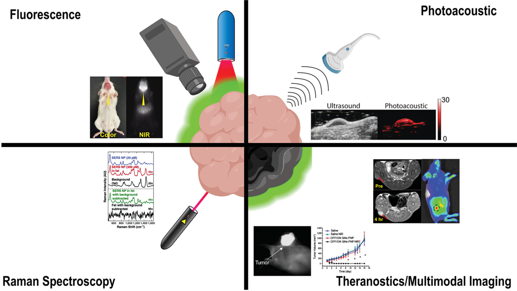
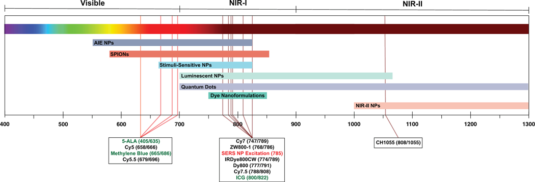
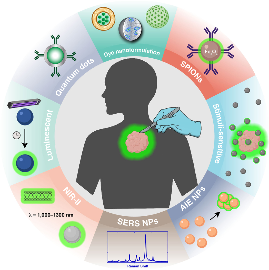
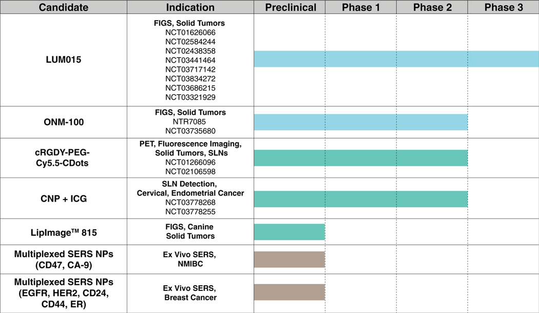
Surgical resection is a mainstay treatment for solid tumors. Yet, methods to distinguish malignant from healthy tissue are primarily limited to tactile and visual cues as well as the surgeon's experience. As a result, there is a possibility that a positive surgical margin (PSM) or the presence of residual tumor left behind after resection may occur. It is well-documented that PSMs can negatively impact treatment outcomes and survival, as well as pose an economic burden. Therefore, surgical tumor imaging techniques have emerged as a promising method to decrease PSM rates. Nanoparticles (NPs) have unique characteristics to serve as optical contrast agents during image-guided surgery (IGS). Recently, there has been tremendous growth in the volume and types of NPs used for IGS, including clinical trials. Herein, we describe the most recent contributions of nanotechnology for surgical tumor identification. This article is categorized under: Therapeutic Approaches and Drug Discovery > Nanomedicine for Oncologic Disease Implantable Materials and Surgical Technologies > Nanoscale Tools and Techniques in Surgery Diagnostic Tools > in vivo Nanodiagnostics and Imaging.

Keywords: fluorescence-guided surgery; image-guided surgery; imaging; nanoparticle; optical; surgical oncology.


Conflict of interest statement

CONFLICT OF INTEREST

The authors have declared no conflicts of interest for this article.

Figures

FIGURE 1
FIGURE 1
Optical imaging modalities used for IGS with examples of surgical tumor detection from the literature: fluorescence (Reprinted with permission from Zhao et al., 2017. Copyright 2016 Springer Nature), photoacoustic (Reprinted with permission from Thawani et al., 2017. Copyright 2017 Wiley-VCH Verlag GmbH & Co. KGaA, Weinheim), Raman spectroscopy (Reprinted with permission from Mohs et al., 2010. Copyright 2010 American Chemical Society), theranostics (Reprinted with permission from Li et al., 2018. Copyright Ivy Spring International Publisher), and multimodal imaging (Reprinted with permission from Jin, Li, Yang, & Tian, 2019. Copyright Acta Materialia Inc. published by Elsevier Ltd.)
FIGURE 2
FIGURE 2
Emission profiles of commonly used dyes and NPs for surgical tumor detection and the emission wavelengths of NPs covered in this review. The majority of dyes fall into the NIR range. Fluorophores in the green text indicate FDA-approved compounds. Red text indicates the typical excitation wavelength for SERS NPs. The excitation and emission wavelength (nm) are reported in parenthesis after each dye
FIGURE 3
FIGURE 3
Types of nanoparticles used for surgical image guidance
FIGURE 4
FIGURE 4
Nanoparticles currently undergoing clinical investigation

References

    1. Agarwal V, Bajpai M, & Sharma A. (2018). Patented and approval scenario of Nanopharmaceuticals with relevancy to biomedical application, manufacturing procedure and safety aspects. Recent Patents on Drug Delivery & Formulation, 12(1), 40–52. 10.2174/1872211312666180105114644 - DOI - PubMed
    1. Agdeppa ED, & Spilker ME (2009). A review of imaging agent development. The AAPS Journal, 11(2), 286–299. 10.1208/s12248-009-9104-5 - DOI - PMC - PubMed
    1. Ai T, Shang W, Yan H, Zeng C, Wang K, Gao Y, … Tian J. (2018). Near infrared-emitting persistent luminescent nanoparticles for hepatocellular carcinoma imaging and luminescence-guided surgery. Biomaterials, 167, 216–225. 10.1016/j.biomaterials.2018.01.031 - DOI - PubMed
    1. Alam IS, Steinberg I, Vermesh O, van den Berg NS, Rosenthal EL, van Dam GM, … Rogalla S. (2018). Emerging intraoperative imaging modalities to improve surgical precision. Molecular Imaging and Biology, 20(5), 705–715. 10.1007/s11307-0181227-6 - DOI - PubMed
    1. Alander JT, Kaartinen I, Laakso A, Pätilä T, Spillmann T, Tuchin VV, … Välisuo P. (2012). A review of Indocyanine green fluorescent imaging in surgery. International Journal of Biomedical Imaging, 2012, 1–26. 10.1155/2012/940585 - DOI - PMC - PubMed

FURTHER READING

    1. Wang P, Fan Y, Lu L, Liu L, Fan L, Zhao M, … Zhang F. (2018). NIR-II nanoprobes in-vivo assembly to improve image-guided surgery for metastatic ovarian cancer. Nature Communications, 9(1), 2898. 10.1038/s41467-018-05113-8 - DOI - PMC - PubMed
    1. Wang P, Wang X, Luo Q, Li Y, Lin X, Fan L, … Liu X. (2019). Fabrication of red blood cell-based multimodal Theranostic probes for second near-infrared window fluorescence imaging-guided tumor surgery and photodynamic therapy. Theranostics, 9(2), 369–380. 10.7150/thno.29817 - DOI - PMC - PubMed
    1. Wang P, Shi Y, Zhang S, Huang X, Zhang J, Zhang Y, … Dong X. (2019). Hydrogen peroxide responsive iron-based nanoplatform for multimodal imaging-guided cancer therapy. Small, 15(4), 1803791. 10.1002/smll.201803791 - DOI - PubMed
    1. Wang YW, Reder NP, Kang S, Glaser AK, Yang Q, Wall MA, … Liu JTC (2017). Raman-encoded molecular imaging with topically applied SERS nanoparticles for intraoperative guidance of lumpectomy. Cancer Research, 77(16), 4506–4516. 10.1158/0008-5472.CAN-17-0709 - DOI - PMC - PubMed

Publication types