. 2020 May;34(5):6086-6098.
doi: 10.1096/fj.201902426R. Epub 2020 Mar 12.

Regulation of stanniocalcin-1 secretion by BeWo cells and first trimester human placental tissue from normal pregnancies and those at increased risk of developing preeclampsia

Affiliations

Regulation of stanniocalcin-1 secretion by BeWo cells and first trimester human placental tissue from normal pregnancies and those at increased risk of developing preeclampsia

Naila Abid et al. FASEB J. 2020 May.

Abstract

Stanniocalcin-1 (STC-1) is a multi-functional glycosylated peptide present in the plasma of healthy women postpartum and increased further in pregnancies complicated by preeclampsia. Although the STC-1 gene is expressed by the placenta what regulates its secretion and from which cells at the feto-maternal interface is unknown. Here, we demonstrate for the first time that the syncytiotrophoblast and cytotrophoblast are a major site of STC-1 protein expression in first trimester placental tissue. Further, in response to low oxygen, first trimester chorionic villous tissue from pregnancies at increased risk of developing preeclampsia secreted significantly more STC-1 than normal tissue under the same conditions. Using the human trophoblast cell line BeWo we have shown that low oxygen increased the secretion of STC-1 but it required co-stimulation with the Adenosine-3', 5'-cyclic monophosphate (cAMP) analogue, 8-Bromo adenosine-3', 5'-cyclic monophosphate cAMP (8 Br-cAMP) to reach significance. Inhibition of Hypoxia inducible factor 2α (HIF-2α) and the Phosphatidylinositol-3 kinase (PI3 -Kinase)/AKT/Serum and glucocorticoid-induced kinase-1(SGK-1) pathway resulted in significant inhibition of STC-1 secretion. As both low oxygen and cAMP are known to play a central role in placental function, their regulation of STC-1 points to a potentially important role in the maintenance of a normal healthy pregnancy and we would hypothesize that it may act to protect against prolonged placental hypoxia seen in preeclampsia.

Keywords: first trimester; hypoxia; placenta; stanniocalcin-1; trophoblasts.


Conflict of interest statement

The authors declare no conflicts of interest.

Figures
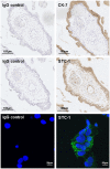
Figure 1
Figure 1
Histological and cytochemical analysis of first trimester placental tissue sections and BeWo cells grown in culture. Top Left: Treated with non‐immune IgG negative control stained purple. Top Right: Treated with mouse monoclonal anti‐CK‐7 antibody, brown highlights positive expression of CK‐7. Middle Left: Treated with non‐immune IgG negative control, Middle Right: Treated with mouse anti‐STC‐1 antibody, brown highlights positive expression STC‐1. The data are representative of results from three different patient samples performed on separate occasions. BeWo cells were grown 1% O2 and stimulated with 100 µM 8Br‐cAMP for 24 h. Bottom Left: Treated with non‐immune IgG negative control, Bottom Right: Treated with mouse anti‐STC‐1 antibody (Green) and the nuclei stained with Dapi (Blue)
Figure 2
Figure 2
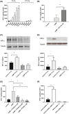
Concentration of STC‐1 in serum obtained from pregnancies in the third trimester with known outcomes. A, Serum was obtained from normal and preeclamptic pregnancies the third trimester. The concentration of STC‐1 was determined by ELISA and the data log‐transformed. The data shown are expressed as mean ± SEM of n = 19 normal pregnancies and n = 11 preeclamptic pregnancies. Significance was determined using an unpaired student t test *P < .05. Secretion of STC‐1 by first trimester chorionic tissue from nRI and hRI pregnancies incubated at 1% and 21% O2. B, First trimester chorionic villous tissue was incubated at either 1% or 21% O2 for 72 h. The medium was then collected and the tissue weighed. STC‐1 was determined by ELISA and the amount produced was corrected for the weight of tissue. The number of pregnancies in each group were nRI 1% (n = 14), hRI 1% (n = 12), nRI 21% (n = 13) hRI 21% (n = 12). The data were tested for normality using the D'Agostino & Pearson normality test before analysis using a one‐way ANOVA followed by a Holm‐Sidak's multiple comparisons test, **P < .01
Figure 3
Figure 3
The effect of cAMP and oxygen on production of STC‐1 by BeWo cells. A, BeWo cells (3 × 105 per well) were incubated in either 1% or 21% oxygen with increasing concentration of 8Bromo‐cAMP. The data were expressed as mean + sem, of n = 4 independent expts. B, BeWo cells were cultured in 21% O2 and stimulated with 100 µM 8 Bromo‐cAMP in the presence and absence of 50 µM DFO (n = 4 independent expts). The effect of the low oxygen and cAMP on the expression of HIF‐1, and HIF‐2α. BeWo cells were cultured in 1% and 21% O2 in presence and absence of 100 µM 8 Bromo‐cAMP for 48 h and cell lysates were examined by western blot analysis. C, HIF‐1α (n = 4 independent expts) and D, HIF‐2α (n = 7 independent expts) were detected by chemiluminescence, using tubulin as the loading control. The effect of inhibiting HIF‐2α on the secretion of STC‐1 was determined using (E) PT2385, (n = 3 independent expts) or (F) RNA interference, (n = 4 independent expts) in the presence and absence of 100 µM 8 Bromo‐cAMP. Stimulation in all experiments was for 48 h. The concentration of STC‐1 in the medium was determined by ELISA. Total cellular protein was determined by Bradford assay. The results are expressed as pg of STC‐1/mg of protein. The level of significance was determined using either, a Student t test or a one‐way ANOVA and a Sidak's multiple comparisons test where appropriate, *P < .05, **P < .01, P < .005(**), P < .0005 (***), P < .0001 (****)
Figure 4
Figure 4
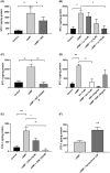
The effect of the inhibiting PI3kinase, Akt and mTORC 1 and 2 on the secretion of STC‐1 by BeWo cells. BeWo cells were cultured in 1% O2 in 100 µM 8 Bromo‐cAMP in the presence and absence of (A) the PKA inhibitor H89 (10 µM), (B) the PI3kinase inhibitor Ly 294002 (LY) at 1, 5 and 10 µM (n = 3) (C) Akt inhibitor IV (2.5 µM) or (D) either the mTORC1 inhibitor rapamycin (RAP) or the mTORC1 and 2 inhibitor KU 0063794 (KU). (E) 10 µM (n = 5) and 100 µM (n = 3) GSK650394 in 1% O2 or, (F) 1 µM Kenpaullone. After 48 h, the medium was removed and the STC‐1 secreted measured by ELISA. Total cellular protein was determined by Bradford assay. The results are expressed as pg of STC‐1/mg of protein. All data were expressed as mean + SEM. The level of significance was determined using a one‐way ANOVA and a Sidak's multiple comparisons test, or an unpaired t‐test *P < .5, **P < .01 and ***P < .001
Figure 5
Figure 5
Summary of the pathway intermediates involved in the secretion STC‐1 by BeWo cells. Solid arrows indicate demonstrated connections, dashed arrows are connections based on the literature

References

    1. Chang AC, Janosi J, Hulsbeek M, et al. A novel human cDNA highly homologous to the fish hormone stanniocalcin. Mol Cell Endocrinol. 1995;112:241‐247. - PubMed
    1. Trindade DM, Silva JC, Navarro MS, Torriani IC, Kobarg J. Low‐resolution structural studies of human stanniocalcin‐1. BMC Struct Biol. 2009;9:57. 10.1186/1472-6807-9-57. - DOI - PMC - PubMed
    1. Mohammadipoor A, Lee RH, Prockop DJ, Bartosh TJ. Stanniocalcin‐1 attenuates ischemic cardiac injury and response of differentiating monocytes/macrophages to inflammatory stimuli. Transl Res. 2016;177:127‐142. - PMC - PubMed
    1. Ellard JP, McCudden CR, Tanega C, et al. The respiratory effects of stanniocalcin‐1 (STC‐1) on intact mitochondria and cells: STC‐1 uncouples oxidative phosphorylation and its actions are modulated by nucleotide triphosphates. Mol Cell Endocrinol. 2007;264:90‐101. - PubMed
    1. Sheikh‐Hamad D, Bick R, Wu GY, et al. Stanniocalcin‐1 is a naturally occurring L‐channel inhibitor in cardiomyocytes: relevance to human heart failure. Am J Physiol Heart Circ Physiol. 2003;285:H442‐H448. - PubMed

Publication types