Clinical Trial
. 2020 Apr 9;5(7):e134838.
doi: 10.1172/jci.insight.134838.

Multimodal immune phenotyping of maternal peripheral blood in normal human pregnancy

Affiliations
Clinical Trial

Multimodal immune phenotyping of maternal peripheral blood in normal human pregnancy

Richard Apps et al. JCI Insight. .

Abstract

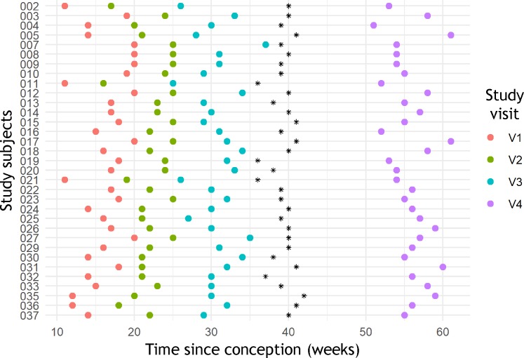
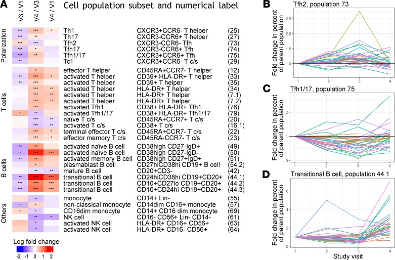
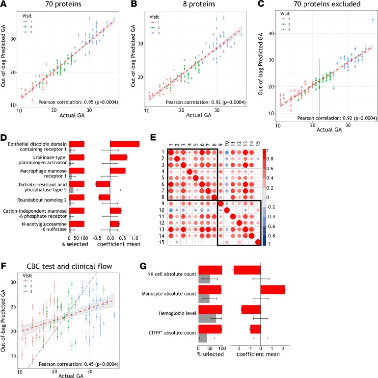
Changes in maternal immunity during pregnancy can result in an altered immune state, and as a natural perturbation, this provides an opportunity to understand functional interactions of the immune system in vivo. We report characterization of maternal peripheral immune phenotypes for 33 longitudinally sampled normal pregnancies, using clinical measurements of complete blood counts and major immune cell populations, as well as high parameter flow cytometry for 30 leukocyte antigens characterizing 79 cell populations, and monitoring of 1305 serum proteins using the SomaLogic platform. Cellular analyses characterized transient changes in T cell polarization and more persistent alterations in T and B cell subset frequencies and activation. Serum proteomic analysis identified a potentially novel set of 7 proteins that are predictive of gestational age: DDR1, PLAU, MRC1, ACP5, ROBO2, IGF2R, and GNS. We further show that gestational age can be predicted from the parameters obtained by complete blood count tests and clinical flow cytometry characterizing 5 major immune cell populations. Inferring gestational age from this routine clinical phenotyping data could be useful in resource-limited settings that lack obstetric ultrasound. Overall, both the cellular and proteomic analyses validate previously reported phenotypic immunological changes of pregnancy and uncover potentially new alterations and predictive markers.

Keywords: Cellular immune response; Immunology; Obstetrics/gynecology; Proteomics; Reproductive Biology.


Conflict of interest statement

Conflict of interest: The authors have declared that no conflict of interest exists.

Figures

Figure 1
Figure 1. Distribution of time points sampled in the study population.
For all 33 individuals the time points sampled are shown for 3 visits during gestation, and 1 visit after parturition, with parturition marked as a black asterisk.
Figure 2
Figure 2. Changes in peripheral blood immune populations throughout pregnancy.
(A) Using all 33 subjects in our study, longitudinal comparisons between visits 1 and 3 (early and late gestation), visits 3 and 4 (late gestation and postpartum), or visits 1 and 4 (early gestation and postpartum) identified 32 subsets of immune cell populations with fold change greater than 1.2 or less than 0.8 and significant differences for at least 1 comparison using Wilcoxon’s signed-rank paired tests (*FDR < 0.05, **FDR < 0.01, and ***FDR < 0.001). T c/s, CD8+ T cells (cytotoxic/suppressors). (B) For representative populations that demonstrated different patterns of change, fold changes compared with visit 1 are shown for all 4 visits studied. Type 2 CD4+ Tfh cells increased during gestation and declined after parturition. (C) Type 1/17 Tfh cells decreased during gestation and rebounded after parturition. (D) Transitional B cells declined significantly during gestation but increased much more strongly after parturition. See A for FDR-adjusted P values.
Figure 3
Figure 3. Serum proteins and cell populations predictive of GA.
(A and B) EN models generated from both a set of 70 proteins, and a subset of 8 proteins previously identified to predict GA, demonstrated significant correlation between observed and predicted GA when using serum proteomic data from 3 time points during pregnancy for the 33 women in our study. Predicted GA is shown with mean and standard deviation of 100 model iterations randomly sampling training and test sets, with red dashed line indicating a linear regression with 95% confidence interval. (C) Excluding these 70 proteins and selecting from the remaining 1124 serum proteins measured, EN models still showed significant correlation between observed and predicted GA. (D) For the 7 proteins newly identified to predict GA, frequency of selection and average weights differed significantly from null models and are plotted in red showing mean and standard deviation from the 100 model iterations randomly sampling training and test sets. In gray are shown null models with mean and standard deviation from random permutation of sample labels 25 times and 100 random samplings of the training and test sets. (E) The relationship between new and previously identified proteins predicting GA was assessed by pairwise Pearson’s correlations of protein relative intensities during gestation in our data set. Eight proteins previously identified to predict GA by Aghaeepour et al., and the 7 proteins identified in this study, are numbered 1–8 and 9–15, respectively, and defined as follows: 1, chorionic somatomammotropin hormone; 2, prolactin; 3, macrophage colony-stimulating factor 1 receptor; 4, polymeric immunoglobulin receptor; 5, proto-oncogene tyrosine-protein kinase receptor Ret; 6, glypican-3; 7, granulins; 8, α-fetoprotein; 9, macrophage mannose receptor 1; 10, tartrate-resistant acid phosphatase type 5; 11, N-acetylglucosamine-6-sulfatase; 12, cation-independent mannose-6-phosphate receptor; 13, epithelial discoidin domain-containing receptor 1; 14, urokinase-type plasminogen activator; and 15, roundabout homolog 2. (F) Standard clinical phenotyping measurements from CBC tests and clinical flow cytometry can also be used to predict GA. For the 33 subjects sampled at 3 time points during pregnancy, 19 routinely acquired clinical parameters were used to generate EN models for which predicted and observed GA correlated significantly, with black dashed line marking equal GA. (G) Frequency of selection and average weights are shown for the significant model features, plotted as in D.

References

    1. Avelino MM, Campos D, Parada JB, Castro AM. Risk factors for Toxoplasma gondii infection in women of childbearing age. Braz J Infect Dis. 2004;8(2):164–174. - PubMed
    1. Shulman CE, Dorman EK. Importance and prevention of malaria in pregnancy. Trans R Soc Trop Med Hyg. 2003;97(1):30–35. doi: 10.1016/S0035-9203(03)90012-5. - DOI - PubMed
    1. Harger JH, et al. Risk factors and outcome of varicella-zoster virus pneumonia in pregnant women. J Infect Dis. 2002;185(4):422–427. doi: 10.1086/338832. - DOI - PubMed
    1. Schlaudecker EP, Ambroggio L, McNeal MM, Finkelman FD, Way SS. Declining responsiveness to influenza vaccination with progression of human pregnancy. Vaccine. 2018;36(31):4734–4741. doi: 10.1016/j.vaccine.2018.05.117. - DOI - PMC - PubMed
    1. Klein SL, Passaretti C, Anker M, Olukoya P, Pekosz A. The impact of sex, gender and pregnancy on 2009 H1N1 disease. Biol Sex Differ. 2010;1(1):5. doi: 10.1186/2042-6410-1-5. - DOI - PMC - PubMed

Publication types