. 2020 Mar 12;16(3):e1008323.
doi: 10.1371/journal.ppat.1008323. eCollection 2020 Mar.

Structural basis of Fusarium myosin I inhibition by phenamacril

Affiliations

Structural basis of Fusarium myosin I inhibition by phenamacril

Yuxin Zhou et al. PLoS Pathog. .

Abstract

Fusarium is a genus of filamentous fungi that includes species that cause devastating diseases in major staple crops, such as wheat, maize, rice, and barley, resulting in severe yield losses and mycotoxin contamination of infected grains. Phenamacril is a novel fungicide that is considered environmentally benign due to its exceptional specificity; it inhibits the ATPase activity of the sole class I myosin of only a subset of Fusarium species including the major plant pathogens F. graminearum, F. asiaticum and F. fujikuroi. To understand the underlying mechanisms of inhibition, species specificity, and resistance mutations, we have determined the crystal structure of phenamacril-bound F. graminearum myosin I. Phenamacril binds in the actin-binding cleft in a new allosteric pocket that contains the central residue of the regulatory Switch 2 loop and that is collapsed in the structure of a myosin with closed actin-binding cleft, suggesting that pocket occupancy blocks cleft closure. We have further identified a single, transferable phenamacril-binding residue found exclusively in phenamacril-sensitive myosins to confer phenamacril selectivity.


Conflict of interest statement

The authors have declared that no competing interests exist.

Figures

Fig 1
Fig 1. FgMyoI ATPase activity.
(A) Myosin/actin catalytic cycle (modified from [13]). ATP-bound myosin is dissociated from actin and becomes rapidly hydrolyzed in the catalytic site. Release of the hydrolyzed phosphate is slow and requires actin binding for acceleration. Initial weak electrostatic actin binding partially closes the actin-binding cleft and stabilizes a switch loop movement. This allows concerted phosphate release, full cleft closure, actin binding and lever arm movement (power stroke). This is followed by ADP release, which allows rapid ATP rebinding, actin dissociation, and a lever arm recovery stroke. (B) Calmodulin dependence of the FgMyoI activity measured by ATP-Glo ATPase luminescence assay. (C) Phenamacril inhibits the ATPase activity of FgMyoI; n = 3, error bars = SD.
Fig 2
Fig 2. FgMyoI structure.
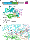
(A) Domain map of F. graminearum FgMyoI encoded by the myo5 gene in complex with F. graminearum calmodulin (CaM). (B) Structure overview. (C) Chemical structure of phenamacril. (D) Catalytic center and phenamacril pocket with ligand Fo-Fc omit map contoured at 3.0 σ. The ligand phenamacril shows strong electron density at 3 σ except the ethyl ester tail that is partially disordered due to its flexibility. Pocket residues are shown in thick line presentation, the Mg2+ ion as green sphere, and water molecules as red spheres. *: pocket residues that have been found mutated in highly resistant (K216E/R, S217L/P, E420G/K/D) or moderately resistant (S418R, I424R, A577G) Fusarium strains.
Fig 3
Fig 3. Allosteric myosin motor domain binding sites.
Allosteric modulators of myosin motor domains overlaid onto the structure of FgMyoI. Switch 2 is shown in pink. Myosin inhibitors: Pentachloropseudilin (PDB 2XEL [18]), pentabromopseudilin (PDB 2JHR [24]), blebbistatin (PDB 1YV3 [37]), tribromodichlorpseudilin (PDB 2XO8 [38]), ammosamide 272 (PDB 4AE3); positive cardiac inotrope: omecamtiv mecarbil (PDB 4PA0 [39]). The insert illustrates the steric clash between Switch 2 Y409 and blebbistatin.
Fig 4
Fig 4. Localization of mutant residues that cause mild phenamacril resistance.
(A) A577, R580, and I581 are pocket residues. A577 directly interacts with phenamacril, whereas R580 and I581 interact with pocket residues L213 from U50 (cyan) and C423, D540, and A577 from L50 (green). (B) A135 is a P-loop (yellow) residue of the ATP-pocket that interacts with the α- and β-phosphates of ATP. (C) I434 of helix HP (helix 12) interacts with L588, V589, L592, and M593 of helix HW (helix 19). These interactions likely determine the orientation of the N-terminus of HP, which leads into Switch 2 and directly interacts with phenamacril. (D) V151 and P204 are residues in the Transducer (purple), which communicates conformational changes between motor and converter domains. The Transducer consists of the last three β-strands of the U50 β-sheet and Loop 1 (flanked by V151) and the P204-containing β-bulge.
Fig 5
Fig 5. Phenamacril blocks closure of the actin-binding cleft.
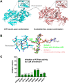
(A) Side-by-side structural alignment of the myosin motor domain in closed (ATP-γS/phenamacril-bound FgMyoI) and open (nucleotide-free chicken myosin V) conformation overlaid with the position of phenamacril from the FgMyoI structure. The inserts show the phenamacril pocket as mesh in the open conformation and the corresponding pocket in the closed conformation. (B) Switch 2 Y409 binds the phenamacril pocket. Fg Switch 2 in the open (pre-powerstroke) conformation is shown in green, the Switch 2 conformation of chicken myosin V in closed conformation is shown in pink. (C) Phenamacril pocket mutations relieve inhibition of FgMyoI ATPase activity. Reactions contained 0.5 μM myosin and 0.1 μM calmodulin. n = 3, error bars = SD. ****: p-value<0.0001 (One-way ANOVA).
Fig 6
Fig 6. Colony morphology of wild-type (PH-1) and pocket mutant Fusarium graminearum.
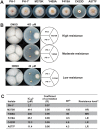
(A) Growth state of F. graminearum isolates on potato dextrose agarose (PDA) at 25 °C for 3 days. * F. graminearum PH-1 transformed with hygromycin resistance cassette. (B) Growth state of F. graminearum isolates on PDA with DMSO and indicated concentration of phenamacril at 25 °C for 3 days. (C) IC50 and RF values. aIC50 = effective concentration for 50% inhibition. bRF, Resistance factor (ratio of IC50 for a resistant isolate relative to IC50 for the original isolate. cRF>100 (high resistance, HR);10<RF<100 (intermediate resistance, MR); RF<10 (low resistance, LR).
Fig 7
Fig 7. Alignment of myosin domains in nucleotide-bound and nucleotide-free states.
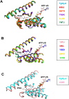
(A) Alignment of FgMyoI with five structures of myosin motor domains in nucleotide-bound state with relatively more open actin-binding clefts. (B) Alignment of five structures of myosin motor domains in nucleotide-free state with closed actin-binding clefts. The positions of phenamacril (PHA) and ATP-γS from FgMyoI are shown for orientation. Parts of the motor domains that are not close to the phenamacril-binding pocket have been removed for clarity. (C) Close-up of structure overlay of FgMyoI with that of chicken myosin in nucleotide-free, closed cleft conformation. (4ANJ [44]/2BKH [45]: Sus scrofa class 6 myosin; 3MYH [46]/1W9K: Dictyostelium discoideum class 2 myosin; 1LKX [36]: Dictyostelium discoideum class 1 myosin; 1W7J [40]/1W8J [40]/1OE9 [41]: Gallus gallus class 5 myosin; 3I5G [42]: Doryteuthis pealeii class 2 myosin; 2OS8 [42]: Placopecten magellanicus class 2 myosin).
Fig 8
Fig 8. M375 is a major phenamacril specificity determinant.
(A) Conservation among the phenamacril pocket residues in myosin I motor domains from phenamacril-sensitive strains (Fg, Fo, Fv) and phenamacril-insensitive strains (Fs, Mg, Bc, Af, Sc). Fg: Fusarium graminearum, Fo: F. oxysporin, Fv: F. verticillioides, Fs: F. solani, Mg: Magnaporthe grisea, Bc: Botrytis cinerea, Af: Aspergillus flavus, Sc: Saccharomyces cerevisiae. (B) Overlay of FgMyoI phenamacril pocket residues (cyan) and their corresponding residues from Dictyostelium discoideum myosin I (pink, PDB 1LKX [36]). For clarity, only those residues are shown that have different side chain positions among the two myosins. (C) Mutation of MgMyoI K375 to M confers phenamacril sensitivity. ATPase activity (relative luminescence units) of purified wildtype and K375M MgMyoI/CaM in the presence of the indicated concentrations of phenamacril; n = 3, error bars = SD.
Fig 9
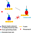
Fig 9. Simplified model of the proposed phenamacril resistance mechanism.
Binding of phenamacril in the actin-binding cleft blocks cleft closure. The open cleft myosin motor domain is in equilibrium between free and phenamacril-bound states.

Similar articles

Cited by

References

    1. Goswami RS, Kistler HC. Heading for disaster: Fusarium graminearum on cereal crops. Mol Plant Pathol. 2004;5(6):515–25. Epub 2004/11/01. 10.1111/j.1364-3703.2004.00252.x . - DOI - PubMed
    1. Xu X, Nicholson P. Community ecology of fungal pathogens causing wheat head blight. Annu Rev Phytopathol. 2009;47:83–103. Epub 2009/04/24. 10.1146/annurev-phyto-080508-081737 . - DOI - PubMed
    1. Foth BJ, Goedecke MC, Soldati D. New insights into myosin evolution and classification. Proc Natl Acad Sci USA. 2006;103(10):3681–6. Epub 2006/03/01. 10.1073/pnas.0506307103 . - DOI - PMC - PubMed
    1. Zheng Z, Hou Y, Cai Y, Zhang Y, Li Y, Zhou M. Whole-genome sequencing reveals that mutations in myosin-5 confer resistance to the fungicide phenamacril in Fusarium graminearum. Sci Rep. 2015;5:8248 10.1038/srep08248 https://www.nature.com/articles/srep08248#supplementary-information. - DOI - PMC - PubMed
    1. Ross JL, Ali MY, Warshaw DM. Cargo transport: molecular motors navigate a complex cytoskeleton. Curr Opin Cell Biol. 2008;20(1):41–7. Epub 2008/01/30. 10.1016/j.ceb.2007.11.006 . - DOI - PMC - PubMed

Publication types

MeSH terms