Review
. 2020 Oct 1;100(4):1415-1454.
doi: 10.1152/physrev.00027.2019. Epub 2020 Mar 12.

Ion Channels Controlling Circadian Rhythms in Suprachiasmatic Nucleus Excitability

Affiliations
Review

Ion Channels Controlling Circadian Rhythms in Suprachiasmatic Nucleus Excitability

Jenna R M Harvey et al. Physiol Rev. .

Abstract

Animals synchronize to the environmental day-night cycle by means of an internal circadian clock in the brain. In mammals, this timekeeping mechanism is housed in the suprachiasmatic nucleus (SCN) of the hypothalamus and is entrained by light input from the retina. One output of the SCN is a neural code for circadian time, which arises from the collective activity of neurons within the SCN circuit and comprises two fundamental components: 1) periodic alterations in the spontaneous excitability of individual neurons that result in higher firing rates during the day and lower firing rates at night, and 2) synchronization of these cellular oscillations throughout the SCN. In this review, we summarize current evidence for the identity of ion channels in SCN neurons and the mechanisms by which they set the rhythmic parameters of the time code. During the day, voltage-dependent and independent Na+ and Ca2+ currents, as well as several K+ currents, contribute to increased membrane excitability and therefore higher firing frequency. At night, an increase in different K+ currents, including Ca2+-activated BK currents, contribute to membrane hyperpolarization and decreased firing. Layered on top of these intrinsically regulated changes in membrane excitability, more than a dozen neuromodulators influence action potential activity and rhythmicity in SCN neurons, facilitating both synchronization and plasticity of the neural code.

Keywords: action potential; circadian rhythm; excitability; ion channel; suprachiasmatic nucleus.


Conflict of interest statement

No conflicts of interest, financial or otherwise, are declared by the authors.

Figures

None
Graphical abstract
FIGURE 1.
FIGURE 1.
The suprachiasmatic nucleus (SCN) is the central circadian pacemaker. Top: sagittal view of rodent brain illustrating light input to the SCN via the retinohypothalamic tract (RHT). Rhythmic SCN output regulates the daily behavioral state via multi-relay neural and humoral projections. Major SCN outputs are predominantly local: medial preoptic nucleus (MPN), dorsomedial hypothalamic nucleus (DMH), paraventricular nucleus (PVN), and dorsomedial hypothalamic nucleus (DMH). These hypothalamic relays convey temporal information from the SCN to neuroendocrine (pituitary/hypothalamus-pituitary-adrenal axis and pineal) and brainstem autonomic nuclei (IML, interomediolateral nucleus; SCG, superior cervical ganglion; NTS, nucleus tractus solitary; DMV, dorsal motor nucleus of the vagus nerve). Bottom left (SCN neuron): simplified scheme for the core mammalian transcription-translation feedback loop (TTFL). Clock and Bmal1 are basic helix-loop-helix transcriptional activators, forming a heterodimer which regulates expression of clock-controlled genes as well as the repressor proteins encoded by Period and Cryptochome. The PER/CRY heterodimer repressor complex, in turn, inhibits CLK/BMAL1 activity at E-box target sequences, on both clock-controlled genes and for CLK/BMAL1 autoregulation. The repressor complexes undergo phosphorylation-regulated translocation and degradation, reducing inhibition of Clock and Bmal1 synthesis to reinitiate the transcriptional cycle. A second component generated by retinoid-related orphan receptors (RORs) and REV-ERBα stabilizes the core TTFL (not depicted). The kinetics of this core cycle are ~24 h in constant darkness or in the isolated SCN (234). The TTFL cycle forms the basis for cellular rhythms in gene expression, intracellular Ca2+, action potentials (APs), and neurotransmitters [e.g., arginine vasopressin (AVP)] (121). Light entrains the phase of these rhythms to the ambient light-dark cycle through glutamatergic synaptic input and subsequent Ca2+ influx (FIGURE 6), activation of Ca2+-dependent kinases [Ca2+/calmodulin-dependent protein kinase II (CamKII) and CREB], and Per1 and 2 transcription (100). Light sets the higher Per levels during the day, corresponding to the phase of higher action potential firing. Incongruence between light input and the intrinsic phase of the TTFL is the basis of jet lag. Bottom right box: representative rhythmic outputs from the SCN circuit. In nocturnal rodents, action potential firing rate recorded from individual SCN neurons is higher during the day (light) phase. Multi-electrode array recordings reveal the synchronization of this activity across the SCN circuit. Higher firing is associated with suppression of locomotor wheel running, and decreased firing is observed during the active phase. Shaded portion denotes the dark phase (in LD) or subjective night (in DD). Diurnal rodents exhibit behavioral rhythms in the opposite phase, although the phase of clock gene and action potential rhythms may be similar to nocturnal mammals (273).
FIGURE 2.
FIGURE 2.
Clock-linked cellular processes regulating ion channel expression and function in suprachiasmatic nucleus (SCN) neurons. Mutations in several components of the core clock transcription-translation feedback loop (TTFL) result in alterations in daily action potential rhythmicity (13, 59, 127, 183, 216), providing evidence that there are clock-linked mechanisms regulating ion channel expression and function. Day versus night expression varies for several additional ion channels based on transcripts and/or protein levels, including Na+, K+, and Ca2+ channels (FIGURE 5). Circadian regulation of intracellular signaling pathways, Ca2+, and metabolism provide the possibility for additional posttranslational functional regulation of ion channels via activation of Ca2+-dependent kinases, phosphorylation, and redox modification of channel proteins (98). For example, glycogen synthase kinase 3 (GSK3) contributes to regulation of the persistent Na+ current [INa(P)] in SCN neurons (235). Some SCN K+ currents have been shown to be redox sensitive (303). Multi-mechanism circadian regulation has been demonstrated for Big K+ (BK) channels, which contribute to the day versus night difference in firing frequency in SCN. BK channel activity is varied by clock-linked differences in expression levels, alternative splicing, phosphorylation, beta subunit modulation, and signaling complexes (Ca2+ channel association) (199, 261, 312, 313). Less certain are the mechanisms contributing to circadian downregulation of the number of ion channels at the membrane in SCN, but ubiquitination in particular is central to both core clock protein functions and general ion channel degradation (98, 281). Membrane-transcription coupling has been demonstrated through changes in clock gene expression or reporters with inhibition of several different types of ion channels, including NaV1.1, voltage-gated Ca2+ channel, KV4.1, hyperpolarization-activated cyclic nucleotide-gated channel, and GABAA receptors (17, 74, 119, 124, 187, 214, 223, 225, 319). LTCC, L-type Ca2+ channel; ROS, reactive oxygen species.
FIGURE 3.
FIGURE 3.
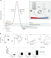
The suprachiasmatic nucleus (SCN) action potential. A: schematic of a spontaneous action potential waveform generalized from SCN neuronal firing. Pharmacological and genetic approaches have defined some of the ionic currents (Ix) or specific channel subtypes contributing to discrete phases of the waveform as annotated. Sun and moon symbols denote currents identified to exhibit larger daytime or nighttime current magnitudes, respectively. Na+ currents: INa(St) is the riluzole-sensitive subthreshold Na+ current (which can include persistent and slowly inactivating components); INa(L) is the voltage-independent current identified by Na+ substitution that is active throughout the interspike interval; INa(F) is the voltage-dependent, fast-activating Na+ current. Ca2+ currents: ICa(L, N, or R). K+ currents: IA, IK(GIRK, BK, Kv12, SK). Intracellular Ca2+ channels: ryanodine receptors (RyR). Inset: daytime SCN firing rate is higher than nighttime, correlated with a reciprocal relationship between input resistance (Ri) and K+ current (IK). Closure of channels during the day, particularly K+ channels, is associated with increased Ri, which would increase membrane sensitivity to depolarizing stimuli. At night, the opening of more K+ channels is associated with decreased Ri, which would decrease membrane sensitivity to depolarizing stimuli and reduce firing. Differences in action potential waveforms are correlated with day and night firing rates. B: ionic currents exhibiting a day versus night difference in current magnitude. The voltage-independent INa(L) and voltage-dependent ICa(L) and IK(FDR) are larger during the day. IK(BK) is larger at night. Open circles (day), filled circles (night). C: relative change in firing rate compared with control with inhibition of each channel subtype. Inhibition of INa(L) affects firing at both times of day, while inhibition of ICa(L) and IK(FDR) decrease daytime firing, and IK(BK) increases nighttime firing. [Schematized panels in B and C reproduced from data in Flourakis et al. (84), with permission from Elsevier; Itri et al. (143), with permission from Springer Nature; Montgomery et al. (210); and Whitt et al. (312).]
FIGURE 4.
FIGURE 4.
Subthreshold currents: a central node for circadian regulation of firing rate. Membrane potential is regulated by a variety of voltage-dependent and -independent currents. Notably, most pacing cells offset Na+- and Ca2+-based depolarizing currents [INa(St), INa(L), and ICa(L)] with K+-based hyperpolarizing currents (IA, IBK, KirGIRK, Kv12, and the TEA-sensitive IK). Changes in the balance of the currents will shift both the resting membrane potential (Vm), measured in the presence of tetrodotoxin, and input resistance (Ri). For circadian regulation of firing rate, ionic currents comprising the central node should show 1) different properties or magnitudes between day and night, 2) activation before threshold where they can regulate initiation of the action potential (AP), and 3) time-of-day-dependent effects on Ri, Vm, and firing rate when inhibited or activated. This evidence has been identified for NALCN cation and BK K+ channels in suprachiasmatic nucleus (SCN) (84, 146, 210, 313). During the day, subthreshold BK current is reduced via inactivation (BKi), mediated by the β2 subunit (313). As a result, daytime excitation exceeds inhibition, leading to increased AP initiation. In contrast, nighttime BK current is sustained (BKs), producing larger subthreshold inhibitory outward current and opposing AP initiation. ΔVm and ΔRi values indicate the average day versus night difference observed in acute SCN slice electrophysiological recordings. Dotted green boxes denote the inter-spike current levels. g, conductance; TTFL, transcription-translation feedback loop. [Action potential and current traces modified from data in Whitt et al. (313) and Flourakis et al. (84), with permission from Elsevier.]
FIGURE 5.
FIGURE 5.
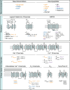
Diurnal differences in expression or function of neuromodulators, receptors, and ion channels. List of subunits or ionic currents that are found in the suprachiasmatic nucleus. Variation was assessed by comparing either day versus night expression of individual subunits, or by application of pharmacology to isolate specific ionic currents (denoted as Ix). Color of text represents increased daytime (orange), dusk (purple), or nighttime (blue) expression or current. Black text indicates subunits or currents that either do not vary between day and night or have not been tested. PAC1* exhibits bimodal expression. AVP, arginine vasopressin; EPSC, excitatory postsynaptic current; GPCR, G protein-coupled receptor; GRP, gastrin releasing peptide; 5-HT, 5-hydroxytryptamine; IPSC, inhibitory postsynaptic current; KA, kainic acid; NMDA, N-methyl-d-aspartate; NPY, neuropeptide Y; PACAP, pituitary adenylate cyclase-activating peptide; SP, substance P; VIP, vasoactive intestinal polypeptide.
FIGURE 6.
FIGURE 6.
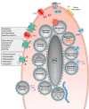
Light evoked changes in excitability. The retinohypothalamic tract (RHT) terminates on suprachiasmatic nucleus (SCN) neurons, releasing glutamate which activates postsynaptic N-methyl-d-aspartate (NMDA) and α-amino-3-hydroxy-5-methyl-4-isoxazolepropionic acid (AMPA) channels and metabotropic glutamate receptors (mGluRs). Depolarization via these receptors leads to action potential (AP) initiation and intracellular Ca2+ influx. The transient increase in Ca2+ via these ionotropic glutamate receptors may be amplified by influx through voltage-gated Ca2+ channels (VGCCs) and ryanodine receptors (RyRs), resulting in a change in action potential excitability (52, 66, 108, 116, 138, 155, 202). The Ca2+ rise activates protein kinase A (PKA) and Ca2+/calmodulin-dependent protein kinase II (CaMKII), leading to phospho-dependent activation of cAMP response element-binding protein (CREB) (87, 288, 289, 318). CaMKII may achieve this through nitric oxide synthase and protein kinase G (PKG) (67, 68, 83, 99, 289, 306, 308). Mitogen-activated protein kinase (MAPK) can be activated by a Ca2+-dependent mechanism, also activating CREB (70, 226). Most ion channels exhibit phosphorylation-dependent changes in activity (320), but the mechanisms by which CaMKII, PKA, PKG, and MAPK exert potential effects on channels that regulate intrinsic excitability in the SCN have not yet been comprehensively addressed. Phosphorylated CREB binds to the CRE element on Per1 and Per2 genes to activate transcription. Metabotropic receptors also influence excitability via Ca2+-dependent signaling (108, 233). Finally, clock genes have been shown to regulate the periodic expression of some ion channels involved in the daily regulation of action potential frequency. ER, endoplasmic reticulum; LTCC, L-type Ca2+ channel; TTCC, T-type Ca2+ channel; TTFL, transcription-translation feedback loop; Vm, membrane potential.
FIGURE 7.
FIGURE 7.
Phase relationships for intracellular Ca2+, action potential firing, and PER2 rhythms. Schematized rhythms in GCaMP6s fluorescence (intracellular Ca2+), PER2::LUC bioluminescence (PER2), and spontaneous firing rate recorded on a multielectrode array from an organotypic suprachiasmatic nucleus slice. [Modified from data in Enoki et al. (78).]
FIGURE 8.
FIGURE 8.
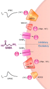
GABA-evoked inhibitory versus excitatory postsynaptic currents and circadian variation in Cl potential (ECl). Suprachiasmatic nucleus (SCN) neurons are among several types of central neurons that respond with variable action potential firing to GABA application. Normally, GABA produces an inward Cl current, leading to membrane hyperpolarization and decreased firing (105, 182). However, several studies have shown that SCN neurons can respond to GABA with increased activity (62, 299). One mechanism that could produce a direct excitatory effect of GABA on firing is a shift in ECl toward more depolarizing potentials, observed in three studies of spontaneous and GABA-evoked synaptic currents recorded from SCN neurons (11, 268, 300). The direction of response would depend on the Cl gradient, which is determined by expression of cation chloride co-transporters (CCCs). Na+-K+-Cl cotransporters (NKCC) mediate chloride uptake with 2Cl:1Na+:1K+ stoichiometry. K+-Cl cotransporters (KCCs) mediate chloride extrusion with 1Cl:1K+ stoichiometry. NKCC1 expression is significantly higher during the night than during the day in the dorsal SCN (48, 213). Inhibiting NKCC1 hyperpolarized the GABA reversal potential (48). All 4 types of KCCs (KCC1–4) are expressed in SCN neurons, with a cell-type specific distribution (21, 22), but an inhibitor of KCC2 has been shown to shift the direction of the GABA response in SCN neurons (160, 229). In the schematic, transporter size indicates the expected function under each condition that would be required to explain the bidirectional effects of GABA. Neurons that can be excited by GABA usually have increased expression of the NKCC1 compared with KCC2, leading to an increased intracellular chloride concentration. Decreased extrusion of Cl by KCC2 and increased uptake by NKCC1 would be responsible for shunting the Cl reversal potential enough to change GABA’s action from inhibitory to excitatory, under certain conditions. EPSC, excitatory postsynaptic current; IPSC, inhibitory postsynaptic current.

References

    1. Abe M, Herzog ED, Block GD. Lithium lengthens the circadian period of individual suprachiasmatic nucleus neurons. Neuroreport 11: 3261–3264, 2000. doi:10.1097/00001756-200009280-00042. - DOI - PubMed
    1. Abrahamson EE, Moore RY. Suprachiasmatic nucleus in the mouse: retinal innervation, intrinsic organization and efferent projections. Brain Res 916: 172–191, 2001. doi:10.1016/S0006-8993(01)02890-6. - DOI - PubMed
    1. Acuna-Goycolea C, Obrietan K, van den Pol AN. Cannabinoids excite circadian clock neurons. J Neurosci 30: 10061–10066, 2010. doi:10.1523/JNEUROSCI.5838-09.2010. - DOI - PMC - PubMed
    1. Adelman JP, Maylie J, Sah P. Small-conductance Ca2+-activated K+ channels: form and function. Annu Rev Physiol 74: 245–269, 2012. doi:10.1146/annurev-physiol-020911-153336. - DOI - PubMed
    1. Aggelopoulos NC, Meissl H. Responses of neurones of the rat suprachiasmatic nucleus to retinal illumination under photopic and scotopic conditions. J Physiol 523: 211–222, 2000. doi:10.1111/j.1469-7793.2000.t01-1-00211.x. - DOI - PMC - PubMed

Publication types

LinkOut - more resources