. 2020 May;159(5):627-641.
doi: 10.1530/REP-19-0435.

Low protein diet and methyl-donor supplements modify testicular physiology in mice

Affiliations

Low protein diet and methyl-donor supplements modify testicular physiology in mice

Hannah L Morgan et al. Reproduction. 2020 May.

Abstract

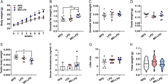
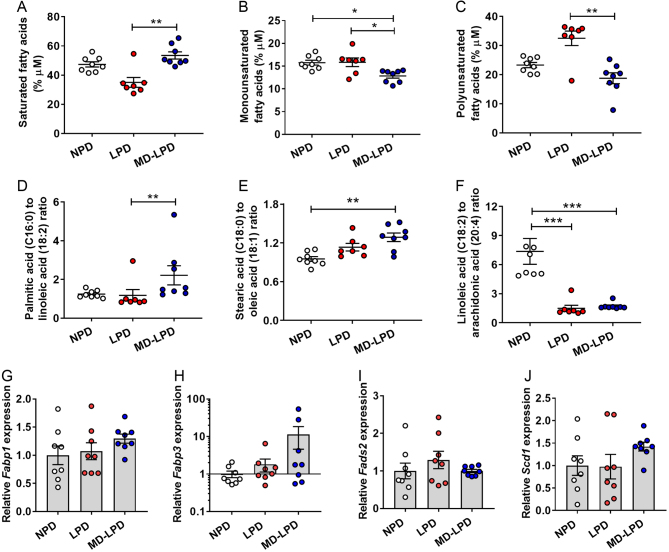
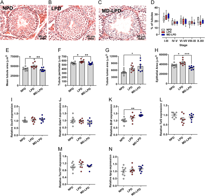
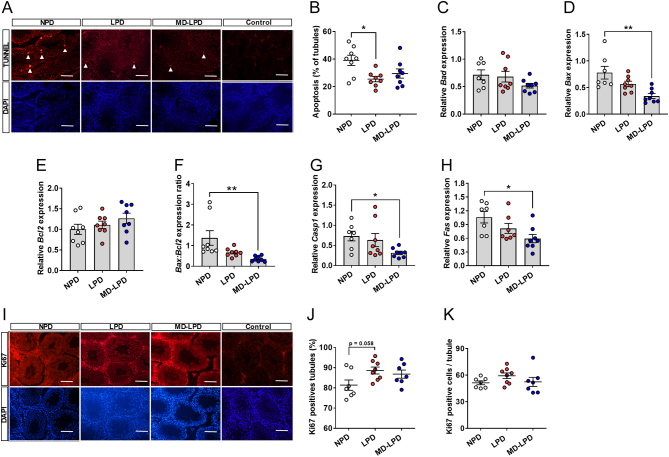
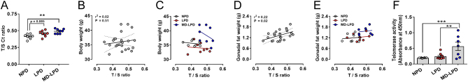
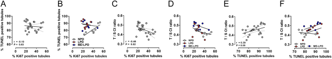
The link between male diet and sperm quality has received significant investigation. However, the impact diet and dietary supplements have on the testicular environment has been examined to a lesser extent. Here, we establish the impact of a sub-optimal low protein diet (LPD) on testicular morphology, apoptosis and serum fatty acid profiles. Furthermore, we define whether supplementing a LPD with specific methyl donors abrogates any detrimental effects of the LPD. Male C57BL6 mice were fed either a control normal protein diet (NPD; 18% protein; n = 8), an isocaloric LPD (LPD; 9% protein; n = 8) or an LPD supplemented with methyl donors (MD-LPD; choline chloride, betaine, methionine, folic acid, vitamin B12; n = 8) for a minimum of 7 weeks. Analysis of male serum fatty acid profiles by gas chromatography revealed elevated levels of saturated fatty acids and lower levels of mono- and polyunsaturated fatty acids in MD-LPD males when compared to NPD and/or LPD males. Testes of LPD males displayed larger seminiferous tubule cross section area when compared to NPD and MD-LPD males, while MD-LPD tubules displayed a larger luminal area. Furthermore, TUNNEL staining revealed LPD males possessed a reduced number of tubules positive for apoptosis, while gene expression analysis showed MD-LPD testes displayed decreased expression of the pro-apoptotic genes Bax, Csap1 and Fas when compared to NPD males. Finally, testes from MD-LPD males displayed a reduced telomere length but increased telomerase activity. These data reveal the significance of sub-optimal nutrition for paternal metabolic and reproductive physiology.


Conflict of interest statement

The authors declare that there is no conflict of interest that could be perceived as prejudicing the impartiality of the research reported.

Figures

Figure 1
Figure 1
Impact of NPD, LPD and MD-LPD on male growth (A), gonadal fat weight (B) gonadal fat:body weight ratio (C), testis weight (D) testis:body weight ratio (E), serum testosterone (F), late gestation litter size (G) and fetal weight. Data are mean ± s.e.m. in A–G and mean (solid line) with 25 and 75% quartiles (dashed lines) in H. n = 8 males per dietary group in A–G and eight litters (each from separate males) representing an n of 52–59 fetuses per treatment group in H. Data were analysed by one-way ANOVA followed by Bonferroni post-hoc test, or Kruskal–Wallis test with Dunns multiple comparison test where appropriate. *P < 0.05, **P < 0.01, ***P < 0.001.
Figure 2
Figure 2
Impact of NPD, LPD and MD-LPD on serum saturated fatty acids (A), monounsaturated fatty acids (B), polyunsaturated fatty acids (C), palmitic:linoleic acid ratio (D), stearic:oleic acid ratio (E) and linoleic:arachidonic acid ratio (F). Relative hepatic expression of fatty acid binding protein 1 (Fabp1) (G), fatty acid binding protein 3 (Fabp3) (H), fatty acid desaturase 2 (Fads2) (I) and stearoyl-Coenzyme A desaturase 1 (Scd1) (J). Data are mean ± s.e.m. n = 8 males per dietary group. Data were analysed by one-way ANOVA followed by Bonferroni post-hoc test, or Kruskal–Wallis test with Dunns multiple comparison test where appropriate. *P < 0.05, **P < 0.01, ***P < 0.001.
Figure 3
Figure 3
Impact of NPD, LPD and MD-LPD on testicular morphology. Representative cross section image of NPD (A), LPD (B) and MD-LPD (C) seminiferous tubule morphology. Quantification of seminiferous tubule stage in NPD, LPD and MD-LPD testes (D). Mean seminiferous tubule cross section area (E), perimeter (F), lumen area (G) and area of the epithelium (H). Relative testicular expression of SRY sex determining region Y-box 9 (Sox9) (I), GATA binding protein 1(Gata1) (J), bromodomain, testis-specific (Brdt) (K), TATA-Box Binding Protein Associated Factor 2 (Taf2) (L), testis Expressed 101 (Tex101) (M) and IQ Motif Containing G (Iqcg) (n). Data are mean ± s.e.m. n = 8 males per dietary group with a minimum of 50 tubules per testis analysed in A–H. Data were analysed by one-way ANOVA followed by Bonferroni post-hoc test, or Kruskal–Wallis test with Dunns multiple comparison test where appropriate. *P < 0.05, **P < 0.01.
Figure 4
Figure 4
Impact of NPD, LPD and MD-LPD on testicular apoptosis. Representative TUNEL and DAPI stained sections of NPD, LPD and MD-LPD testis and negative control (bar = 100 µm) (A) showing apoptotic cells (white arrow heads) and the percentage of tubules showing apoptotic cells (B). Relative testicular expression of BCL2-associated agonist of cell death (Bad) (C), BCL2-associated X protein (Bax) (D), B cell leukemia/lymphoma (Bcl2) (E), Bax:Bcl2 expression ratio (F), caspase 1 (Casp1) (G) and TNF receptor superfamily member 6 (Fas) (H). Representative Ki67 and DAPI stained sections of NPD, LPD and MD-LPD testis and negative control (bar = 100 µm) (I) with the percentage of tubules showing positive stained cells (J) and mean number of Ki67 positive cells per tubule (K). Data are mean ± s.e.m. n = 8 males per dietary group. Data were analysed by one-way ANOVA followed by Bonferroni post-hoc test, or Kruskal–Wallis test with Dunns multiple comparison test where appropriate. *P < 0.05, **P < 0.01.
Figure 5
Figure 5
Impact of NPD, LPD and MD-LPD on testicular telomere length (as determined by the T/S ratio) (A). Correlation of body weight to T/S ratio in all males (B) and individual dietary groups (C). Correlation of gonadal fat weight to T/S ratio in all males (D) and individual dietary groups (E). Testicular telomerase activity (F). Data are mean ± s.e.m. n = 8 males per dietary group. Data were analysed by one-way ANOVA followed by Bonferroni post-hoc test, or Kruskal–Wallis test with Dunns multiple comparison test where appropriate. Correlations were analysed using Pearsons correlation. **P < 0.01, ***P < 0.001.
Figure 6
Figure 6
Impact of NPD, LPD and MD-LPD on correlation between the percentage of Ki67 positive stained tubules to percentage of TUNEL positive tubules in all males (A) and individual dietary groups (B); correlation between the percentage of Ki67 positive stained tubules and T/S ratio in all males (C) and individual dietary groups (D) and correlation between percentage of TUNEL positive tubules and T/S ration in all males (E) and individual dietary groups (F). Data are mean ± s.e.m. n = 8 males per dietary group. Data were analysed by Pearsons correlation.

Similar articles

Cited by

References

    1. Ables GP, Johnson JE. 2017. Pleiotropic responses to methionine restriction. Experimental Gerontology 94 83–88. (10.1016/j.exger.2017.01.012) - DOI - PubMed
    1. Aitken RJ, Findlay JK, Hutt KJ, Kerr JB. 2011. Apoptosis in the germ line. Reproduction 141 139–150. (10.1530/REP-10-0232) - DOI - PubMed
    1. Aparecida de Franca S, Dos Santos MP, Garofalo MA, Navegantes LC, Kettelhut Ido C, Lopes CF, Kawashita NH. 2009. Low protein diet changes the energetic balance and sympathetic activity in brown adipose tissue of growing rats. Nutrition 25 1186–1192. (10.1016/j.nut.2009.03.011) - DOI - PubMed
    1. Appasamy M, Muttukrishna S, Pizzey AR, Ozturk O, Groome NP, Serhal P, Jauniaux E. 2007. Relationship between male reproductive hormones, sperm DNA damage and markers of oxidative stress in infertility. Reproductive Biomedicine Online 14 159–165. (10.1016/s1472-6483(10)60783-3) - DOI - PubMed
    1. Asadi N, Bahmani M, Kheradmand A, Rafieian-Kopaei M. 2017. The impact of oxidative stress on testicular function and the role of antioxidants in improving it: a review. Journal of Clinical and Diagnostic Research 11 IE01–IE05. (10.7860/JCDR/2017/23927.9886) - DOI - PMC - PubMed

Publication types