Review
. 2020 Mar 10;11(3):290.
doi: 10.3390/genes11030290.

Claudins in Renal Physiology and Pathology

Affiliations
Review

Claudins in Renal Physiology and Pathology

Caroline Prot-Bertoye et al. Genes (Basel). .

Abstract

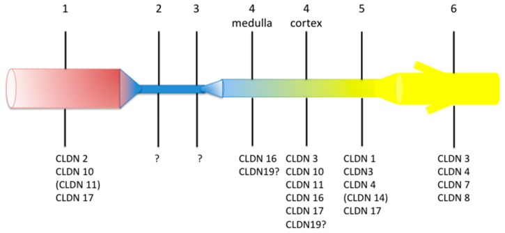
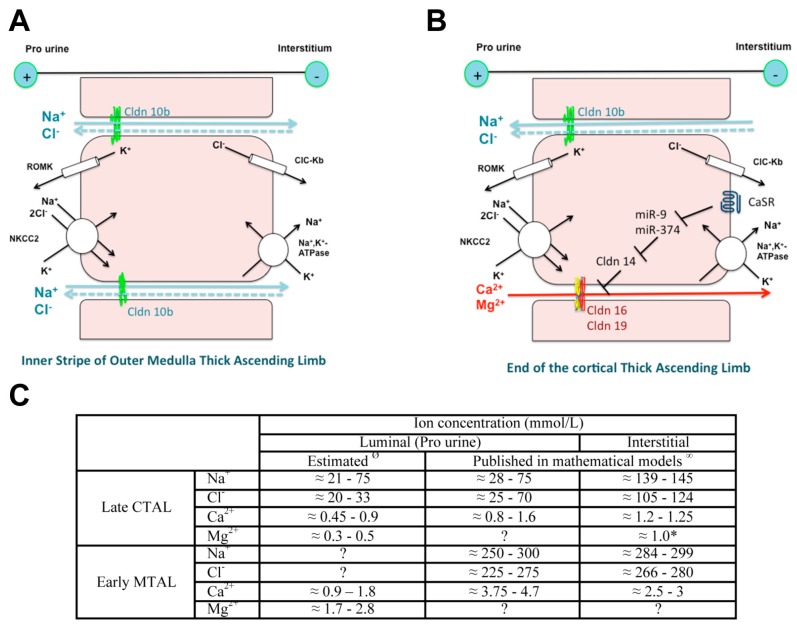
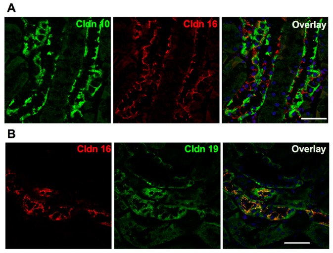
Claudins are integral proteins expressed at the tight junctions of epithelial and endothelial cells. In the mammalian kidney, every tubular segment express a specific set of claudins that give to that segment unique properties regarding permeability and selectivity of the paracellular pathway. So far, 3 claudins (10b, 16 and 19) have been causally traced to rare human syndromes: variants of CLDN10b cause HELIX syndrome and variants of CLDN16 or CLDN19 cause familial hypomagnesemia with hypercalciuria and nephrocalcinosis. The review summarizes our current knowledge on the physiology of mammalian tight junctions and paracellular ion transport, as well as on the role of the 3 above-mentioned claudins in health and disease. Claudin 14, although not having been causally linked to any rare renal disease, is also considered, because available evidence suggests that it may interact with claudin 16. Some single-nucleotide polymorphisms of CLDN14 are associated with urinary calcium excretion and/or kidney stones. For each claudin considered, the pattern of expression, the function and the human syndrome caused by pathogenic variants are described.

Keywords: HELIX syndrome; claudin 10b; claudin 14; claudin 16; claudin 19; divalent cations; familial hypomagnesemia with hypercalciuria and nephrocalcinosis; kidney; sodium; tight junction.


Conflict of interest statement

C.P.-B. and P.H. have no conflict of interest to declare regarding the content of the present review.

Figures

Figure 1
Figure 1
Pattern of expression of claudin (CLDN) proteins along the human renal tubule and collecting duct. The figure is a summary of data published in References [26,27,28,29] (the expression pattern in rodent kidney has recently been summarized in several reviews [2,30,31]). Question mark means that direct evidence of the expression of the considered claudin in human is not available but that indirect evidence suggests that it should be expressed there. Brackets mean that the expression is low. The numbers above the various segments are: 1, proximal convoluted and straight tubule; 2, descending thin limb; 3, ascending thin limb; 4, distal straight tubule (thick ascending limb); 5, distal convoluted tubule; 6, cortical and outer medullary collecting duct. Colors illustrate the fact that composition and/or volume of tubular fluid changes along the renal tubule. Readers interested in mRNA expression pattern can visit https://hpcwebapps.cit.nih.gov/ESBL/Database/NephronRNAseq/All_transcripts.html.
Figure 2
Figure 2
Model of ion transport in the inner stripe of outer medulla (IS-) (A) and in the cortical thick ascending limb of the loop of Henle (C-TAL) (B) and estimated ion concentration (C). The original works used to build the model have been published in References [108,109,110]. NaCl is reabsorbed via the apical cotransporter NKCC2. Most of the potassium that enters the cell recycles back to the lumen via the potassium channel ROMK, thereby hyperpolarizing the apical membrane, while most of the chloride leaves the cell across the basolateral chloride channel CLCKB, resulting in a depolarization of the membrane. Sodium exits the cell via the Na+,K+-ATPase at the basolateral membrane. The difference in voltage of the two membranes accounts for the lumen positive transepithelial potential difference, the driving force for the paracellular diffusion of divalent cations in the C-TAL and of sodium in the IS-TAL. Claudin (Cldn)16 and Cldn19 may confer a paracellular permeability and selectivity to cations. Cldn14 may interact with Cldn16 and inhibit the Cldn16/Cldn19 complex. It is suggested that Cldn10b drives paracellular NaCl back flux in cortical TAL, adding to the lumen-positive voltage as the paracellular pathway is more permeable to sodium than to chloride. High Ca2+ diet and allosteric agonists of calcium-sensing receptor (CaSR) may trigger the expression of Cldn14 via the inhibition of the transcription of two microRNAs miR-9 and miR-374 suppressing Cldn14 gene expression. Ø Direct measurement of luminal ion concentration in the TAL is not possible because the segment is inaccessible to micropuncture. Luminal concentrations of Na+, chloride (Cl), Mg2+ and Ca2+ in the early distal convoluted tubule, the segment just downstream the late cortical thick ascending limb (CTAL) have been measured during micropuncture experiments in rodents [111,112,113,114,115,116,117,118,119]. In most studies, concentrations are expressed as a ratio between tubular fluid and plasma ultrafilterable ion concentration. Plasma NaCl is freely filtered, whereas around 60% of Ca and 80% of Mg are ultrafilterable. Luminal ion concentrations in the early medullary thick ascending limb (MTAL) can be estimated based on a Ca2+ and Mg2+ reabsorption equaling 20–25% and 60–70% of filtered load, respectively and the lack of substantial water reabsorption in the TAL. Values of ion concentrations in the cortical interstitial fluid are similar to those in plasma, due to the dense capillary network and high blood flow in the cortex. Those concentrations have been reported in mathematical models except for magnesium (*). Estimates of the luminal and interstitial concentrations of Na+, Cl and Ca2+ in the early medullary thick ascending limbs have been published in mathematical models [120,121,122,123,124,125,126].
Figure 3
Figure 3
Expression of claudin 10, claudin 16 and claudin 19 in the murine cortical thick ascending limb in immunofluorescence. (A) Pattern of expression of claudin 10 and claudin 16 in the mouse C-TAL. Either claudin 10 (Cldn 10, in green) or claudin 16 (Cldn 16, in red) are expressed at tight junction. (B) Pattern of expression of claudin 16 and claudin 19 in the mouse C-TAL. Claudin 16 (Cldn 16, in red) and claudin 19 (Cldn 19, in green) are colocalized at tight junction. Bar = 20 µm.

References

    1. Pei L., Solis G., Nguyen M.T., Kamat N., Magenheimer L., Zhuo M., Li J., Curry J., McDonough A.A., Fields T.A., et al. Paracellular epithelial sodium transport maximizes energy efficiency in the kidney. J. Clin. Investig. 2016;126:2509–2518. doi: 10.1172/JCI83942. - DOI - PMC - PubMed
    1. Muto S. Physiological roles of claudins in kidney tubule paracellular transport. Am. J. Physiol. Ren. Physiol. 2017;312:F9–F24. doi: 10.1152/ajprenal.00204.2016. - DOI - PubMed
    1. Powell D.W. Barrier function of epithelia. Am. J. Physiol. 1981;241:G275–G288. doi: 10.1152/ajpgi.1981.241.4.G275. - DOI - PubMed
    1. Van Itallie C.M., Anderson J.M. The molecular physiology of tight junction pores. Physiology. 2004;19:331–338. doi: 10.1152/physiol.00027.2004. - DOI - PubMed
    1. Reuss L. Tight junction permeability to ions and water. In: Cereijido M., Anderson J.M., editors. Tight Junctions. 2nd ed. CRC Press; Boca Raton, FL, USA: 2001. pp. 61–88.

Publication types

Supplementary concepts

LinkOut - more resources