

Latest Insights into Marek's Disease Virus Pathogenesis and Tumorigenesis
- PMID: 32164311
- PMCID: PMC7139298
- DOI: 10.3390/cancers12030647
Latest Insights into Marek's Disease Virus Pathogenesis and Tumorigenesis
Abstract
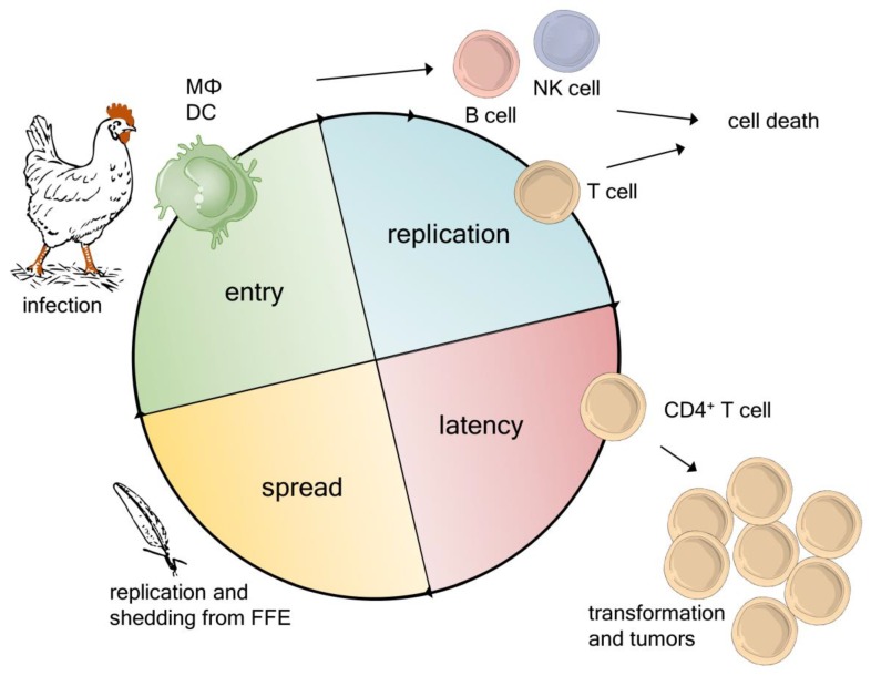
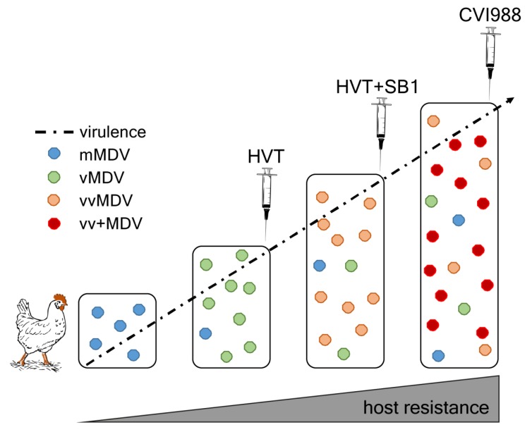
Marek's disease virus (MDV) infects chickens and causes one of the most frequent cancers in animals. Over 100 years of research on this oncogenic alphaherpesvirus has led to a profound understanding of virus-induced tumor development. Live-attenuated vaccines against MDV were the first that prevented cancer and minimized the losses in the poultry industry. Even though the current gold standard vaccine efficiently protects against clinical disease, the virus continuously evolves towards higher virulence. Emerging field strains were able to overcome the protection provided by the previous two vaccine generations. Research over the last few years revealed important insights into the virus life cycle, cellular tropism, and tumor development that are summarized in this review. In addition, we discuss recent data on the MDV transcriptome, the constant evolution of this highly oncogenic virus towards higher virulence, and future perspectives in MDV research.
Keywords: Marek’s disease virus; avian cancer; cell tropism; herpesvirus; life cycle; lymphoma; vaccine.
Conflict of interest statement
The authors declare no conflict of interest.
Figures
References
-
- Purchase H.G. Clinical Disease and Its Economic Impact. In: Payne L.N., editor. Marek’s Disease. Springer; Boston, MA, USA: 1985. pp. 17–42.
-
- Davison T.F., Nair V. Marek’s Disease: An Evolving Problem. Elsevier Academic Press; London, UK: 2004.
-
- Marek J. Multiple Nervenentzündung (Polyneuritis) bei Hühnern. Dtsch. Tierärztl. Wochenschr. 1907;15:417–421.
Publication types
Grants and funding
LinkOut - more resources
Full Text Sources
