. 2020 Mar 10;12(3):638.
doi: 10.3390/cancers12030638.

c-MYC Expression Is a Possible Keystone in the Colorectal Cancer Resistance to EGFR Inhibitors

Affiliations

c-MYC Expression Is a Possible Keystone in the Colorectal Cancer Resistance to EGFR Inhibitors

Antonia Strippoli et al. Cancers (Basel). .

Abstract

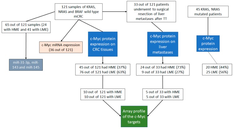
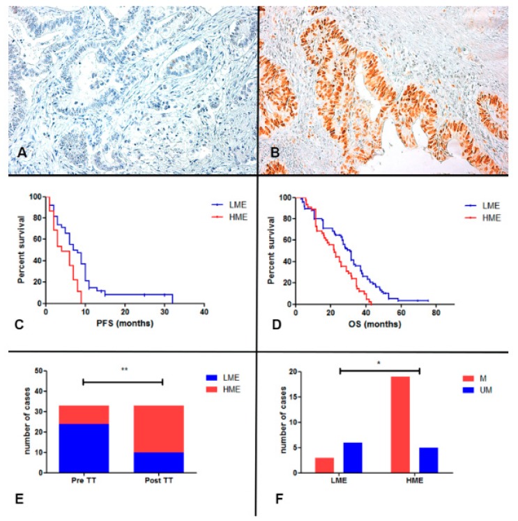
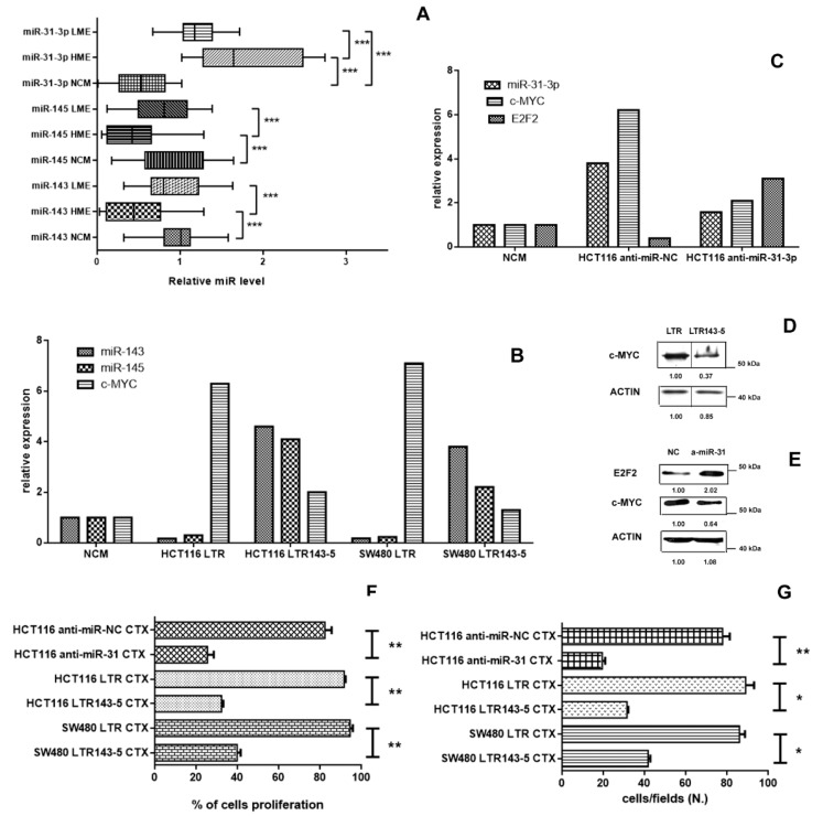
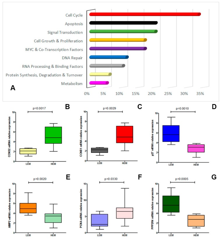
Alterations in the transcriptional factor c-MYC could be involved in the anti-EGFR resistance in metastatic colorectal cancer (mCRC). The c-MYC expression was evaluated in 121 RAS and BRAF wild-type mCRC before treatment with anti-EGFR+Folfiri therapy and in 33 subsequent metastases collected during target therapy (TT) or in TT resistance phase. We analyzed the expression and the functional role of some c-MYC linked miRNAs (miR-31-3p, miR-143 and miR-145) in our patient group and in two CRC cell lines, also performing a c-MYC target PCR array. Patients with higher c-MYC expression (HME) showed a significant lower PFS and OS when compared to those with low c-MYC expression (LME). HME pattern was significantly more frequent in the metastases after TT and significantly associated to anti-EGFR molecular resistance alterations. We also found a significant correlation between the expression of the above-mentioned c-MYC linked miRNAs, c-MYC level and anti-EGFR resistance. Moreover, expression gene profiling pointed out the pivotal role of c-MYC in CRC-related cell-cycle, apoptosis, signal transduction and cell-growth pathways. c-MYC expression might distinguish patients with a lower PFS and OS in anti-EGFR treated mCRC. The individuation of some miRNAs involved in the c-MYC pathway regulation and the downstream c-MYC effector genes could provide a new possible target to overcome the anti-EGFR resistance in mCRC.

Keywords: EGFR inhibitor resistance; c-MYC; colorectal cancer; targeted therapy.


Conflict of interest statement

The authors declare that they have no conflict of interest. Written informed consent was provided by the patient prior to the first study specific intervention. Ethical approval for study was provided by the ethics committee of Università Cattolica del Sacro Cuore, Roma (Roma PROT. OM-2009-1 and 200603000). The report does not present identifying images or other personal or clinical details of participants that compromise anonymity.

Figures

Figure 1
Figure 1
The figure shows the flow chart of the different analyses performed in this work in the different patient subgroups.
Figure 2
Figure 2
Panel (A,B). Immunohistochemical analysis of c-MYC protein expression. The figure shows two representative cases of CRC with positive (panel (B); score 8) and negative staining (panel (A); score 1) for c-MYC (Original magnification 200×); Panel (C,D). Kaplan-Meier curves for PFS and OS of RAS-BRAF wild-type anti-EGFR mCRC patients stratified by c-MYC expression. LME patients (blue-line) was significantly associated to a better PFS (p < 0.0001) and OS (p = 0.0016) respect to HME (red-line). Panel (E) The figure shows that the HME cases were significantly higher in the metastatic liver samples after TT (post-TT) in comparison to the correspondent primary CRC (Pre-TT; p = 0.0012; Fisher’s exact test); Panel (F) The HME metastases after TT had a significant higher molecular alterations (M) respect to LME cases (p = 0.0334; M vs. UM, liver metastasis without molecular alterations after TT; Fisher’s exact test). * p < 0.05, ** p < 0.01.
Figure 3
Figure 3
Panel (A). The figure shows the significant low expression of miR-143 and miR-145 in the HME cases in comparison to the LME cases (p = 0.0001 and p = 0.0006, respectively; Mann Whitney t test) and normal colonic mucosa (NCM; p = 0.0002 and p = 0.0005, respectively; Mann Whitney t test); conversely, miR-31-3p had a high expression both in HME and LME cases respect to the normal colonic mucosa (p < 0.0001 and p < 0.0001, respectively; Mann Whitney t test). Panel (B). MiR-143 and miR-145 expression show low level in HCT116 and SW480 cell lines transduced with LTR, while c-MYC expression was upregulated in both cancer cell lines. Conversely, LTR143-5 transduction cell lines showed an upregulation of the two miRs and a c-MYC downregulation. Panel (C) Mir-31-3p and c-MYC expression were downregulated in HCT116 cell lines transduced with anti-miR-31-3p in comparison with the controls, while E2F2 transcription factor was upregulated. Panel (D) Western-blot analysis for c-MYC expression in HCT116 cells transduced with LTR and with LTR143-5 (a representative image with, below, the relative expression ratio below). Panel (E) Western-blot analysis for c-MYC and E2F2 expression in HCT116 cells transduced with NC and with anti-miR31-3p (a representative image with, below, the relative expression ratio below). Panel (F,G). The figures show the significant cytotoxicity (panel F) and the significant reduction of the number of migrated cell (panel G) of cetuximab-treated (CTX) LTR143-5-transduced HCT116 and SW480 cell lines and cetuximab-treated (CTX) anti-miR-31-3p HCT116 respect to the those transduced with LTR and NC (p < 0.05; Mann Whitney t test). * p < 0.05, ** p < 0.01, *** p < 0.001.
Figure 4
Figure 4
Panel (A). The figure shows the effect of c-MYC overexpression on several cellular pathways (expressed as altered genes percentage/pathway); Panel (BG). Real-time confirmed expression of altered genes comparing HME and LME patients (CRCND2, p = 0.0017; CRCNB1, p = 0.0029; p27, p = 0.0010; AIMP2, p = 0.0020; PCNA, p = 0.0330; PPP2R4, p = 0.0005).

References

    1. Fakih M.G. Metastatic colorectal cancer: Current state and future directions. J. Clin. Oncol. 2015;33:1809–1824. doi: 10.1200/JCO.2014.59.7633. - DOI - PubMed
    1. Leichsenring J., Koppelle A., Reinacher-Schick A. Colorectal Cancer: Personalized. Ther. Gastrointest Tumors. 2014;1:209–220. doi: 10.1159/000380790. - DOI - PMC - PubMed
    1. Van-Cutsem E., Lenz H.J., Kohne C.H., Heinemann V., Tejpar S., Melezínek I., Beier F., Stroh C., Rougier P., Van Krieken J.H., et al. Fluorouracil, leucovorin, and irinotecan plus cetuximab treatment and RAS mutations in colorectal cancer. J. Clin. Oncol. 2015;33:692–700. doi: 10.1200/JCO.2014.59.4812. - DOI - PubMed
    1. Peeters M., Karthaus M., Rivera F., Terwey J.H., Douillard J.Y. Panitumumab in metastatic colorectal cancer: The importance of tumour RAS Status. Drugs. 2015;75:731–748. doi: 10.1007/s40265-015-0386-x. - DOI - PMC - PubMed
    1. Bertotti A., Papp E., Jones S., Adleff V., Anagnostou V., Lupo B., Sausen M., Phallen J., Hruban C.A., Tokheim C., et al. The genomic landscape of response to EGFR blockade in colorectal cancer. Nature. 2015;526:263–267. doi: 10.1038/nature14969. - DOI - PMC - PubMed