Review
. 2020 Mar 10;9(3):674.
doi: 10.3390/cells9030674.

The Intersection of Serine Metabolism and Cellular Dysfunction in Retinal Degeneration

Affiliations
Review

The Intersection of Serine Metabolism and Cellular Dysfunction in Retinal Degeneration

Tirthankar Sinha et al. Cells. .

Abstract

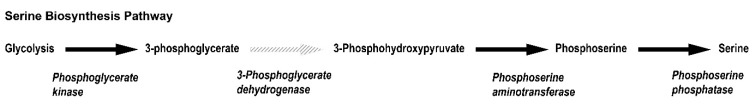
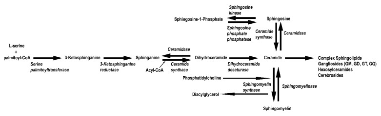
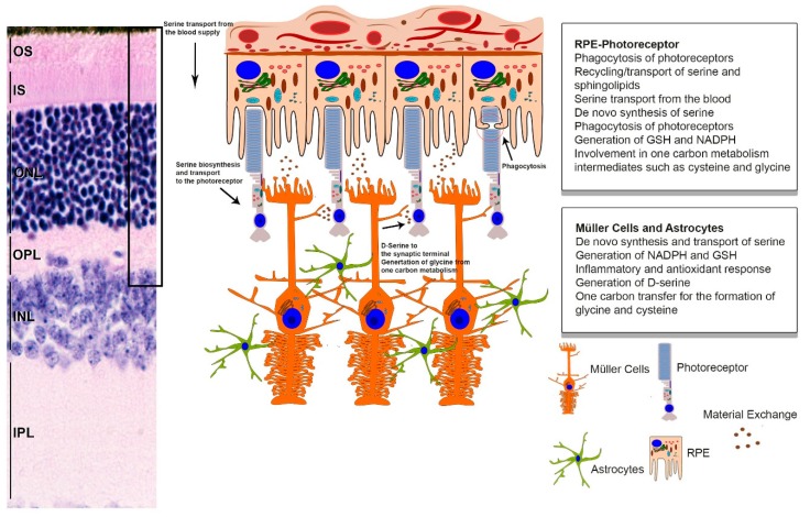
In the past, the importance of serine to pathologic or physiologic anomalies was inadequately addressed. Omics research has significantly advanced in the last two decades, and metabolomic data of various tissues has finally brought serine metabolism to the forefront of metabolic research, primarily for its varied role throughout the central nervous system. The retina is one of the most complex neuronal tissues with a multitude of functions. Although recent studies have highlighted the importance of free serine and its derivatives to retinal homeostasis, currently few reviews exist that comprehensively analyze the topic. Here, we address this gap by emphasizing how and why the de novo production and demand for serine is exceptionally elevated in the retina. Many basic physiological functions of the retina require serine. Serine-derived sphingolipids and phosphatidylserine for phagocytosis by the retinal pigment epithelium (RPE) and neuronal crosstalk of the inner retina via D-serine require proper serine metabolism. Moreover, serine is involved in sphingolipid-ceramide balance for both the outer retina and the RPE and the reductive currency generation for the RPE via serine biosynthesis. Finally and perhaps the most vital part of serine metabolism is free radical scavenging in the entire retina via serine-derived scavengers like glycine and GSH. It is hard to imagine that a single tissue could have such a broad and extensive dependency on serine homeostasis. Any dysregulation in serine mechanisms can result in a wide spectrum of retinopathies. Therefore, most critically, this review provides a strong argument for the exploration of serine-based clinical interventions for retinal pathologies.

Keywords: Müller cells; RPE; diabetic retinopathy; macular degeneration; macular telangiectasia; oxidative stress; retina; retinal degeneration; serine; sphingolipids.


Conflict of interest statement

The authors declare no conflict of interest.

Figures

Figure 1
Figure 1
Pathway depicting serine biosynthesis from glycolysis. Metabolic intermediates involved in the enzymatic synthesis of L-serine from glycolysis is shown here with the rate limiting step marked with a dashed grey arrow. The enzymes involved in respective steps are shown in bold italics below the arrow for the individual reaction.
Figure 2
Figure 2
Pathway depicting sphingolipid biosynthesis from L-serine. Metabolic intermediates involved in the enzymatic synthesis of sphingolipids from L-serine are shown here. The enzymes involved in respective steps are shown in bold italics below the arrow for the individual reaction.
Figure 3
Figure 3
Graphical summary of serine metabolism in the retina. Serine homeostasis is primarily maintained by the RPE and retinal glia. The RPE transports and generates serine, which is ultimately transported to the photoreceptors. Additionally, important serine metabolic products such as glycine and cysteine are transported or catabolized to be used as fuel, as the energetic requirements of the RPE are very high. Photoreceptors also receive serine from the Müller glia and astrocytes. Glial cells are vital to the macula and generate serine from glycolysis, which is crucial in maintaining the redox balance in the photoreceptors, controlling neurotransmission, and mediating inflammation response elements. (IS, inner segment; OS, outer segment; ONL, outer nuclear layer; OPL, outer plexiform layer; INL, inner nuclear layer; IPL, inner plexiform layer).

References

    1. El-Hattab A.W. Serine biosynthesis and transport defects. Mol. Genet. Metab. 2016;118:153–159. doi: 10.1016/j.ymgme.2016.04.010. - DOI - PubMed
    1. Kalhan S.C., Hanson R.W. Resurgence of serine: An often neglected but indispensable amino Acid. J. Biol. Chem. 2012;287:19786–19791. doi: 10.1074/jbc.R112.357194. - DOI - PMC - PubMed
    1. Simon M.V., Spalm F.H.P., Vera M.S., Rotstein N.P. Sphingolipids as Emerging Mediators in Retina Degeneration. Front. Cell Neurosci. 2019;13 doi: 10.3389/fncel.2019.00246. - DOI - PMC - PubMed
    1. Metcalf J.S., Dunlop R.A., Powell J.T., Banack S.A., Cox P.A. L-Serine: A Naturally-Occurring Amino Acid with Therapeutic Potential. Neurotox. Res. 2018;33:213–221. doi: 10.1007/s12640-017-9814-x. - DOI - PubMed
    1. Mattaini K.R., Sullivan M.R., Vander Heiden M.G. The importance of serine metabolism in cancer. J. Cell Biol. 2016;214:249–257. doi: 10.1083/jcb.201604085. - DOI - PMC - PubMed

Publication types

LinkOut - more resources