. 2020 Mar 10;21(5):1900.
doi: 10.3390/ijms21051900.

Cockle Shell-Derived Aragonite CaCO3 Nanoparticles for Co-Delivery of Doxorubicin and Thymoquinone Eliminates Cancer Stem Cells

Affiliations

Cockle Shell-Derived Aragonite CaCO3 Nanoparticles for Co-Delivery of Doxorubicin and Thymoquinone Eliminates Cancer Stem Cells

Kehinde Muibat Ibiyeye et al. Int J Mol Sci. .

Abstract

Cancer stem cells CSCs (tumour-initiating cells) are responsible for cancer metastasis and recurrence associated with resistance to conventional chemotherapy. This study generated MBA MD231 3D cancer stem cells enriched spheroids in serum-free conditions and evaluated the influence of combined doxorubicin/thymoquinone-loaded cockle-shell-derived aragonite calcium carbonate nanoparticles. Single loaded drugs and free drugs were also evaluated. WST assay, sphere forming assay, ALDH activity analysis, Surface marker of CD44 and CD24 expression, apoptosis with Annexin V-PI kit, cell cycle analysis, morphological changes using a phase contrast light microscope, scanning electron microscopy, invasion assay and migration assay were carried out; The combination therapy showed enhanced apoptosis, reduction in ALDH activity and expression of CD44 and CD24 surface maker, reduction in cellular migration and invasion, inhibition of 3D sphere formation when compared to the free drugs and the single drug-loaded nanoparticle. Scanning electron microscopy showed poor spheroid formation, cell membrane blebbing, presence of cell shrinkage, distortion in the spheroid architecture; and the results from this study showed that combined drug-loaded cockle-shell-derived aragonite calcium carbonate nanoparticles can efficiently destroy the breast CSCs compared to single drug-loaded nanoparticle and a simple mixture of doxorubicin and thymoquinone.

Keywords: breast cancer; cancer stem cell; doxorubicin; nanoparticle; thymoquinone.


Conflict of interest statement

The authors declare no conflict of interest. The funders had no role in the design of the study; in the collection, analyses, or interpretation of data; in the writing of the manuscript, or in the decision to publish the results.

Figures

Figure 1
Figure 1
Graph shows there is no significant change in the number mammosphere formed at 3 different passages.
Figure 2
Figure 2
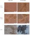
Microphotographs of mammospheres show no obvious changes in morphology at passage 1 to 2 cultured in ultra-low attachment plates.
Figure 3
Figure 3
Graphical representation and flow cytometry analysis results of expression of CD44 and CD24 surface markers in parental and 3D mammosphere cells: 92% 3D mammosphere cells are CD44+CD24-/low and 70% parental cells.
Figure 4
Figure 4
Graphical representation of ADLH activity in parental and 3D mammosphere cells.
Figure 5
Figure 5
Graphs showing cell viability of (A) monolayer, (B) single cell 3D, and (C) 3D mammosphere after various treatments for 10 days.
Figure 5
Figure 5
Graphs showing cell viability of (A) monolayer, (B) single cell 3D, and (C) 3D mammosphere after various treatments for 10 days.
Figure 6
Figure 6
Graphical representation of IC50 data for the three culture conditions upon various treatments for 10 days. * p < 0.05 compared to monolayer.
Figure 7
Figure 7
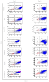
Flow cytometry results of cytopathology in control and treated 3D mammosphere cells; percentages of viable cells (Q3), early apoptosis (Q4), late apoptosis (Q2) and necrosis (Q1) at day 3 and day 10.
Figure 8
Figure 8
Estimation of percentage cytopathology in MDA MB 231 cells that were untreated and treated with free and drug-loaded nanoparticles. Percentages of viable cells (Q3), early apoptosis (Q4), late apoptosis (Q2) and necrosis (Q1) at day 3 (A) and day 10 (B).
Figure 8
Figure 8
Estimation of percentage cytopathology in MDA MB 231 cells that were untreated and treated with free and drug-loaded nanoparticles. Percentages of viable cells (Q3), early apoptosis (Q4), late apoptosis (Q2) and necrosis (Q1) at day 3 (A) and day 10 (B).
Figure 9
Figure 9
Photomicrographs of light microscopy showing the morphology of the mammosphere at days 0 and 10 after treatments.
Figure 9
Figure 9
Photomicrographs of light microscopy showing the morphology of the mammosphere at days 0 and 10 after treatments.
Figure 9
Figure 9
Photomicrographs of light microscopy showing the morphology of the mammosphere at days 0 and 10 after treatments.
Figure 10
Figure 10
Scanning electron micrograph of 3D MDA MB 231 cells mammosphere after 10 days of treatment.
Figure 11
Figure 11
Self-renewal efficiency of MDA MB 231 CSCs after treatment at first passage through to the third passage. P1 = first passage; P2 = second passage; P3 = third passage. * p < 0.05 compared to control.
Figure 12
Figure 12
Graphical representation of CD44+CD24- cells after treatment at days 3 and 10. * p < 0.05 compared to control.
Figure 13
Figure 13
Flow cytometry representation of CD44+CD24- cells after treatment at days 3 and 10.
Figure 14
Figure 14
Graphical representation of ADLH activity after treatment at days 3 and 10. * p < 0.05 compared to control.
Figure 15
Figure 15
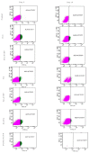
Flow cytometry result of ADLH activity after treatment at days 3 and 10. Purple and green indicate viable cells and ALDH activity, respectively.
Figure 16
Figure 16
Graphical representation of wound closure in untreated and treated 3D mammosphere at 6 and 24 h post scratching. * p < 0.05 compared to control.
Figure 17
Figure 17
Graphical representation of the percentage of 3D mammosphere cell invasion across the basement membrane compared to control. * p < 0.05 compared to control.
Figure 18
Figure 18
Flow cytometry data showing the effect of various treatments on the cell cycle distribution of 3D mammosphere cells at 48 h.
Figure 19
Figure 19
Graphical representation of the effect of various treatments on the cell cycle distribution of 3D mammosphere cells at 48 h. * p < 0.05 significant increase compared to control.

Similar articles

Cited by

References

    1. Ginestier C., Liu S., Diebel M.E., Korkaya H., Luo M., Brown M., Wicinski J., Cabaud O., Charafe-Jauffret E., Birnbaum D., et al. CXCR1 blockade selectively targets human breast cancer stem cells in vitro and in xenografts. J. Clin. Investig. 2010;120:485–497. doi: 10.1172/JCI39397. - DOI - PMC - PubMed
    1. Croker A.K., Allan A.L. Inhibition of aldehyde dehydrogenase (ALDH) activity reduces chemotherapy and radiation resistance of stem-like ALDHhiCD44+ human breast cancer cells. Breast Cancer Res. Treat. 2012;133:75–87. doi: 10.1007/s10549-011-1692-y. - DOI - PubMed
    1. Paula A.D.C., Lopes C. Implications of Different Cancer Stem Cell Phenotypes in Breast Cancer. Anticancer Res. 2017;37:2173–2183. doi: 10.21873/anticanres.11552. - DOI - PubMed
    1. Liu S., Wicha M.S. Targeting Breast Cancer Stem Cells. J. Clin. Oncol. 2010;28:4006–4012. doi: 10.1200/JCO.2009.27.5388. - DOI - PMC - PubMed
    1. McDermott S.P., Wicha M.S. Targeting breast cancer stem cells. Mol. Oncol. 2010;4:404–419. doi: 10.1016/j.molonc.2010.06.005. - DOI - PMC - PubMed

MeSH terms