. 2020 Mar 10;8(1):123.
doi: 10.3390/vaccines8010123.

mRNA Vaccines Encoding the HA Protein of Influenza A H1N1 Virus Delivered by Cationic Lipid Nanoparticles Induce Protective Immune Responses in Mice

Affiliations

mRNA Vaccines Encoding the HA Protein of Influenza A H1N1 Virus Delivered by Cationic Lipid Nanoparticles Induce Protective Immune Responses in Mice

Xinyu Zhuang et al. Vaccines (Basel). .

Abstract

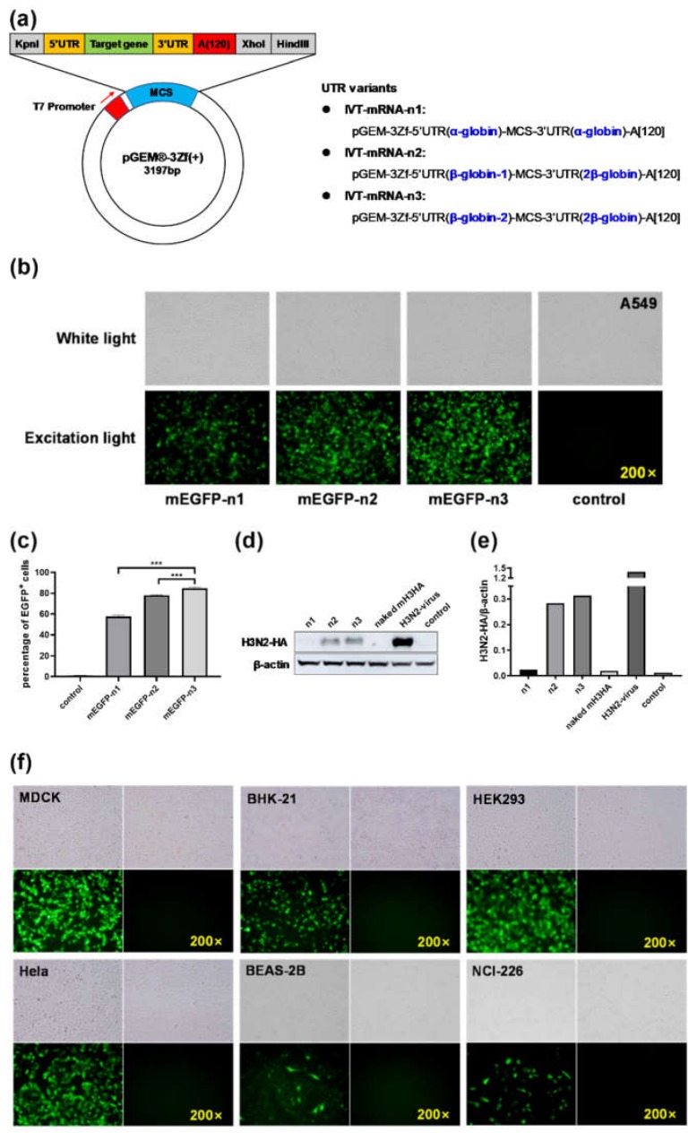
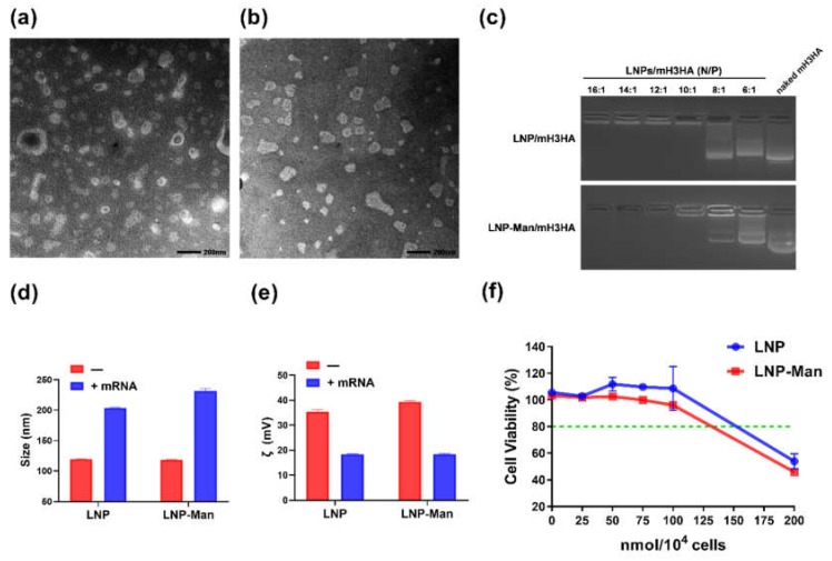
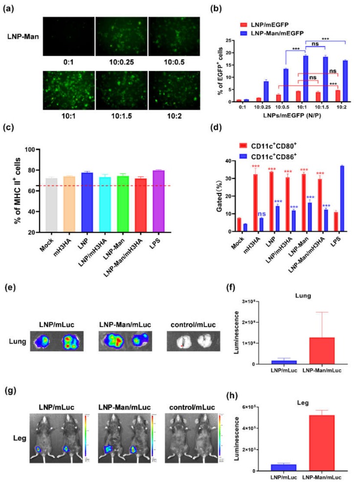
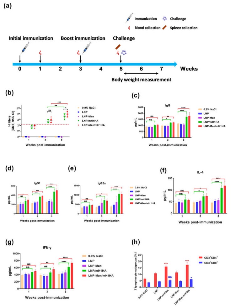
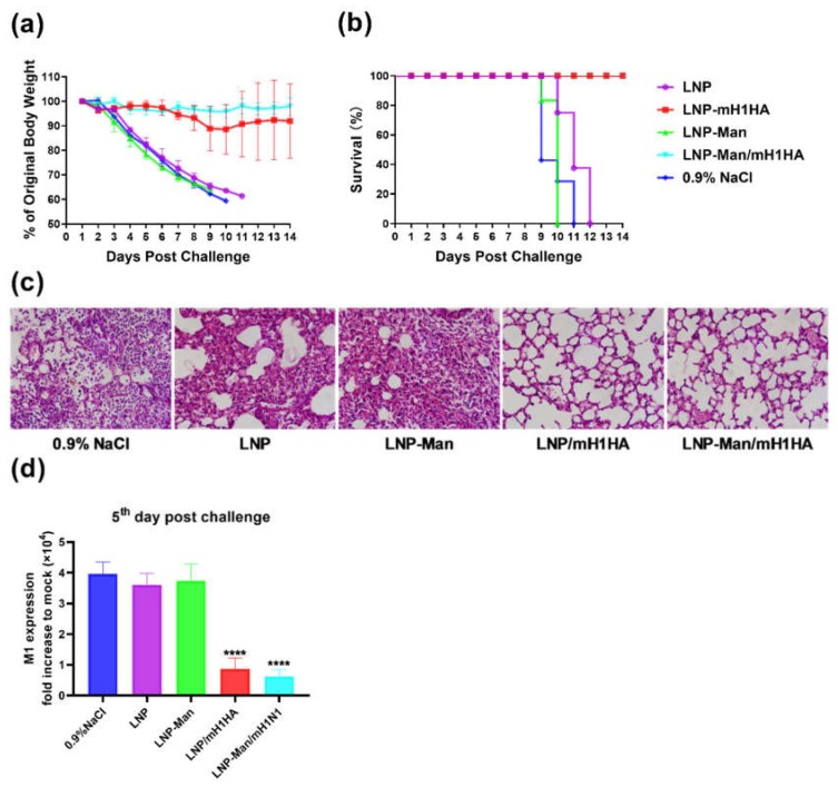
The design of the mRNA vaccine involves the selection of in vitro transcription (IVT) systems and nonviral delivery vectors. This study aimed to verify the effect of 5' and 3' untranslated region (UTR) sequences on the translation efficiency of mRNA. Three modes of IVT-mRNA systems (IVT-mRNA-n1/n2/n3) with diverse UTRs were constructed, and EGFP (enhanced green fluorescent protein) and HA (hemagglutinin) gene of H3N2 influenza virus were introduced into each of them. The results showed that the mode of 5' and 3' UTRs originating from human β-globulin was better than the mode of UTRs from human α-globulin, and the n3 mode was the best. mEGFP-n3, mH3HA-n3, and mLuciferease-n3 were prepared to compare the effect of cationic lipid nanoparticle (LNP) with that of mannose-conjugated LNP (LNP-Man) on the efficiency of gene delivery. The results showed that the effect of LNP-Man was better than that of LNP both in vitro and in vivo. Choosing appropriate ligands might help in vaccine design. After selecting the IVT-mRNA-n3 system and delivery vectors, mRNA vaccines were constructed against the H1N1 influenza virus, and C57BL/6 mice were immunized through intranasal administration. The results showed that mRNA vaccines could elicit both humoral and cellular immune responses and completely protect mice from the tenfold LD50 H1N1 influenza virus challenge.

Keywords: cationic lipid nanoparticles; influenza A H1N1 virus; intranasal administration; mRNA vaccine; mannose.


Conflict of interest statement

The authors declare no conflicts of interest. The sponsors had no role in the design, execution, interpretation, or writing of the study.

Figures

Figure 1
Figure 1
(a) Pattern diagram of the three modes of in vitro transcription systems. A549 cells transfected with enhanced green fluorescent protein (mEGFP)-n1/n2/n3 were observed 12 h after transfection under a fluorescence microscope (b), and the positive rates were detected by flow cytometry (c). Data are shown as means ± SDs (standard deviation) and analyzed using one-way ANOVA (analysis of variance) (n = 3, mEGFP-n3 vs. mEGFP-n1, *** p < 0.0001; mEGFP-n3 vs. mEGFP-n2, *** p < 0.0001). (d,e) Western blot analysis. A549 cells were harvested 12 h after transfection. The H3N2-HA protein was detected using rabbit anti-influenza A virus HA Mab (Sino Biological, Beijing, China). The gray value of the strips was analyzed using ImageJ software, and the bar chart was drawn using GraphPad Prism 8.0. (f) Fluorescence microscope images of various cell lines transfected with mEGFP-n3 after 12 h.
Figure 2
Figure 2
Characterization of lipid nanoparticles (LNPs) and LNPs/mRNA. (a,b) TEM images of LNP and LNP-Man. (c) Gel retardation assay. LNPs/mRNA were run in the 1.2% nuclease-free agarose gel. LNPs/mRNA complexes were prepared at different N/P molar ratios. Naked mRNA was used as the negative control without any complexation. (d) Size and (e) zeta potential of LNPs and LNPs/mH3HA (N/P = 10:1). (f) Cytotoxicity of LNP and LNP-Man was tested on A549 cells. Untreated cells were defined as 100% viability cells. Data are shown as means ± SDs (n = 3).
Figure 3
Figure 3
Functional verification of LNPs/mRNA. (a) Fluorescence microscope imaging. A549 cells transfected with LNP-Man/mEGFP at indicated N/P molar ratios were observed under a fluorescence microscope 12 h after transfection. (b) The EGFP positivity rates of cells transfected with LNPs/mEGFP at indicated N/P molar ratios were detected by flow cytometry. Data are shown as means ± SDs and were analyzed by two-way ANOVA (n = 3, ns p > 0.05; *** p < 0.0001). (c,d) Flow cytometric analysis of the dendritic cells’ (DC) maturation levels. Data are shown as means ± SDs and were analyzed by two-way ANOVA. (n = 3, ns p > 0.05; ***p < 0.0001; compared with Mock). (e,f) In vivo imaging. Images of lungs were acquired using an IVIS Lumina S5, and bioluminescence intensity from the region of interest was quantified using Living Image software. (g,h) In vivo imaging. Images of legs and the bioluminescence intensity.
Figure 4
Figure 4
Construction and verification of mRNA vaccine encoding the H1N1-HA protein. (a) Agarose gel electrophoresis of XhoI enzyme digestion products. 1 represents the intact plasmids of pGEM-H1N1-HA-n3 and 2 represents its linearized product of 5355 bp. M1: DL 5,000 DNA Marker (TaKaRa, Tokyo, Japan); M2: DL 2000 DNA Marker (TaKaRa, Tokyo, Japan). (b,c) Western blot and indirect immunofluorescence analyses. A549 cells were harvested 12 h and 48 h after transfection. The H1N1-HA protein was detected using rabbit anti-influenza A virus HA Mab. The H1N1-virus group was used as the positive control. Mock represents the negative control. DAPI was used to dye the nuclei.
Figure 5
Figure 5
Immune responses in mice induced by LNPs/mH1HA vaccines. Serum samples were collected in the first, third, and fifth weeks after the initial immunization. (a) The flow chart of the animal experiment. (b) Hemagglutination inhibition (HI) assay. Data are shown as the geometric mean with 95% CI and were analyzed by two-way ANOVA (n = 6, ns p > 0.05; ** p < 0.01; *** p < 0.001). (c–e) Total IgG/IgG1/IgG2a levels in the serum were detected using ELISA kits. Data are shown as means ± SDs and were analyzed by two-way ANOVA (n = 3, ns p > 0.05; * p < 0.05; ** p < 0.01; *** p < 0.001; **** p < 0.0001). (f,g) The IL-4 and IFN-γ levels in sera were determined by ELISA. Data are shown as means ± SDs and were analyzed by two-way ANOVA (n = 3, ns p > 0.05; ** p < 0.01; **** p < 0.0001). (h) The percentages of CD3+CD4+ and CD3+CD8+ T lymphocyte subgroups were tested by flow cytometry. Data are shown as means ± SDs and were analyzed by two-way ANOVA (n = 3, ns p > 0.05; * p < 0.05; *** p < 0.0001, compared with the 0.9% NaCl group).
Figure 6
Figure 6
Evaluation of the protective effects after the challenge. (a) Body weight changes and (b) Kaplan–Meier survival curves of H1N1-infected C57BL/6 mice (n = 6) treated with vaccines or vehicles by intranasal administration. Two weeks after the boost immunization, mice were infected with 10 × LD50 A/Jilin/JYT-01/2018(H1N1) influenza virus. Lungs were collected at 5th day post-infection. (c) Representative images of lung pathology in hematoxylin-and-eosin-stained sections from H1N1-infected C57BL/6 mice (n = 3). (d) Viral loads expressed as the fold change compared to noninfected lungs. Data are shown as means ± SDs and were analyzed by one-way ANOVA (n = 3, **** p < 0.0001, compared to 0.9% NaCl, LNP, and LNP-Man groups).

Similar articles

Cited by

References

    1. Krammer F. The human antibody response to influenza A virus infection and vaccination. Nat. Rev. Immunol. 2019;19:383–397. doi: 10.1038/s41577-019-0143-6. - DOI - PubMed
    1. Petrova N.V., Russell C.A. The evolution of seasonal influenza viruses. Nat. Rev. Microbiol. 2018;16:47–60. doi: 10.1038/nrmicro.2017.118. - DOI - PubMed
    1. Yamayoshi S., Kawaoka Y. Current and future influenza vaccines. Nat. Med. 2019;25:212–220. doi: 10.1038/s41591-018-0340-z. - DOI - PubMed
    1. Brazzoli M., Magini D., Bonci A., Buccato S., Giovani C., Kratzer R., Zurli V., Mangiavacchi S., Casini D., Brito L.M., et al. Induction of Broad-Based Immunity and Protective Efficacy by Self-amplifying mRNA Vaccines Encoding Influenza Virus Hemagglutinin. J. Virol. 2016;90:332–344. doi: 10.1128/JVI.01786-15. - DOI - PMC - PubMed
    1. Murray K.A., Preston N., Allen T., Zambrana-Torrelio C., Hosseini P.R., Daszak P. Global biogeography of human infectious diseases. Proc. Natl. Acad. Sci. USA. 2015;112:12746–12751. doi: 10.1073/pnas.1507442112. - DOI - PMC - PubMed