. 2020 Mar 12;18(1):43.
doi: 10.1186/s12951-020-00604-7.

Docetaxel-loaded solid lipid nanoparticles prevent tumor growth and lung metastasis of 4T1 murine mammary carcinoma cells

Affiliations

Docetaxel-loaded solid lipid nanoparticles prevent tumor growth and lung metastasis of 4T1 murine mammary carcinoma cells

Márcia Cristina Oliveira da Rocha et al. J Nanobiotechnology. .

Abstract

Background: Metastasis causes the most breast cancer-related deaths in women. Here, we investigated the antitumor effect of solid lipid nanoparticles (SLN-DTX) when used in the treatment of metastatic breast tumors using 4T1-bearing BALB/c mice.

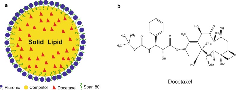
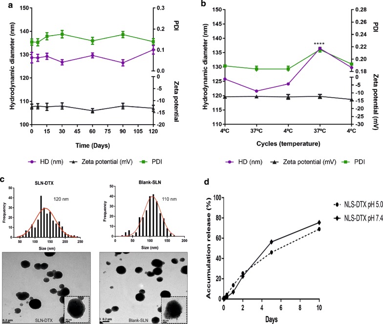
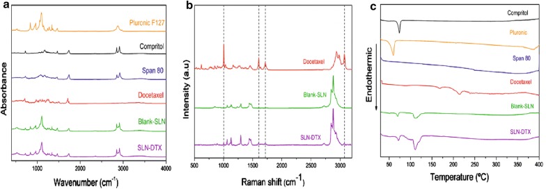
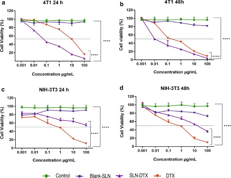
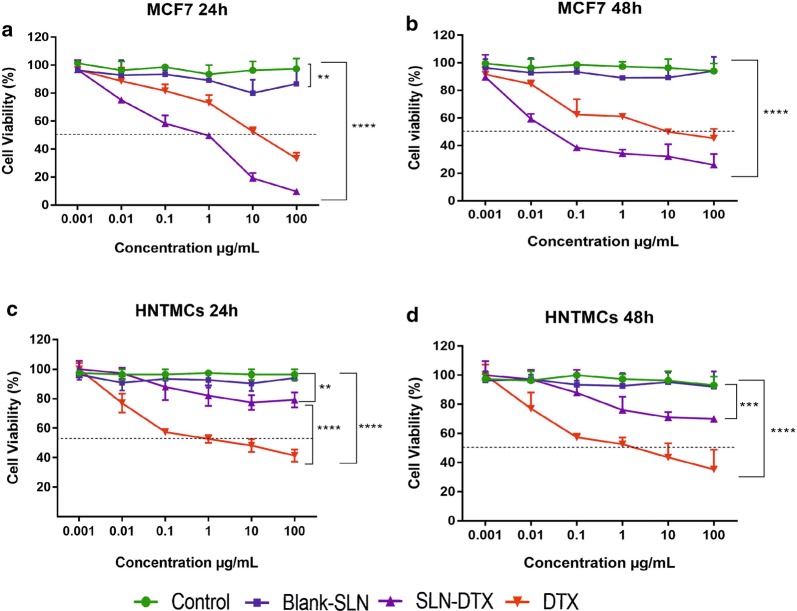
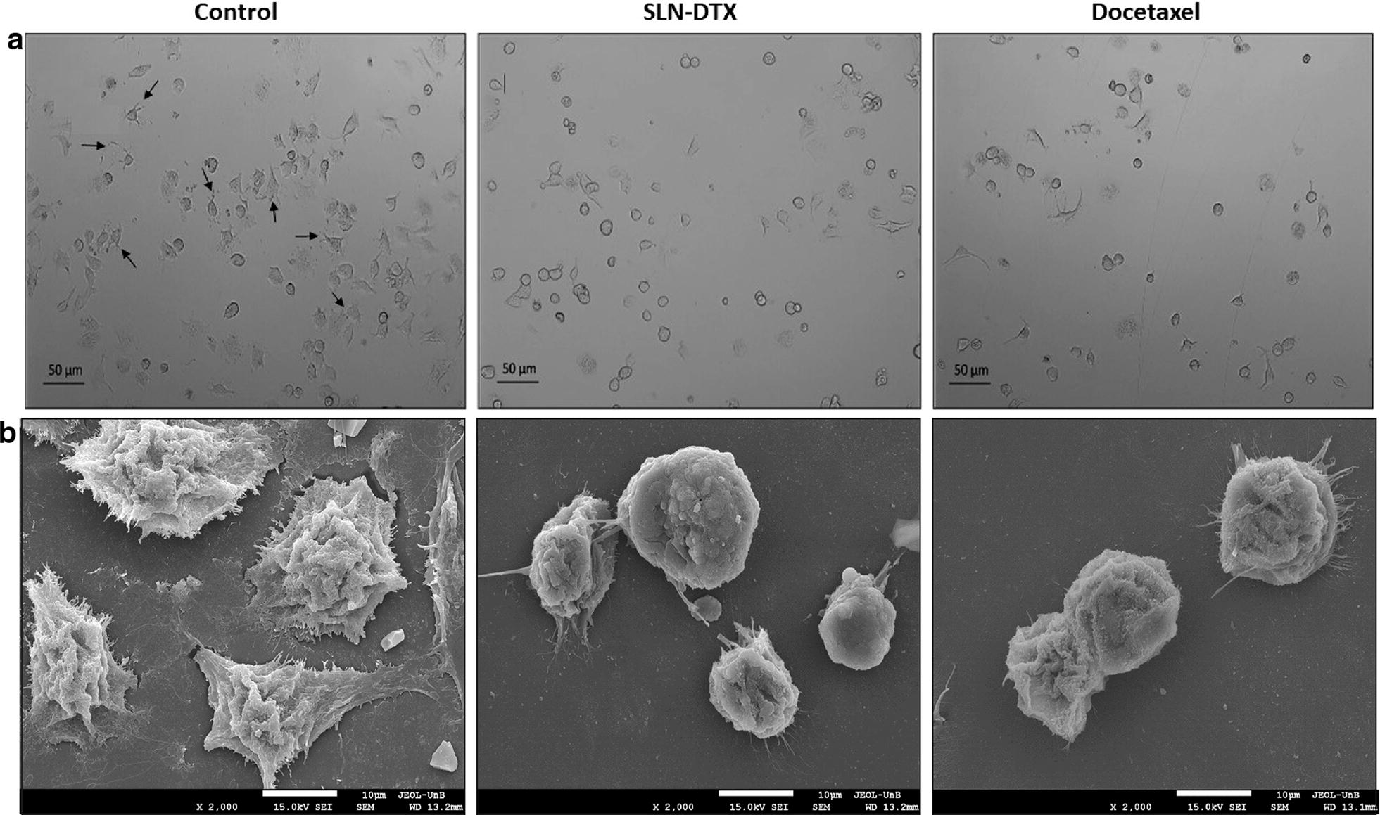
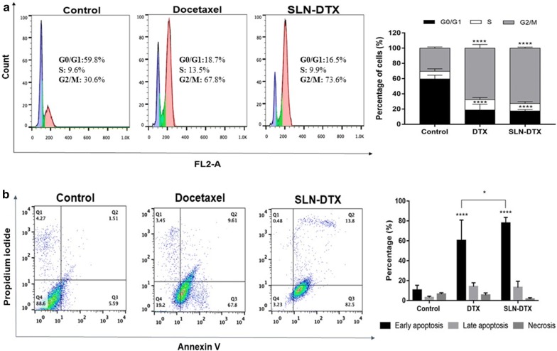
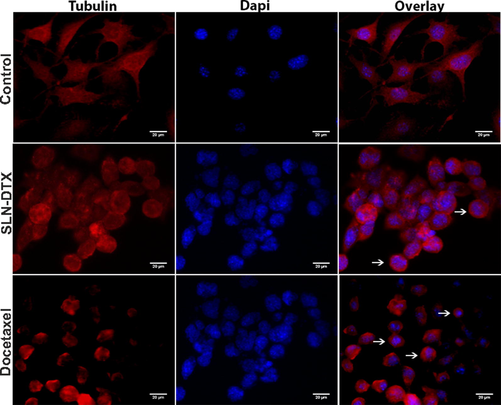
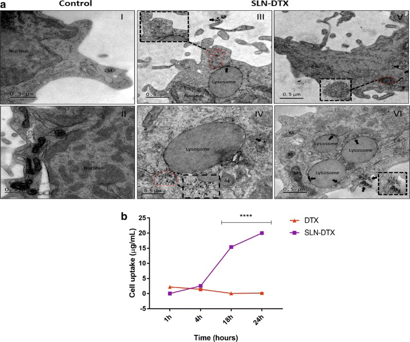
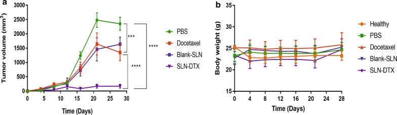
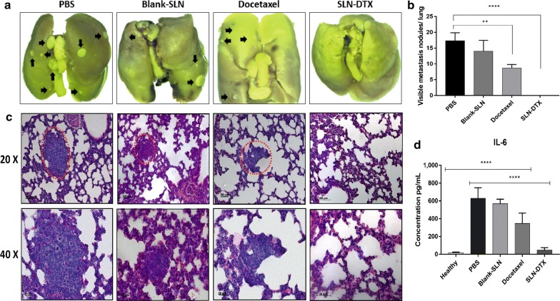
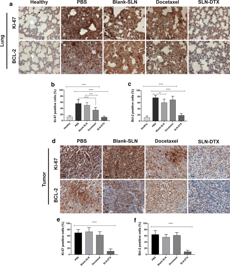
Results: Solid lipid nanoparticles (SLNs) were produced using the high-energy method. Compritol 888 ATO was selected as the lipid matrix, and Pluronic F127 and Span 80 as the surfactants to stabilize nanoparticle dispersion. The particles had high stability for at least 120 days. The SLNs' dispersion size was 128 nm, their polydispersity index (PDI) was 0.2, and they showed a negative zeta potential. SLNs had high docetaxel (DTX) entrapment efficiency (86%), 2% of drug loading and showed a controlled drug-release profile. The half-maximal inhibitory concentration (IC50) of SLN-DTX against 4T1 cells was more than 100 times lower than that of free DTX after 24 h treatment. In the cellular uptake test, SLN-DTX was taken into the cells significantly more than free DTX. The accumulation in the G2-M phase was significantly higher in cells treated with SLN-DTX (73.7%) than in cells treated with free DTX (23.0%), which induced subsequent apoptosis. TEM analysis revealed that SLN-DTX internalization is mediated by endocytosis, and fluorescence microscopy showed DTX induced microtubule damage. In vivo studies showed that SLN-DTX compared to free docetaxel exhibited higher antitumor efficacy by reducing tumor volume (p < 0.0001) and also prevented spontaneous lung metastasis in 4T1 tumor-bearing mice. Histological studies of lungs confirmed that treatment with SLN-DTX was able to prevent tumor. IL-6 serum levels, ki-67 and BCL-2 expression were analyzed and showed a remarkably strong reduction when used in a combined treatment.

Conclusions: These results indicate that DTX-loaded SLNs may be a promising carrier to treat breast cancer and in metastasis prevention.

Keywords: 4T1; BCL-2; Cellular uptake; IL-6; Ki-67 and antitumor effect; NIH-3T3.


Conflict of interest statement

The authors declare that they have no conflict of interests.

Figures

Fig. 1
Fig. 1
a Schematic illustration of SLN-DTX. b Chemical structure of docetaxel (DTX)
Fig. 2
Fig. 2
a Colloidal stability of SLN-DTX over 120 days. Hydrodynamic diameter (HD), zeta potential and polydispersity index (PDI) measured by dynamic light scattering. b Storage stability study of SLN-DTX. HD, zeta potential and PDI index were analyzed after samples were incubated for 24 h at 4 °C or 37 °C (n = 3). c Nanoparticle size distribution of SLN-DTX had an average size of 120 nm and blank-SLN around 110 nm; measurements were performed using ImageJ software. Morphology of SLN-DTX and blank-SLN was assessed by transmission electron microscopy (TEM) at 100 K and in dashed line 250 K magnification. d Drug release profiles from SLN-DTX at pH 7.4 and pH 5.0 PBS at 10 days. Data are expressed as mean ± standard error of the mean (****p < 0.0001)
Fig. 3
Fig. 3
a Vibrational spectra in the infrared region of Pluronic F127, Compritol, Span 80, docetaxel, Blank-SLN and SLN-DTX. b Raman spectra of docetaxel. The highlights represent docetaxel bands in SLN-DTX spectra. c DSC thermograms
Fig. 4
Fig. 4
Viability of 4T1 (a, b) and NIH-3T3 (c, d) cells after 24 and 48 h treated with free DTX, blank-SLN and SLN-DTX at 0.001, 0.01, 0.1, 1, 10 and 100 µg/mL (equivalent DTX concentrations), evaluated by MTT assay. Data are expressed as mean ± standard error of the mean (****p < 0.0001)
Fig. 5
Fig. 5
Viability of MCF7 (a, b) and HNTMCs (c, d) cells after 24 and 48 h treated with free DTX, blank-SLN and SLN-DTX at 0.001, 0.01, 0.1, 1, 10 and 100 µg/mL (equivalent DTX concentrations), evaluated by MTT assay. Data are expressed as mean ± standard error of the mean (**p < 0.01; ***p < 0.001; ****p < 0.0001)
Fig. 6
Fig. 6
Morphology of murine breast adenocarcinoma cells (4T1). a Light microscopy (phase contrast). b Scanning electron micrograph (SEM). Control cells (without treatment), cells treated with SLN-DTX or DTX. The arrows show the cytoplasmic projection in cells. Cells were treated at 10 µg/mL for 24 h. Objective: 10×. The micrographs (SEM) were performed at 2 k
Fig. 7
Fig. 7
a Representative flow cytometry profiles of the cell cycle phase distribution of 4T1 cells. The first peak indicates 2n DNA content in the G0/G1 phase (purple), the second peak indicates 4n DNA content in the G2/M phase (pink), and in-between the two peaks is the S phase (green). b Dot plots representing the distribution of apoptotic cells after dual staining with Annexin V-FITC (horizontal axis) and propidium iodide (vertical axis). Annexin V-FITC and propidium iodide tagged cells symbolize early apoptosis (lower right quadrant) and late apoptosis (upper right quadrant), respectively. On the right side the percentage of the cells is presented. Groups: untreated cells, SLN-DTX and DTX. Cell were treated with 10 µg/mL for 24 h. Data are presented as mean ± standard error from three experiments (*p < 0.05; ****p < 0.0001)
Fig. 8
Fig. 8
Immunofluorescence assay of 4T1 cells. Microtubules and nuclear staining are shown separately (Tubulin and DAPI, respectively) and combined (Overlay), for 4T1 cells untreated or treated with SLN-DTX or DTX 10 µg/mL for 24 h. The arrows show bundling of microtubules. (Objective 63×)
Fig. 9
Fig. 9
a Ultrastructural images of 4T1 cells. (I–II) Control cells (untreated). (III–VI) Cells treated with 10 µg/mL of SLN-DTX for 24 h. (III–V) SLN-DTX internalized (dashed line); (V) cluster of SLN-DTX in cytoplasm (dashed line); (VI) internalization of SLN-DTX into lysosomes (arrows). Dashed lines, arrows (into lysosomes) and arrow heads indicate nanoparticles. ER Endoplasmic reticulum, M Mitochondria. Magnification: 5 K; 15 K (black dashed lines). b In vitro cellular uptake studies of DTX in 4T1 cells. Cells were exposed to SLN-DTX or DTX for 1 h, 4 h, 18 h and 24 h at 100 µg/mL. Data are expressed as mean ± standard error of the mean (****p < 0.0001)
Fig. 10
Fig. 10
Antitumoral effect of SLN-DTX, Docetaxel, Blank-SLN and PBS on 4T1 tumor-bearing Balb/c mice. a Variation of tumor volume and b mice weight. ***p < 0.001; ****p < 0.0001, compared with PBS group
Fig. 11
Fig. 11
a Photo of lung. b Statistical analysis of numbers of tumor nodules on the lungs. c Histopathological examination of the lungs. d IL-6 serum levels of mice were measured by ELISA. The metastasis was marked by red dotted circle and black arrows. Data represent mean values ± standard error (**p < 0.01; ****p < 0.0001)
Fig. 12
Fig. 12
Immunohistochemistry analysis. The brown dye-stained cells represent positive cells. a Lung and d tumor slides were immunostained with anti-Ki-67 and BCL-2. b, e Quantification of Ki-67 and c, f BCL-2 staining. Data represent mean values ± standard error (**p < 0.01; ***p < 0.001; ****p < 0.0001)

References

    1. Li Y, Jin M, Shao S, Huang W, Yang F, Chen W, et al. Small-sized polymeric micelles incorporating docetaxel suppress distant metastases in the clinically-relevant 4T1 mouse breast cancer model. BMC Cancer. 2014;14:329. doi: 10.1186/1471-2407-14-329. - DOI - PMC - PubMed
    1. Hammadi NI, Abba Y, Hezmee MNM, Razak ISA, Jaji AZ, Isa T, et al. Formulation of a sustained release docetaxel loaded cockle shell-derived calcium carbonate nanoparticles against breast cancer. Pharm Res. 2017;34:1193–1203. doi: 10.1007/s11095-017-2135-1. - DOI - PubMed
    1. Zhang H, Wang K, Zhang P, He W, Song A, Luan Y. Redox-sensitive micelles assembled from amphiphilic mPEG-PCL-SS-DTX conjugates for the delivery of docetaxel. Colloids Surf B Biointerfaces. 2016 doi: 10.1016/j.colsurfb.2016.02.045. - DOI - PubMed
    1. Wang L, Liu Z, Liu D, Liu C, Juan Z, Zhang N. Docetaxel-loaded-lipid-based-nanosuspensions (DTX-LNS): preparation, pharmacokinetics, tissue distribution and antitumor activity. Int J Pharm. 2011;413:194–201. doi: 10.1016/j.ijpharm.2011.04.023. - DOI - PubMed
    1. Cho H, Yoon I. Surface-modified solid lipid nanoparticles for oral delivery of docetaxel: enhanced intestinal absorption and lymphatic uptake. Int J Nanomed. 2014;9:495–504. - PMC - PubMed