. 2020 Mar;41(3):522-528.
doi: 10.3174/ajnr.A6482. Epub 2020 Mar 12.

4D-Dynamic Contrast-Enhanced MRI for Preoperative Localization in Patients with Primary Hyperparathyroidism

Affiliations

4D-Dynamic Contrast-Enhanced MRI for Preoperative Localization in Patients with Primary Hyperparathyroidism

J L Becker et al. AJNR Am J Neuroradiol. 2020 Mar.

Abstract

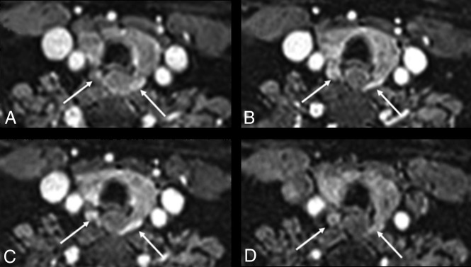
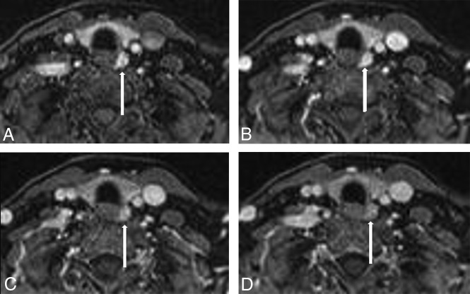
Background and purpose: Our aim was to test the hypothesis that our recently introduced 4D-dynamic contrast-enhanced MR imaging with high spatial and temporal resolution has equivalent accuracy to 4D-CT for preoperative gland localization in primary hyperparathyroidism without requiring exposure to ionizing radiation.

Materials and methods: Inclusion criteria were the following: 1) confirmed biochemical diagnosis of primary hyperparathyroidism, 2) preoperative 4D-dynamic contrast-enhanced MR imaging, and 3) surgical cure with >50% decrease in serum parathyroid hormone intraoperatively. 4D-dynamic contrast-enhanced studies were reviewed independently by 2 neuroradiologists to identify the side, quadrant, and number of abnormal glands, and compared with surgical and pathologic results.

Results: Fifty-four patients met the inclusion criteria: 37 had single-gland disease, and 17, multigland disease (9 with double-gland hyperplasia; 3 with 3-gland hyperplasia; and 5 with 4-gland hyperplasia). Interobserver agreement (κ) for the side (right versus left) was 0.92 for single-gland disease and 0.70 for multigland disease. Interobserver agreement for the quadrant (superior versus inferior) was 0.70 for single-gland disease and 0.69 for multigland disease. For single-gland disease, the gland was correctly located in 34/37 (92%) patients, with correct identification of the side in 37/37 (100%) and the quadrant in 34/37 (92%) patients. For multigland disease, the glands were correctly located in 35/47 (74%) patients, with correct identification of the side in 35/47 (74%) and the quadrant in 36/47 (77%).

Conclusions: The proposed high spatial and temporal resolution 4D-dynamic contrast-enhanced MR imaging provides excellent diagnostic performance for preoperative localization in primary hyperparathyroidism, with correct gland localization of 92% for single-gland disease and 74% in multigland disease, superior to 4D-CT studies.


Figures

Fig 1.
Fig 1.
A 39-year-old woman with PHPT and MGD with bilateral glands imaged in the lower quadrants. A, Axial early arterial phase. B, Early venous phase. C, Midvenous phase. D, Later venous phase. The contour of the glands is different in A and B due to interval patient motion without image degradation. Images show bilateral MGD glands (white arrows) with early arterial enhancement and increased signal intensity compared with the adjacent thyroid gland (A). Signal intensity increases into the early venous phase, with MGD glands having signal intensity similar to that of the carotid artery on the early venous phase and increased signal compared with the thyroid gland (B). At the mid-venous phase, the MGD glands have decreased signal intensity compared with the carotid artery due to early washout but still have increased signal compared with the thyroid gland (C). The MGD gland signal decreases further in the later venous phase, becoming isointense with the thyroid gland (D).
Fig 2.
Fig 2.
A 54-year-old woman with PHPT and SGD in the left lower quadrant. A, Axial early arterial phase. B, Early venous phase. C, Midvenous phase. D, Later venous phase. Images show a left lower quadrant parathyroid adenoma (white arrow) with early arterial enhancement (A), increasing with signal intensity similar to that of the internal carotid artery (B), with mildly decreased signal intensity compared with the carotid artery due to early venous washout (C), and decreased signal intensity compared with the adjacent internal carotid artery and jugular vein in the later venous phase (D).

References

    1. Ruda JM, Hollenbeak CS, Stack BC Jr.. A systematic review of the diagnosis and treatment of primary hyperparathyroidism from 1995 to 2003. Otolaryngol Head Neck Surg 2005;132:359–72 10.1016/j.otohns.2004.10.005 - DOI - PubMed
    1. Heath H 3rd, Hodgson SF, Kennedy MA. Primary hyperparathyroidism: incidence, morbidity, and potential economic impact in a community. N Engl J Med 1980;302:189–93 10.1056/NEJM198001243020402 - DOI - PubMed
    1. Ghandur-Mnaymneh L, Kimura N. The parathyroid adenoma: a histopathologic definition with a study of 172 cases of primary hyperparathyroidism. Am J Pathol 1984;115:70–83 - PMC - PubMed
    1. Kazaure HS, Thomas S, Scheri RP, et al. The devil is in the details: assessing treatment and outcomes of 6,795 patients undergoing remedial parathyroidectomy in the Collaborative Endocrine Surgery Quality Improvement Program. Surgery 2019;165:242–49 10.1016/j.surg.2018.03.026 - DOI - PubMed
    1. Kunstman JW, Udelsman R. Superiority of minimally invasive parathyroidectomy. Adv Surg 2012;46:171–89 10.1016/j.yasu.2012.04.004 - DOI - PubMed