. 2020 Jun;105(6):e315-e317.
doi: 10.3324/haematol.2020.246744. Epub 2020 Mar 12.

IGH rearrangement in myeloid neoplasms

Affiliations

IGH rearrangement in myeloid neoplasms

Amy W Xiao et al. Haematologica. 2020 Jun.
No abstract available


Figures

Figure 1.
Figure 1.
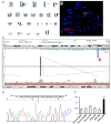
Acute myeloid leukemia with an IGH-CCNG1 rearrangement. (A) Chromosome analysis of the bone marrow aspirate showed a complex karyotype, including an unbalanced translocation involving chromosomes 5, 14 and 19. Arrows indicate clonal aberrations. (B) Fluorescence in situ hybridization (FISH) on abnormal metaphase showed the 3′IGH (red) remaining on the derivative chromosome 14 and the 5′IGH lost, consistent with an unbalanced IGH rearrangement. (C) A mate-pair next-generation sequencing (NGS) found an IGH-CCNG1 rearrangement (3IGH-V3::5CCNG1). (D) IGH-CCNG1 breakpoints were confirmed by PCR followed by Sanger sequencing. (E) The CCNG1 expression level was evaluated by a real-time quantitative RT-PCR with the patient’s bone marrow, a normal blood sample, two normal bone marrow samples, a myelodysplastic syndrome (MDS) with a normal karyotype and an acute myeloid leukemia (AML) with a complex karyotype without 14q (IGH) rearrangement. CCNG1 expression was normalized to ACTB. ***: P<0.001.
Figure 2.
Figure 2.
Myelodysplastic syndrome with an IGH-MECOM rearrangement. (A) Chromosome analysis of the bone marrow aspirate showed monosomy 7 and a balanced translocation t(3;14). Arrows indicate clonal aberrations. (B) Fluorescence in situ hybridization (FISH) on an abnormal metaphase with a MECOM (EVI1) probe showed the centromeric MECOM (aqua) remaining on chromosome 3 and both the MECOM (green) and the telomeric MECOM (red) translocated to chromosome 14. (C) FISH on the same abnormal metaphase (after probe stripping) with a IGH probe showed the 3′IGH (red) remaining on chromosome 14 and the 5′IGH translocated to chromosome 3.

References

    1. Vasmatzis G, Johnson SH, Knudson RA, et al. Genome-wide analysis reveals recurrent structural abnormalities of TP63 and other p53-related genes in peripheral T-cell lymphomas. Blood. 2012; 120(11):2280–2289. - PMC - PubMed
    1. Roy AL, Sen R, Roeder RG. Enhancer-promoter communication and transcriptional regulation of Igh. Trends Immunol. 2011;32(11):532–539. - PMC - PubMed
    1. Rovigatti U, Mirro J, Kitchingman G, Dahl G. Heavy chain immunoglobulin gene rearrangement in acute nonlymphocytic leukemia. Blood. 1984;63(5):1023–1027. - PubMed
    1. Qiu Y, Korteweg C, Chen Z, et al. Immunoglobulin G expression and its colocalization with complement proteins in papillary thyroid cancer. Mod Pathol. 2012;25(1):36–45. - PubMed
    1. Qiu X, Sun X, He Z, et al. Immunoglobulin gamma heavy chain gene with somatic hypermutation is frequently expressed in acute myeloid leukemia. Leukemia. 2013;27(1):92–99. - PubMed