. 2020 Apr 17;295(16):5427-5448.
doi: 10.1074/jbc.RA119.010002. Epub 2020 Mar 12.

Cell-specific expression of the transcriptional regulator RHAMM provides a timing mechanism that controls appropriate wound re-epithelialization

Affiliations

Cell-specific expression of the transcriptional regulator RHAMM provides a timing mechanism that controls appropriate wound re-epithelialization

Cornelia Tolg et al. J Biol Chem. .

Abstract

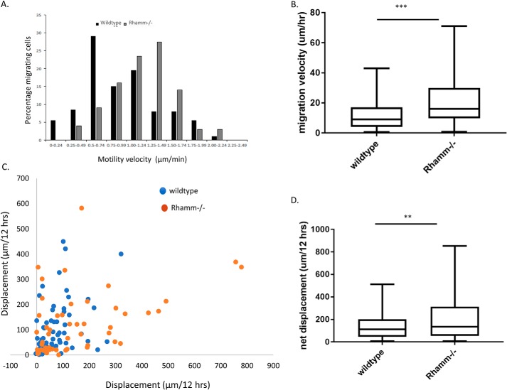
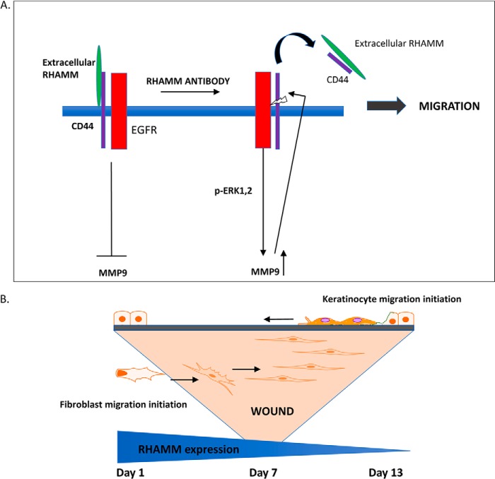
Prevention of aberrant cutaneous wound repair and appropriate regeneration of an intact and functional integument require the coordinated timing of fibroblast and keratinocyte migration. Here, we identified a mechanism whereby opposing cell-specific motogenic functions of a multifunctional intracellular and extracellular protein, the receptor for hyaluronan-mediated motility (RHAMM), coordinates fibroblast and keratinocyte migration speed and ensures appropriate timing of excisional wound closure. We found that, unlike in WT mice, in Rhamm-null mice, keratinocyte migration initiates prematurely in the excisional wounds, resulting in wounds that have re-surfaced before the formation of normal granulation tissue, leading to a defective epidermal architecture. We also noted aberrant keratinocyte and fibroblast migration in the Rhamm-null mice, indicating that RHAMM suppresses keratinocyte motility but increases fibroblast motility. This cell context-dependent effect resulted from cell-specific regulation of extracellular signal-regulated kinase 1/2 (ERK1/2) activation and expression of a RHAMM target gene encoding matrix metalloprotease 9 (MMP-9). In fibroblasts, RHAMM promoted ERK1/2 activation and MMP-9 expression, whereas in keratinocytes, RHAMM suppressed these activities. In keratinocytes, loss of RHAMM function or expression promoted epidermal growth factor receptor-regulated MMP-9 expression via ERK1/2, which resulted in cleavage of the ectodomain of the RHAMM partner protein CD44 and thereby increased keratinocyte motility. These results identify RHAMM as a key factor that integrates the timing of wound repair by controlling cell migration.

Keywords: RHAMM; cell migration; cell signaling; extracellular matrix; hyaluronan; keratinocyte; keratinocytes; wound healing; wound repair.


Conflict of interest statement

The authors declare that they have no conflicts of interest with the contents of this article

Figures

Figure 1.
Figure 1.
Rhamm−/− keratinocytes re-surface excisional skin wounds more rapidly than WT comparators. A, cross-sections of the center of excisional skin wounds were stained for pan-keratin (brown) and counterstained with hematoxylin. Red arrows indicate the leading edge of the migrating keratinocyte layer. Dotted black lines in day 3 images outline the underlying granulation tissue, and the black arrow indicates the clot. Subcutaneous fat (labeled dermal adipocytes) has uniquely expanded into the granulation tissue of Rhamm−/− mice. Rhamm−/− wounds are delayed in granulation tissue formation, but resurfacing of wounds by keratinocytes is accelerated relative to WT wounds. B, quantification of wound re-epithelialization shows that Rhamm−/− wounds have completed re-surfacing by day 7, and WT wounds complete this process by day 14. Values are the mean and S.E. n = 3 mice. *, p < 0.01.
Figure 2.
Figure 2.
Rhamm-loss causes aberrant epidermal differentiation. A, cross-sections of WT and Rhamm−/− excisional skin wounds at day 14 post-wounding were stained for pan-keratin and show that WT epidermis has re-organized into discernable basal, suprabasal, and cornified layers by day 14; however, these layers are not distinguishable in Rhamm−/− epidermis. B, cytokeratin 10 (K10) staining of day 14 Rhamm−/− and WT wounds was used to identify the suprabasal keratinocyte layer. C, Par3 expression was detected by immunohistochemistry and used as an epidermal polarization marker. Loss of RHAMM reduces expression of this protein at day 14 wound centers and edges.
Figure 3.
Figure 3.
RHAMM protein is transiently expressed in excisional wounds. A, strong RHAMM staining appears in wounds and peri-wound areas at day 1 post-excisional injury and occurs in all skin layers. By day 3, staining intensity has notably decreased in the interfollicular epidermis and is absent by day 14. White arrows indicate the wound edge. Staining is negative in Rhamm−/− wounds (day 3 at wound edge shown) to show antibody specificity for RHAMM. B, cytokeratin 10 (K10) and cytokeratin 14 (K14) staining of day 1, 3, and 7 WT wounds.
Figure 4.
Figure 4.
Rhamm-loss does not alter keratinocyte or fibroblast proliferation. Cell proliferation was quantified using Ki67 immunohistochemistry (A–C). A and B, Ki67 staining of keratinocytes (A) or dermal fibroblasts (B) of day 3 and 7 wound tissue sections is not significantly different in WT versus Rhamm−/− mice. Values are the mean and S.E. n = 50 cells/3 mice, p > 0.05. C, Western blot analysis of RHAMM expression in HaCaT keratinocytes and fibroblasts. D, Rhamm-loss (Rh−/−) also did not significantly alter cell survival/proliferation of fibroblasts compared with WT as detected by Ki67 staining. Box and whisker plots of n = 50 cells, p > 0.05. E, function-blocking RHAMM antibody did not significantly change Ki67 staining of HaCaT keratinocytes in culture. Box and whisker plots of n = 100 cells, p > 0.05.
Figure 5.
Figure 5.
Rhamm-loss reduces fibroblast migration. Immortalized Rhamm−/− fibroblasts were transfected with a full-length mouse Rhamm cDNA, and motility of the null and rescued cells was quantified. Rescue of Rhamm−/− fibroblasts significantly increases cell motility velocity (A) and net translocation (B) but migration persistence is not affected (C). D, speed of WT fibroblasts that express RHAMM is significantly greater than Rhamm−/− counterparts in defined medium. WT fibroblast migration is increased by fetal calf serum (FCS) and is strongly reduced by either a RHAMM function–blocking antibody or MEK inhibitor. Rhamm−/− fibroblasts are unresponsive to these stimuli and inhibitors. Results are shown as scatter plots. *, p < 0.05; **, p < 0.01; and ***, p < 0.001.
Figure 6.
Figure 6.
Rhamm-loss promotes keratinocyte migration. A–D, Rhamm−/− primary keratinocytes migrate more rapidly than WT counterparts in random migration assays. Primary keratinocytes were isolated from Rhamm−/− or WT 1–2-day-old mouse pups, and migration was stimulated by EGF. A, velocity range distribution of WT or Rhamm−/− keratinocytes: n = 64 cells/assay, n = 2 assays, B, velocity of WT and Rhamm−/− keratinocytes. Box and whisker plots of n = 64 cells. C, displacement plot of individual WT and Rhamm−/− keratinocytes. D, Box and whisker plot showing net displacement of WT and Rhamm−/− keratinocytes. n = 50 cells. **, p < 0.01; ****, p < 0.001.
Figure 7.
Figure 7.
Rhamm-loss does not affect the directional persistence of keratinocyte migration. The directional persistence of primary WT and Rhamm−/− keratinocytes was calculated as the total translational/net translocation. Rhamm−/− keratinocytes did not detectably differ from WT comparators in this motility function. A, diagram of time-lapse analyses of individual keratinocytes. B, box and whisker plot of each time point shown in A.
Figure 8.
Figure 8.
Blocking extracellular RHAMM with a RHAMM antibody stimulates the migration of WT keratinocytes but has no effect on Rhamm−/− keratinocytes. Primary keratinocytes were isolated from newborn mouse skin, plated onto fibronectin-coated culture surfaces in the presence of RHAMM blocking antibody or nonimmune IgG, used as a control, and then filmed. A, migration velocity profile of individual primary WT keratinocytes. B, box and whisker plot showing that the RHAMM antibody (RHAMM Ab) significantly increases WT keratinocyte motility velocity. **, p < 0.01. C, migration velocity profile of individual primary Rhamm−/− keratinocytes. D, box and whisker plot showing that the RHAMM antibody does not significantly affect the migration velocity of primary Rhamm−/− keratinocytes.
Figure 9.
Figure 9.
Blocking extracellular RHAMM with a RHAMM antibody stimulates migration of HaCaT keratinocytes. A and B, scratch wounds of HaCaT cells were treated with RHAMM antibodies or control IgG. Migration vector length over time was determined by cinemicrography. A shows vector length of n = 100 individual cells. Graph in B shows box and whisker plots of the results in A. **, p < 0.01. C, random motility net translocation of HaCaT keratinocytes treated with RHAMM antibodies or control IgG. *, p < 0.05; ***, p < 0.001.
Figure 10.
Figure 10.
RHAMM suppresses ERK1/2 activity in wounds and EGF-stimulated keratinocytes. A and B, immunohistochemical staining of phospho-ERK1/2 in wound sections at days 3 and 14. Phospho-ERK1/2 staining is higher in Rhamm−/− keratinocytes than WT at both day 3 and day 14. At day 14, phospho-ERK1/2 staining occurs throughout the Rhamm−/− epidermal layers but is restricted to the proliferating basal keratinocytes in WT mice. B, scatter plots of n = 4 slides with five replicate analyses for each slide. **, p < 0.01; ***, p < 0.001. C, cultured primary Rhamm−/− keratinocytes retain higher phospho-ERK1/2 levels than WT keratinocytes. Scatter plots of n = 7 replicates. *, p < 0.05. D, ERK1/2 activity is required for migration of both Rhamm−/− and WT primary keratinocytes, but Rhamm−/− keratinocyte migration is more strongly blocked by pathway inhibition using the MEK inhibitor PD98059 than WT comparators. Values are presented as percentage inhibition. Scatter plot of n = 4; ****, p < 0.0001.
Figure 11.
Figure 11.
RHAMM regulates ERK1/2 motogenic signaling in EGF-stimulated keratinocytes. A, EGFR inhibition (EGFRI) blocks migration of HaCaT cells treated with RHAMM antibody but had no effect on IgG-treated cells. Box and whisker plots of n = 35 cells. B, RHAMM antibody stimulates ERK1/2 activation in HaCaT cells. Western blot analysis of HaCaT cells treated with RHAMM antibody or control IgG. C, addition of a MEK1,2 inhibitor (PD8059) to HaCaT keratinocytes blocks migration stimulated by the RHAMM antibody indicating that extracellular RHAMM regulates this pathway. Box and whisker plot of n = 35 cells. ***, p < 0.001. D, co-localization of RHAMM and EGFR is reduced by RHAMM antibodies. Scatter plots of n = 15 cells. E, EGFR activation is not affected by RHAMM antibodies. Western blot analysis is shown of active EGFR and total EGFR in HaCaT cells treated with RHAMM antibody or control IgG.
Figure 12.
Figure 12.
RHAMM differentially regulates MMP-9 mRNA expression in keratinocytes and fibroblasts. PCR (A) and Affymetrix (B) arrays were used to detect differences in expression of migration-related genes and were performed with mRNA isolated from primary keratinocytes and immortalized fibroblasts. A, RHAMM-loss increases Mmp-9 and PTK2 mRNA expression in keratinocytes. B, RHAMM suppression decreases mRNA expression of these genes in fibroblasts. Values in A and B are mean and S.E. n = 3 replicates (p < 0.05).
Figure 13.
Figure 13.
MMP-9 expression is regulated by ERK1/2 in keratinocytes and fibroblasts and is required for migration of both cell types. A, RHAMM-blocking antibody significantly increases the expression of MMP-9 mRNA in HaCaT keratinocytes as detected by qRT-PCR using GAPDH as a loading control. Expression is reduced by inhibiting ERK1/2 activity with the MEK inhibitor PD98059. Box and whisker plot of n = 9 replicates. B, Rhamm−/− fibroblasts express little MMP-9 mRNA and expression is increased by Rhamm-rescue. The increased in MMP-9 expression stimulated by Rhamm-rescue is blocked by inhibiting ERK1/2 activity using PD98059. Scatter plot of n = 4 replicates. C, keratinocyte migration stimulated by the RHAMM antibody is blocked by inhibiting MMP-9 activity. Box and whisker plot of n = 25. D, fibroblast migration stimulated by Rhamm-rescued is blocked by inhibiting MMP-9 activity. Values are the mean and S.E. n = 60 cells/condition. *, p < 0.05; **, p < 0.01; ***, p < 0.001
Figure 14.
Figure 14.
RHAMM regulates MMP-9 activity. A and B, scratch wounds of HaCaT keratinocyte cultures were treated with either RHAMM antibodies or control IgG. MMP-9 activity in conditioned medium was analyzed by zymogram gels. RHAMM antibodies increased MMP-9 activity. MMP-9 activity was inhibited by an MMP-9 inhibitor, demonstrating its specificity. A, scatter plot of n = 3–4. *, p < 0.05; ***, p < 0.001. B, zymogram images. C and D, MMP-9 activity in fibroblasts is rescued by RHAMM expression. C, images of collagen degradation. D, scatter plot of n = 10 cells. **, p < 0.01.
Figure 15.
Figure 15.
Loss of CD44 promotes keratinocyte migration and RHAMM regulates CD44 shedding. A, CD44 function-blocking antibody increases HaCaT motility to a similar extent as the RHAMM antibody. Addition of both antibodies together does not further increase motility predicting that CD44 and RHAMM are acting on the same motogenic pathway. Box and whisker plots of n = 65 cells/condition. B, RHAMM antibody increases CD44 shedding, which requires MMP-9 activity. Shedding was quantified using a CD44 ELISA that detects all CD44 isoforms. Box and whisker plots of n = 6 replicates. a-MMP-9, activated recombinant MMP-9 protein; MMP-9I, MMP-9 inhibitor. C, a-MMP-9 stimulates HaCaT keratinocytes migration, which was quantified with a scratch-wound assay. Left panel shows migration vectors of individual cells, and right panel shows the averaged motility of HaCaT keratinocytes exposed to active MMP-9 protein or buffer alone. Values are the mean and S.E. n = 120. *, p < 0.05; ***, p < 0.001.
Figure 16.
Figure 16.
Models for RHAMM-regulated motogenic signaling in keratinocytes and temporal-spatial coordination of keratinocyte and fibroblast migration in excisional wounds. A, signaling model for regulation of keratinocyte migration by RHAMM. RHAMM/CD44 interactions block EGFR-regulated ERK1/2 activity and downstream expression of the motogenic target gene, MMP-9. Blocking extracellular RHAMM function releases the pathway and stimulates the expression and release of MMP-9, which promotes CD44 shedding, resulting in increased keratinocyte migration. B, RHAMM expression is ubiquitous in wounds by 24 h after excisional injury and then decreases over time. Maximal expression corresponds to influx of innate immune cells and initiation of fibroblast migration to form granulation tissue. As RHAMM protein levels decrease, keratinocyte migration is initiated. Aberrant RHAMM expression alters the timing of keratinocyte and fibroblast migration resulting in altered rate of wound closure and dysregulated differentiation within the wound site.

Similar articles

Cited by

References

    1. Lindley L. E., Stojadinovic O., Pastar I., and Tomic-Canic M. (2016) Biology and biomarkers for wound healing. Plast. Reconstr. Surg. 138, 18S–28S 10.1097/PRS.0000000000002682 - DOI - PMC - PubMed
    1. Fang R. C., and Mustoe T. A. (2008) Animal models of wound healing: utility in transgenic mice. J. Biomater. Sci. Polym. Ed. 19, 989–1005 10.1163/156856208784909327 - DOI - PubMed
    1. Greaves N. S., Ashcroft K. J., Baguneid M., and Bayat A. (2013) Current understanding of molecular and cellular mechanisms in fibroplasia and angiogenesis during acute wound healing. J. Dermatol. Sci. 72, 206–217 10.1016/j.jdermsci.2013.07.008 - DOI - PubMed
    1. Bielefeld K. A., Amini-Nik S., and Alman B. A. (2013) Cutaneous wound healing: recruiting developmental pathways for regeneration. Cell. Mol. Life Sci. 70, 2059–2081 10.1007/s00018-012-1152-9 - DOI - PMC - PubMed
    1. Ud-Din S., and Bayat A. (2017) Non-animal models of wound healing in cutaneous repair: in silico, in vitro, ex vivo, and in vivo models of wounds and scars in human skin. Wound Repair Regen. 25, 164–176 10.1111/wrr.12513 - DOI - PubMed

Publication types

MeSH terms

Substances