. 2020 May 8;295(19):6629-6640.
doi: 10.1074/jbc.RA119.012023. Epub 2020 Mar 12.

The tethering function of mitofusin2 controls osteoclast differentiation by modulating the Ca2+-NFATc1 axis

Affiliations

The tethering function of mitofusin2 controls osteoclast differentiation by modulating the Ca2+-NFATc1 axis

Anna Ballard et al. J Biol Chem. .

Abstract

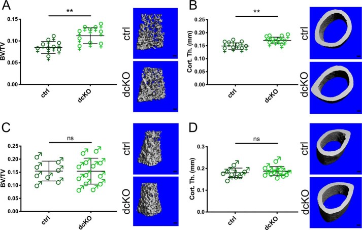
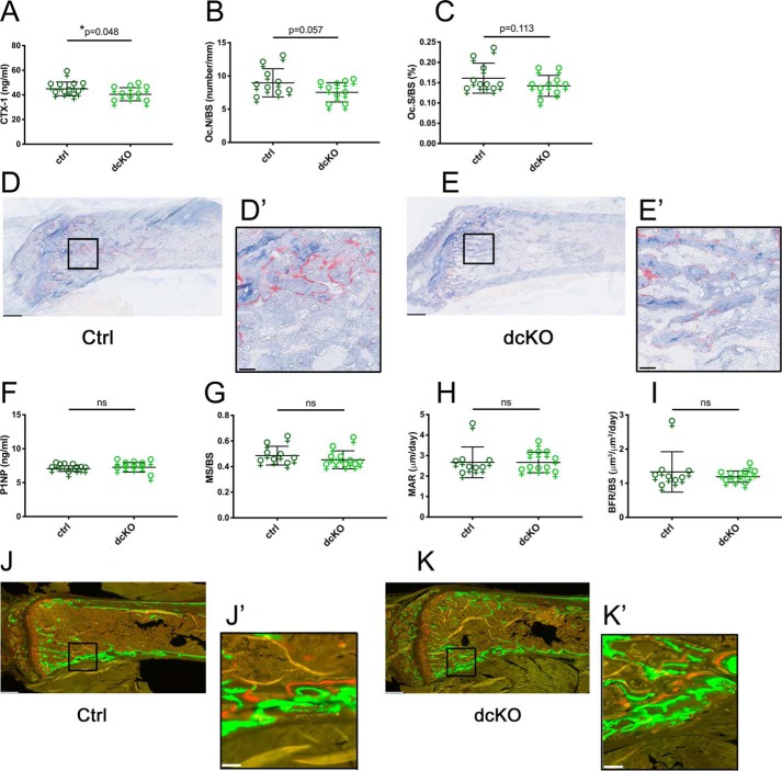
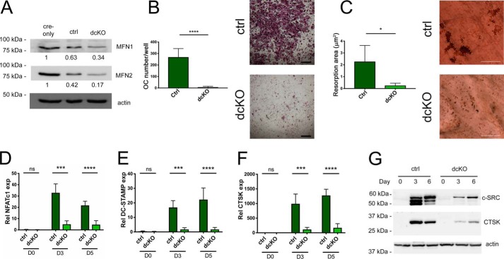
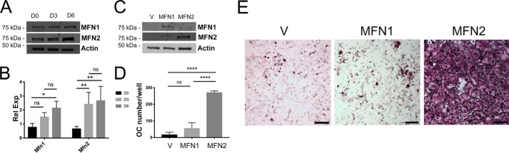
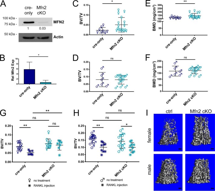
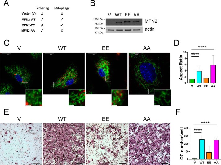
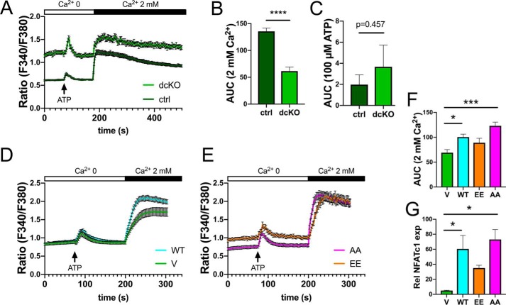
Dynamic regulation of the mitochondrial network by mitofusins (MFNs) modulates energy production, cell survival, and many intracellular signaling events, including calcium handling. However, the relative importance of specific mitochondrial functions and their dependence on MFNs vary greatly among cell types. Osteoclasts have many mitochondria, and increased mitochondrial biogenesis and oxidative phosphorylation enhance bone resorption, but little is known about the mitochondrial network or MFNs in osteoclasts. Because expression of each MFN isoform increases with osteoclastogenesis, we conditionally deleted MFN1 and MFN2 (double conditional KO (dcKO)) in murine osteoclast precursors, finding that this increased bone mass in young female mice and abolished osteoclast precursor differentiation into mature osteoclasts in vitro Defective osteoclastogenesis was reversed by overexpression of MFN2 but not MFN1; therefore, we generated mice lacking only MFN2 in osteoclasts. MFN2-deficient female mice had increased bone mass at 1 year and resistance to Receptor Activator of NF-κB Ligand (RANKL)-induced osteolysis at 8 weeks. To explore whether MFN-mediated tethering or mitophagy is important for osteoclastogenesis, we overexpressed MFN2 variants defective in either function in dcKO precursors and found that, although mitophagy was dispensable for differentiation, tethering was required. Because the master osteoclastogenic transcriptional regulator nuclear factor of activated T cells 1 (NFATc1) is calcium-regulated, we assessed calcium release from the endoplasmic reticulum and store-operated calcium entry and found that the latter was blunted in dcKO cells. Restored osteoclast differentiation by expression of intact MFN2 or the mitophagy-defective variant was associated with normalization of store-operated calcium entry and NFATc1 levels, indicating that MFN2 controls mitochondrion-endoplasmic reticulum tethering in osteoclasts.

Keywords: Mus musculus; animal model; bone; calcium; mitochondria; mitochondrial dynamics; osteoclast; osteoclastogenesis.


Conflict of interest statement

G. W. D. is a founder of Mitochondria in Motion, Inc., a St. Louis–based startup biotech R&D company focused on enhancing mitochondrial trafficking and fitness in neurodegenerative diseases, and may financially benefit if the company is successful in marketing products related to this research. The content is solely the responsibility of the authors and does not necessarily represent the official views of the National Institutes of Health

Figures

Figure 1.
Figure 1.
Trabecular and cortical bone mass are increased in 2-month-old female mice lacking MFN1 and MFN2 in the OC lineage. A and B, BV/TV (A) and Cort.Th (B) are significantly elevated in female dcKOs compared with ctrls in the distal femur at 2 months of age, as shown by μCT. Representative reconstructions of analyzed regions above the growth plate and at mid-shaft are shown in the right panels. Scale bars = 200 μm. C and D, 2-month-old males were evaluated for BV/TV (C) and Cort.Th (D). **, p < 0.01; ns, not significant; unpaired t test with Welch's correction.
Figure 2.
Figure 2.
Female Mfn1/2 dcKOs have decreased OC activity but no change in bone formation. A, serum CTX-1 assay shows decreased bone resorption in 2-month-old dcKO females. B and C, histomorphometry of TRAP stained tibiae reveals a modest decreases in dcKO Oc.N/BS (B) and Oc.S/BS (C). D and E, representative TRAP-stained ctrl and dcKO femora, respectively. F–I, no differences in bone accrual between groups were observed by serum P1NP (F) or dynamic histomorphometric analysis of trabecular bone for mineralizing surface (MS)/BS (G), mineral apposition rate (MAR) (H), and bone formation rate (BFR)/BS (I) in tibiae. J and K, representative fluorescence images of ctrl and dcKO femora, respectively. Scale bars = 400 μm (low power) and 40 μm (insets). *, p < 0.05; ns, not significant; one-tailed unpaired t test with Welch's correction.
Figure 3.
Figure 3.
Osteoclastogenesis is defective in BMMs derived from dcKO bone marrow. A, MFN1 and MFN2 protein levels are progressively decreased in RANKL-treated ctrls and dcKOs compared with RANKL-treated cre-only BMMs. B, BMMs cultured with RANKL for 5 days form OCs in ctrls but not in dcKOs. Scale bars = 400 μm. C, bone resorption is congruently inhibited; resorption pits are outlined in dotted white. Scale bars = 200 μm. D–G, mRNA levels of the OC markers NFATc1 (D), DC-STAMP (E), and CTSK (F) are diminished in dcKOs after 3 and 5 days of differentiation in RANKL, correlating with C-SRC and CTSK protein levels during osteoclastogenesis (G). *, p < 0.05; ***, p < 0.001; ****, p < 0.0001; ns, not significant; unpaired t test with Welch's correction. n = 3 biological replicates.
Figure 4.
Figure 4.
Although both homologs increase during OC formation, addition of MFN2 drives osteoclastogenesis in vitro more efficiently. A and B, protein (A) and mRNA (B) expression through 6 days of osteoclastogenesis in WT BMMs shows parallel increases in MFN1 and MFN2 expression. C, dcKO BMMs were transduced with retroviral pMX-Vector, V, pMX-MFN1, MFN1, or pMX-MFN2, and mitofusin proteins were detected via Western blotting. D and E, following RANKL treatment for 6 days, cultures were stained with TRAP, and OCs were enumerated, revealing robust osteoclastogenesis in MFN2-treated but not MFN1-treated cells. The graphs represent three biological replicates. Scale bars = 400 μm. *, p < 0.05; **, p < 0.01; ****, p < 0.0001; ns, not significant; one-way ANOVA.
Figure 5.
Figure 5.
Female mice lacking MFN2 alone in the OC lineage are protected from bone loss with age and RANKL-induced osteolysis. A and B, MFN2 expression is decreased in Mfn2 cKO OCs in vitro compared with cre-only at the protein (A) and RNA (B) levels. C–F, femora from cre-only and Mfn2 cKO mice at 12 months of age were analyzed by μCT for BV/TV in females (C), BV/TV in males (D), BMD in females (E), and BMD in males (F). Trabecular bone mass is increased in female cKOs but not males. G–I, acute bone loss was induced by intraperitoneal RANKL injection at 2 months of age in cre-only and Mfn2 cKO mice, and assessed by μCT at the distal femur. BV/TV is not decreased with RANKL treatment in female Mfn2 cKO mice (G). BV/TV decreases to a similar degree in male Mfn2 cKO mice compared with cre-only controls (H). I, representative post-RANKL μCT reconstructions from G and H. Scale bars = 200 μm. The untreated cohorts are the same as those listed at 2 months of age in Table 1 and Table S1. *, p < 0.05; **, p < 0.01; ns, not significant; unpaired t test with Welch's correction in B–F and ordinary two-way ANOVA with multiple comparisons in G and H.
Figure 6.
Figure 6.
Restoration of MFN2 tethering function rescues osteoclastogenesis in dcKO BMMs. A, MFN2 functions in each rescue condition. B, dcKO BMMs were transduced with retroviral pMX-Vector (V), pMX-MFN2-WT (WT), pMX-MFN2-EE (EE), and pMX-MFN2-AA (AA), and MFN2 protein was detected via Western blotting in BMMs. C, cultures of transduced BMMs were stained with MitoTracker Green and LysoTracker Red. Scale bars = 10 μm (whole cells) and 2.5 μm (insets). D, quantification of mitochondrial aspect ratio (mitochondrial length/width). E, BMMs were TRAP-stained following 6 days of RANKL exposure, revealing robust osteoclastogenesis with MFN2-AA but not MFN2-EE. Scale bars = 400 μm. F, quantification of OC numbers in three biological replicates. ****, p < 0.0001, ordinary one-way ANOVA.
Figure 7.
Figure 7.
Blunted Ca2+ entry in dcKO preOCs is restored with MFN2-AA overexpression. A–C, Fura-2 staining shows elevated basal intracellular Ca2+ but blunted store-operated calcium entry after addition of extracellular Ca2+ in dcKO pre-OCs. Shown is area under the curve quantification following Ca2+ (B) and ATP (C). D and E, overexpression of MFN2-WT or MFN2-AA restores Ca2+ entry in dcKO pre-OCs whereas MFN2-EE does not. F, quantification of the area under the curve. G, corresponding NFATc1 levels, evaluated by quantitative real-time PCR in mutant OCs, correlate with SOCE responsiveness. *, p < 0.05; ***, p < 0.001; ****, p < 0.0001; unpaired t test (B and C) or ordinary one-way ANOVA (F and G).

References

    1. Schrepfer E., and Scorrano L. (2016) Mitofusins, from mitochondria to metabolism. Mol. Cell 61, 683–694 10.1016/j.molcel.2016.02.022 - DOI - PubMed
    1. Dorn G. W. 2nd, Vega R. B., and Kelly D. P. (2015) Mitochondrial biogenesis and dynamics in the developing and diseased heart. Genes Dev. 29, 1981–1991 10.1101/gad.269894.115 - DOI - PMC - PubMed
    1. Chen H., Detmer S. A., Ewald A. J., Griffin E. E., Fraser S. E., and Chan D. C. (2003) Mitofusins Mfn1 and Mfn2 coordinately regulate mitochondrial fusion and are essential for embryonic development. J. Cell Biol. 160, 189–200 10.1083/jcb.200211046 - DOI - PMC - PubMed
    1. Filadi R., Pendin D., and Pizzo P. (2018) Mitofusin 2: from functions to disease. Cell Death Dis. 9, 330–343 10.1038/s41419-017-0023-6 - DOI - PMC - PubMed
    1. Celsi F., Pizzo P., Brini M., Leo S., Fotino C., Pinton P., and Rizzuto R. (2009) Mitochondria, calcium and cell death: a deadly triad in neurodegeneration. Biochim. Biophys. Acta 1787, 335–344 10.1016/j.bbabio.2009.02.021 - DOI - PMC - PubMed

Publication types