. 2020 Mar 13;367(6483):eaax9554.
doi: 10.1126/science.aax9554.

Liquid-liquid phase separation drives skin barrier formation

Affiliations

Liquid-liquid phase separation drives skin barrier formation

Felipe Garcia Quiroz et al. Science. .

Abstract

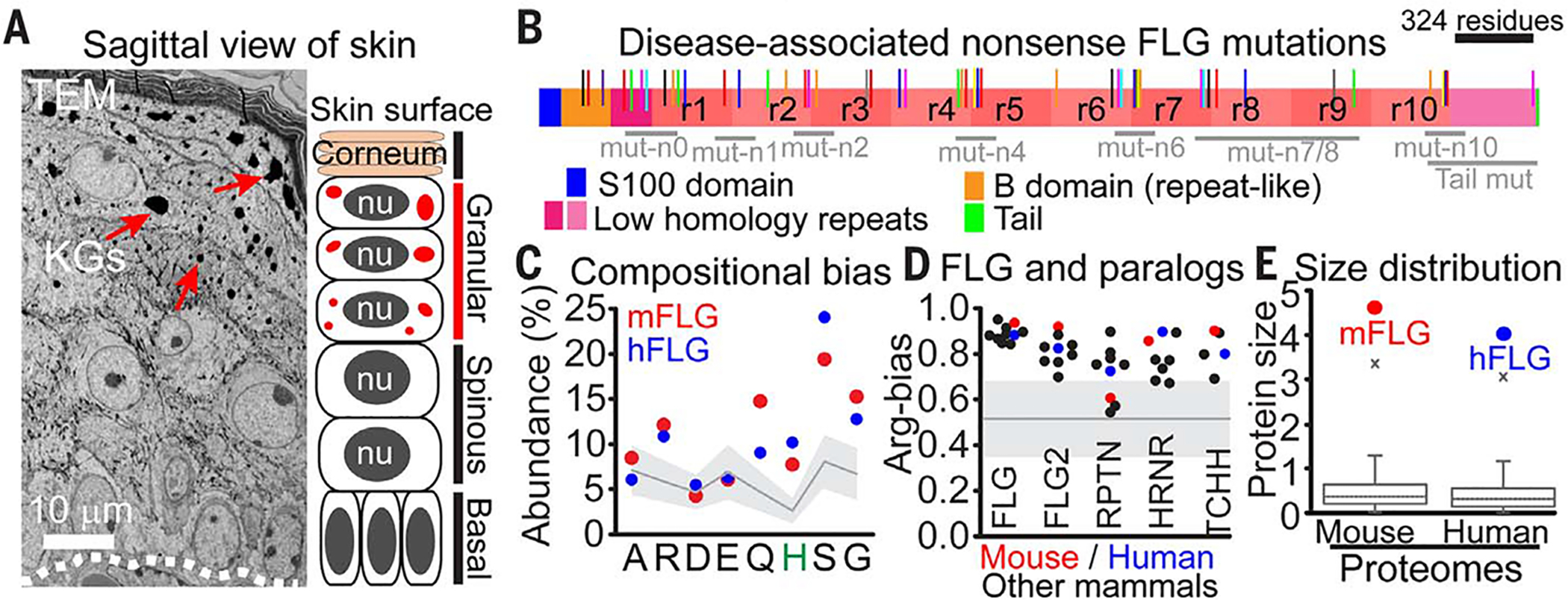
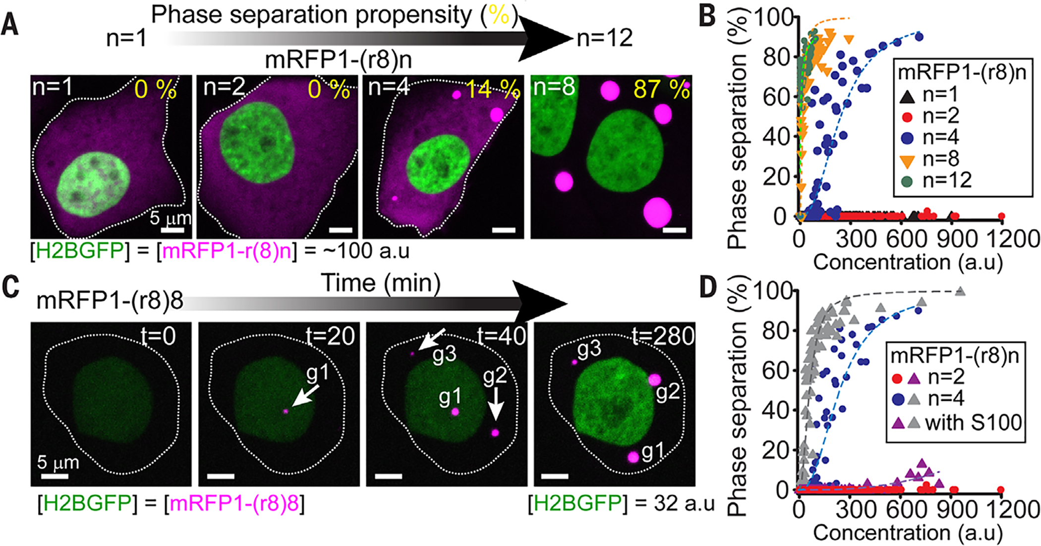
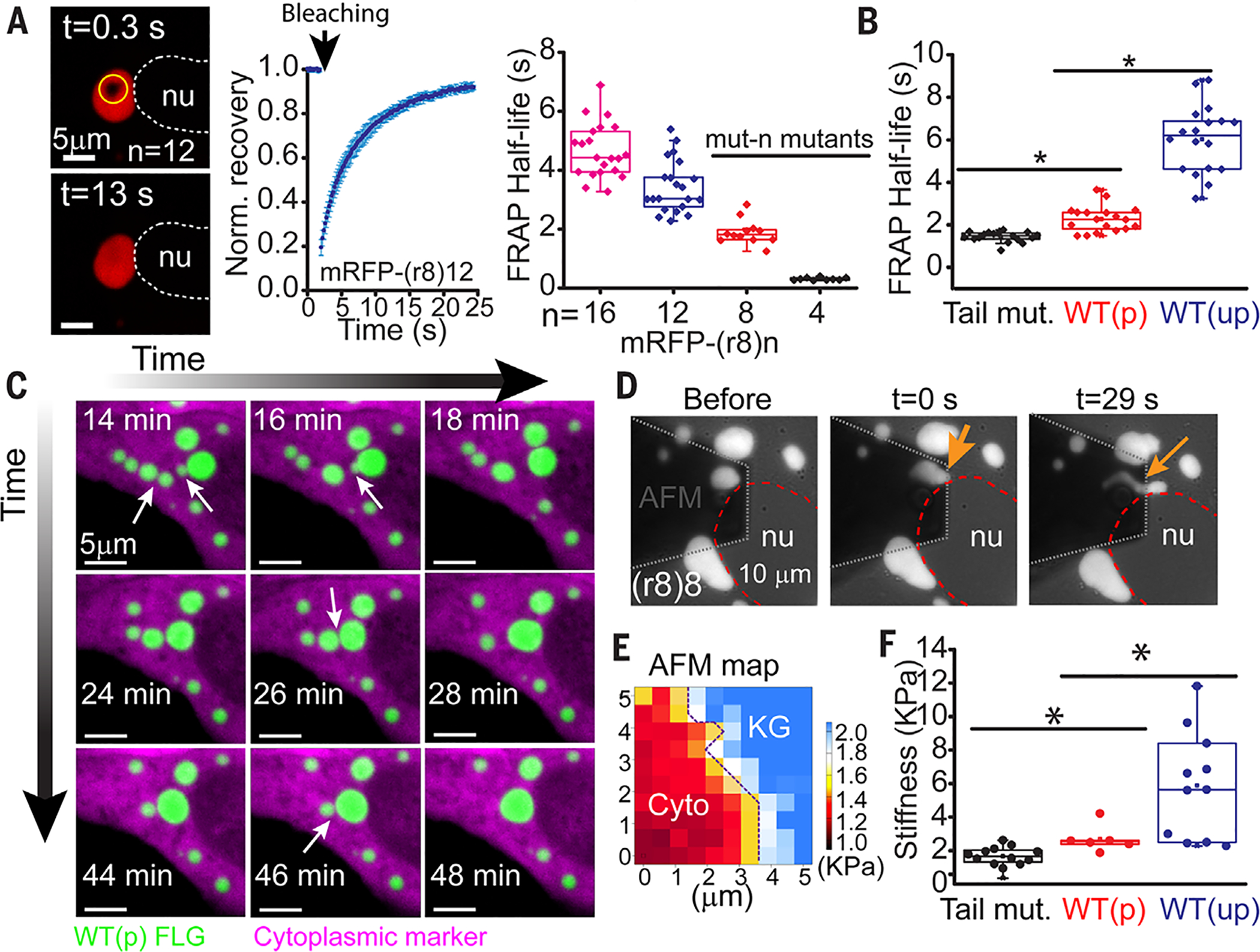
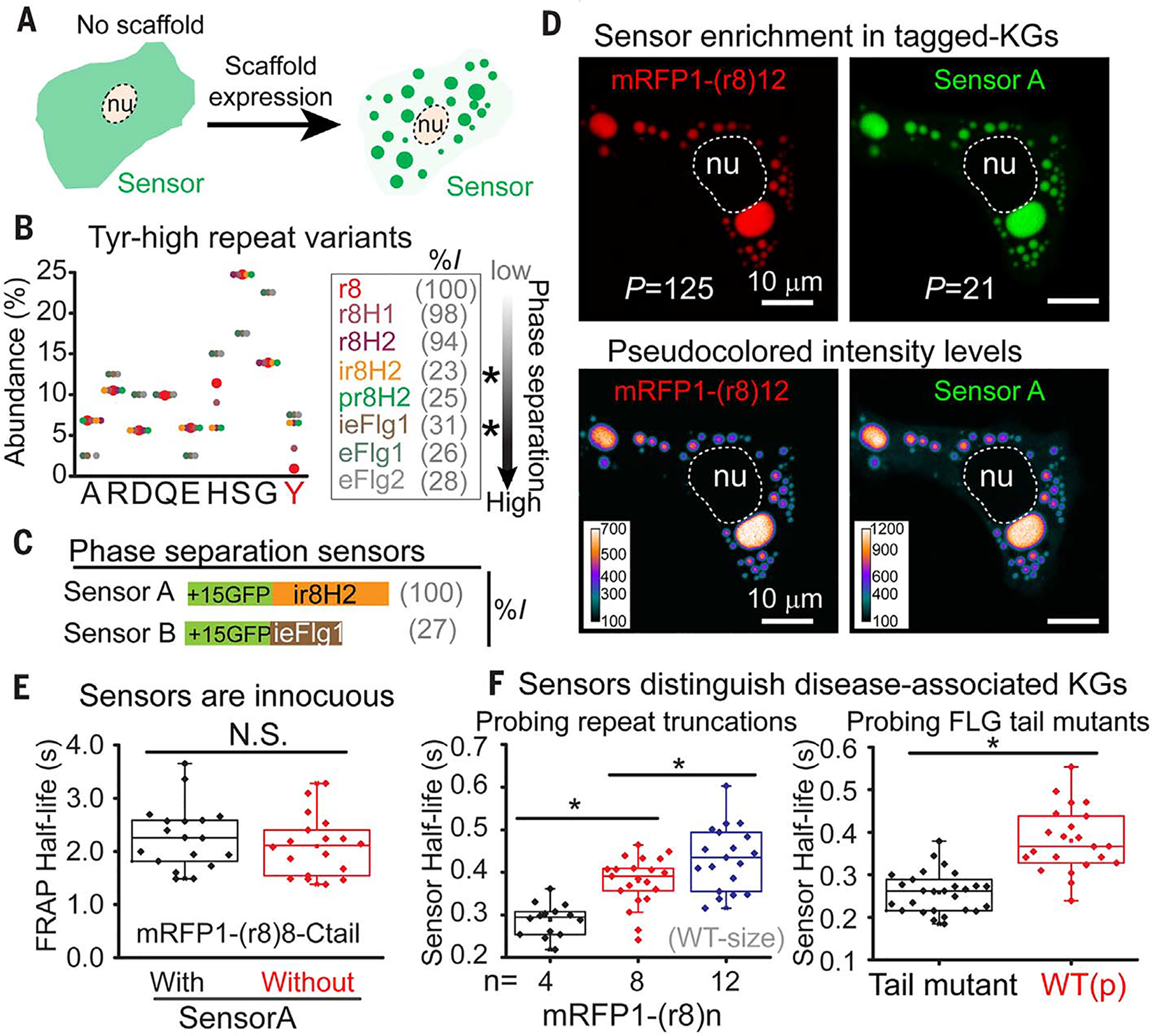
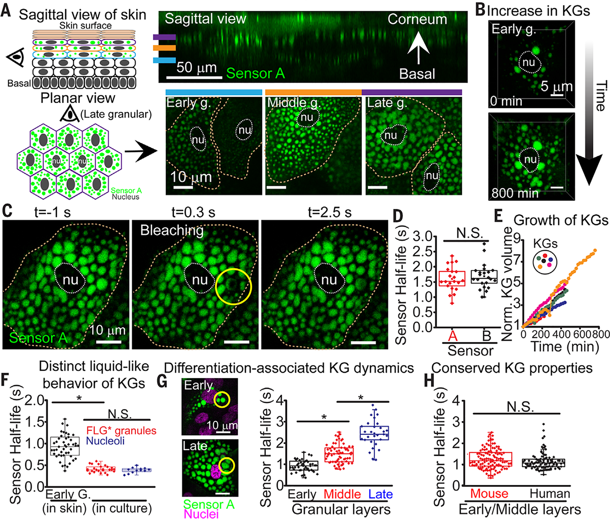
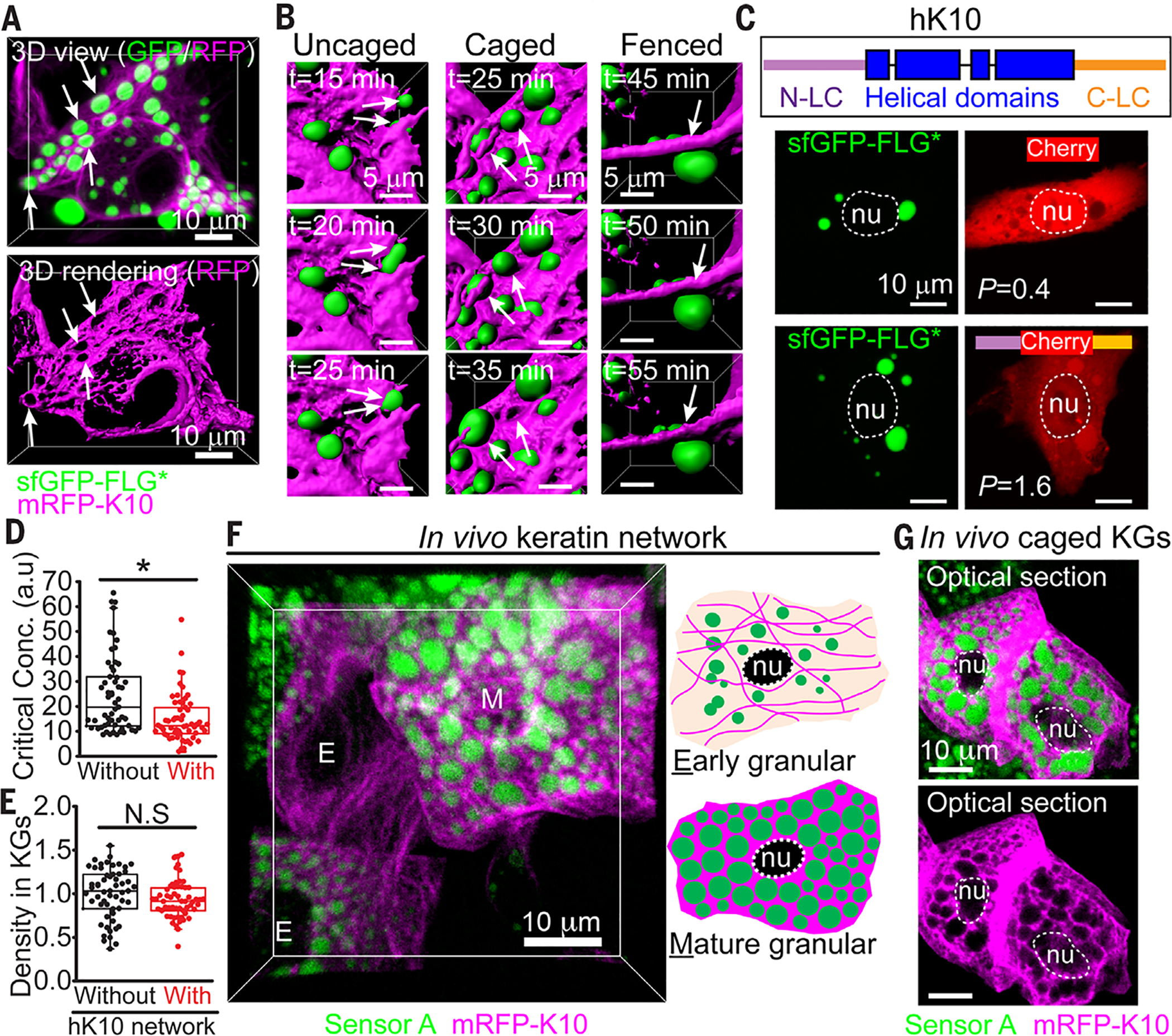
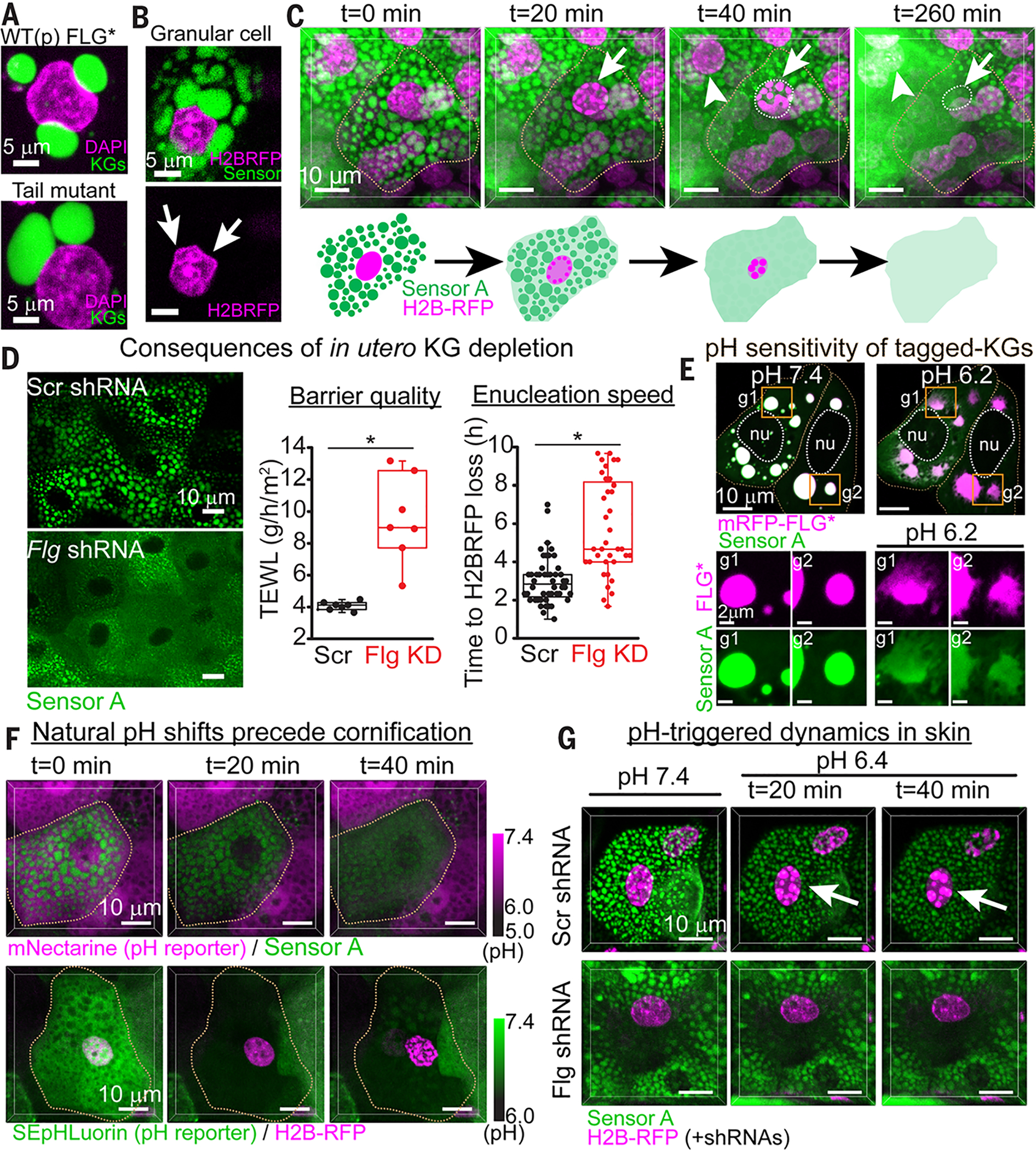
At the body surface, skin's stratified squamous epithelium is challenged by environmental extremes. The surface of the skin is composed of enucleated, flattened surface squames. They derive from underlying, transcriptionally active keratinocytes that display filaggrin-containing keratohyalin granules (KGs) whose function is unclear. Here, we found that filaggrin assembles KGs through liquid-liquid phase separation. The dynamics of phase separation governed terminal differentiation and were disrupted by human skin barrier disease-associated mutations. We used fluorescent sensors to investigate endogenous phase behavior in mice. Phase transitions during epidermal stratification crowded cellular spaces with liquid-like KGs whose coalescence was restricted by keratin filament bundles. We imaged cells as they neared the skin surface and found that environmentally regulated KG phase dynamics drive squame formation. Thus, epidermal structure and function are driven by phase-separation dynamics.


Conflict of interest statement

Competing interests: E.F. and F.G.Q. are inventors on a provisional patent application, filed by Rockefeller University, for the use of phase-separation sensors described in this work. The authors declare no other competing interests.

Figures

Fig. 1.
Fig. 1.. Filaggrin family proteins have phase-separation characteristics, and their mutations are linked to human skin barrier disorders.
(A) Ultrastructure and schematic of mouse skin at E17.5. Dotted lines delineate the basement membrane, where proliferative epidermal progenitors attach (basal layer). Periodically, progenitors initiate terminal differentiation, ceasing to divide, but transcribe the necessary genes for skin barrier formation as they flux upward through keratin filament bundle–rich spinous layers; keratohyalin granule (KGs, arrows)-rich granular layers; and dead, enucleated squames, which continually slough from the skin surface (corneum), replenished by differentiating cells from beneath. nu, nucleus. (B) Domain architecture of human FLG, the major known constituent of KGs, and location of nonsense FLG mutations (colored lines) associated with skin barrier disorders (fig. S1 shows mutants). Many mutations cluster to generate truncated variants in FLG repeat domains (labeled as mut-n0 to mut-n10). (C) Mouse and human FLG are histidine-rich, low-complexity (LC) proteins with identical biases in amino acid composition, but not sequence. Mean amino acid abundance across the human proteome is shown as a gray line (filled area is the standard deviation). Amino acid abbreviations: A, Ala; R, Arg; D, Asp; E, Glu; Q, Gln; H, His; S, Ser; G, Gly. (D) FLG and its paralogs (FLG2, RPTN, HRNR, and TCHH) share a strong preference for arginine over lysine residues [calculated as R/(R+K)], a major determinant of phase separation in LC proteins (22). The gray line marks the mean Arg bias across the mouse and human proteome (filled area is the standard deviation). See fig. S3 for details. (E) Proteome-wide distribution of protein size (unit length 1000 amino acids), underscoring the enormous size of FLG (x marks the 99th percentile).
Fig. 2.
Fig. 2.. Filaggrin proteins undergo liquid-liquid phase transitions that are disrupted by disease-associated filaggrin mutations.
(A) Transfection of synthesized FLG genes into HaCATs reveals that the propensity of FLG repeat proteins to undergo phase separation is governed by the number of FLG repeats. In these experiments, genes encoding tagged-FLG variants [mRFP-(r8)n, where r8 = repeat #8 and n = 1 to 8 of these repeats] were fused C-terminal to a H2B-GFP-(p2a) construct. Cotranslationally, the self-cleavable (p2a) sequence (28) ensures that each construct generates one H2B-GFP molecule for each mRFP-(r8)n molecule. Panels show cells with the same total concentration of mRFP-(8)n. Quantitatively, phase-separation propensity was defined as the percentage of total mRFP signal within phase-separated granules. (B) Phase-separation propensity for FLG variants spanning the repeat distribution of truncated FLG mutants (mut-n0 to mut-n8 in Fig. 1B; WT-size is n =12) and across a wide range of expression levels for each variant. Dashed lines are logistic fits to data with signs of a concentration-dependent phase transition. (C) Time-lapse imaging of HaCATs expressing increasing amounts of mRFP-(r8)8 [related to H2BGFP via (p2a)]. Shown are the initial stages of phase separation through the formation and growth of granules (marked as g1 to g3). (D) The S100 (dimerization) domain of human FLG enhances the phase-separation propensity of FLG repeat proteins but fails to rescue phase behavior in disease-associated variants with ≤2 FLG repeats (mut-n0-n2 in Fig. 1B). Construct design and quantifications are as in (B). Dashed lines are logistic fits to the data. Images are maximum intensity projections.
Fig. 3.
Fig. 3.. Filaggrin-processing and disease-associated mutations alter the liquid-like behavior and material properties of KG-like membraneless compartments.
(A) FRAP half-lives of granules formed de novo in immortalized human keratinocytes after transfection of indicated mRFP1-tagged FLGs with different FLG repeat truncations. Left: Representative images of a recovery event; middle: representative FRAP recovery plot (average ± SD from seven granules); right: quantifications. (B) FRAP half-lives after internal photobleaching of granules formed from a mRFP-FLG [WT(p), mRFP-(r8)8-Tail] in comparison to one that either lacks the 26–amino acid tail domain (Tail mut) or contains the amino (S100) domain of FLG [WT(up)]. Each symbol in (A) and (B) represents an individual FRAP half-life measurement of granules from multiple cells. Data are from ≥2 experiments. (C) Tagged-FLG granules undergo liquid-like fusion events. Live imaging of a cell transfected with a cytoplasmic marker (mCherry) and a WT(p) FLG [sfGFP-r(8) 12-Tail]. Arrows point to granule fusion events over time (movie S1). (D to F) Atomic force microscopy (AFM) reveals liquid-like behaviors of granules. (D) Snapshots of granule (arrows) before and with pressure application reveal liquid-like streaming behavior (movie S3). (E) Representative AFM map shows that even KGs composed of the FLG tail mutant appear to be stiffer than cytoplasm (see fig. S10 for WT-type KGs data). (F) Average stiffness (Young’s modulus) per granule for KGs assembled from the FLG variants described in (B). Each dot corresponds to measurements of a different granule (average of all pixels within the granule domain in the stiffness map) in a different cell. nu, nucleus; asterisks, statistically significant (p < 0.05).
Fig. 4.
Fig. 4.. Phase-separation sensors efficiently enter and detect KGs and accurately report their liquid-like properties.
(A) Concept of a genetically encoded phase-separation sensor. (B) Amino acid composition of LC Tyr-high variants of a FLG repeat (repeat 8, r8), ordered at right according to phase-separation propensity. Variants were generated according to nonpathogenic residues frequently altered in FLG repeats in humans. %I: percent sequence identity to WT FLG repeat. Asterisks denote the two Tyr-high variants used as phase sensors in this study. Y, Tyr. (C) Domain architecture of the two phase-separation sensors. %I: percent sequence identity to sensor A. (D) Sensor partitioning into KGs in HaCATs expressing sensor A and indicated mRFP1-FLG. Partition coefficients (P, ratio of background-corrected signal inside and outside granules) reveal robust ability of sensor A to recognize FLG in its phase-separated granules (bottom row is pseudocolored to reveal the range of fluorescent intensity values). nu, nucleus. (E) Presence of sensor A does not alter FRAP half-life of FLG-assembled KGs in HaCATs. N.S., not statistically significant. (F) Sensor A recovery half-lives after photobleaching granules composed of the indicated mRFP1-tagged FLG variants that model patient mutations. Each symbol in (E) and (F) represents an individual FRAP half-life measurement of granules from multiple cells. Data are from ≥2 experiments. Asterisks, statistically significant (p < 0.05). See also related figs. S11 to S14.
Fig. 5.
Fig. 5.. Skin exhibits pronounced phase separation dynamics during barrier formation.
(A) (Left) Schematics of sagittal and planar views. (Right) Corresponding views of fluorescent sensor A in mouse skin. Planar skin views are through early, middle, and late granular layers. nu, nucleus. Dotted lines denote cell boundaries. (B) Live imaging of an early granular cell over 800 min. (C) Example of photobleaching the sensor A signal within a KG of a granular cell in mouse skin. (D) Sensor recovery half-lives after photobleaching KGs across cells within mid-granular layer of transduced mouse skin (each point is from a different cell; two animals analyzed per sensor). (E) Quantification of changes in KG volume over time in cells as they reach the upper granular layer (related to movie S6). (F) Sensor A reveals distinct liquid phase properties within different biomolecular condensates and contexts [in vivo KGs versus granules generated de novo from S100-mRFP-(r8)8-Tail, expressed in cultured keratinocytes]. For nucleolar measurements, a sensor A variant lacking a nuclear export signal was used. In vivo and in vitro data from ≥2 experiments. (G) Sensor A detects an increase in relative KG viscosity that occurs during granular layer maturation. Shown are FRAP half-lives in KGs within different granular layers (morphological differences at left; data from three animals). (H) Sensor A reveals conserved liquid-like KG properties despite divergence in amino acid sequence of granule-forming proteins. Mouse KG data in (H) are same as in (G). Human KG data are from three skin equivalents and two sources of primary human keratinocytes. Asterisks, statistically significant (p < 0.05). N.S., not significant.
Fig. 6.
Fig. 6.. Keratin-FLG interactions stabilize KGs and structure the cytoplasm in skin.
(A) HaCATs were induced to express mRFP1-K10, which integrates into the endogenous K5/K14 filaments. Cells were then transfected with sfGFP-FLG*, which formed liquid-like KGs (arrows) interspersed within the keratin network. Top: 3D projections of GFP/RFP; bottom: surface rendering of mRFP-K10. (B) Live imaging of cell in (A), showing surface rendering of three different types of keratin-KG interactions (see fig. S18A for maximum intensity projections). Uncaged KGs fuse rapidly; caged KGs fuse rarely or slowly; fenced KGs are impeded from fusing. Double arrows depict temporal fusion events; single arrow denotes keratin cable preventing fusion. (C) When mCherry harbors hK10 LC domains, it partially partitions into KGs (P = 1.6). (D) Phase separation of sfGFP-(r8)4 FLG is promoted in HaCATs displaying mRFP1-K10 fibers. Critical concentrations for phase separation were estimated as in fig. S7C (data from three experiments). (E) FLG density within KGs assembled in (D) is similar ± an hK10 network. (F) Planar 3D view of E18.5 granular layer from skin of an embryo transduced in utero with a suprabasal-specific driver of mRFP1-K10 and sensor A. Accompanying cartoon depicts protein localization patterns seen in early and mature (late) granular cells. (G) Optical sections through mature granular cells show prominent granules encased by thick keratin bundles. Single magenta channel reveals voids where KGs reside, indicative of caged KGs. Asterisks, statistically significant (p < 0.05). N.S., not significant.
Fig. 7.
Fig. 7.. Environmentally regulated KG dynamics drive skin barrier formation.
(A) Nucleus-KG interactions in HaCATs transfected with FLG variants. (B) Nucleus-KG interactions in a granular cell from live imaging of E18.5 mouse skin with resolution of nuclei (H2B-RFP) and KGs (sensor A). Arrows point to KG-associated nuclear deformations. (C) Granular cell–to–squame transition, as depicted by live imaging (3D view) of E18.5 mouse skin (movie S8). Early signs include chromatin compaction (arrows) and diminished partitioning of sensor within KGs. Late signs include KG disassembly and enucleation. (D) In utero Flg knockdown depletes KGs, causes a delay in enucleation, and partially compromises the skin barrier. Enucleation speeds were determined by live imaging of chromatin degradation. Barrier quality was measured as transepidermal water loss (TEWL). Asterisks, statistically significant (p < 0.05). (E) Effects of shifting the intracellular pH on KG dynamics of mRFP1-tagged FLG* and sensor A, as monitored by live imaging of HaCATs (maximum intensity projections). Note the rapid (t = 5 min) pH-triggered dissolution of KG components. g1 and g2 show individual granules. Sensor A mirrored the pH-triggered drop in the phase-separation capacity of FLG, which became increasingly cytoplasmic, reflected by a decrease in its partition coefficient (P = 26 at pH 7.4 to P = 3.6 at pH 6.2). nu, nucleus. (F) Live imaging (3D view) of enucleation and cornification in skin of embryos transduced to express an organelle marker (top: sensorA/KGs; bottom: H2BRFP/nuclei) and a pH reporter whose fluorescence is lost below pH 6.5. mNectarine (top) shows that when the intracellular pH of granular cells drops below pH 6.5, KGs begin to disassemble. SEpHLuorin reports a similar pH drop and shows that it precedes chromatin compaction. (G) Effects of pH-induced KG dynamics in sensor A+ skin explants transduced with H2B-RFP and either Scr-shRNA (top) or Flg-shRNA (bottom). Note that chromatin compaction is not pH-triggered if KGs are missing altogether. See also figs. S19 to S24.

Comment in

  • Liquid droplets in the skin.
    Rai A, Pelkmans L. Rai A, et al. Science. 2020 Mar 13;367(6483):1193-1194. doi: 10.1126/science.abb0060. Science. 2020. PMID: 32165570 No abstract available.

References

    1. Brangwynne CP et al., Germline P granules are liquid droplets that localize by controlled dissolution/condensation Science 324, 1729–1732 (2009). doi: 10.1126/science.1172046 - DOI - PubMed
    1. Banani SF, Lee HO, Hyman AA, Rosen MK, Biomolecular condensates: Organizers of cellular biochemistry. Nat. Rev. Mol. Cell Biol 18, 285–298 (2017). doi: 10.1038/nrm.2017.7 - DOI - PMC - PubMed
    1. Shin Y, Brangwynne CP, Liquid phase condensation in cell physiology and disease. Science 357, eaaf4382 (2017).doi: 10.1126/science.aaf4382 - DOI - PubMed
    1. Feric M et al., Coexisting liquid phases underlie nucleolar subcompartments. Cell 165, 1686–1697 (2016). doi: 10.1016/j.cell.2016.04.047 - DOI - PMC - PubMed
    1. Su X et al., Phase separation of signaling molecules promotes T cell receptor signal transduction. Science 352, 595–599 (2016). doi: 10.1126/science.aad9964 - DOI - PMC - PubMed

Publication types