. 2020 Mar 13;367(6483):1230-1234.
doi: 10.1126/science.aba3526.

Cryo-EM structure of a neuronal functional amyloid implicated in memory persistence in Drosophila

Affiliations

Cryo-EM structure of a neuronal functional amyloid implicated in memory persistence in Drosophila

Ruben Hervas et al. Science. .

Abstract

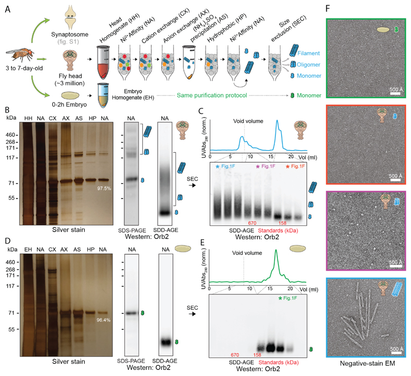
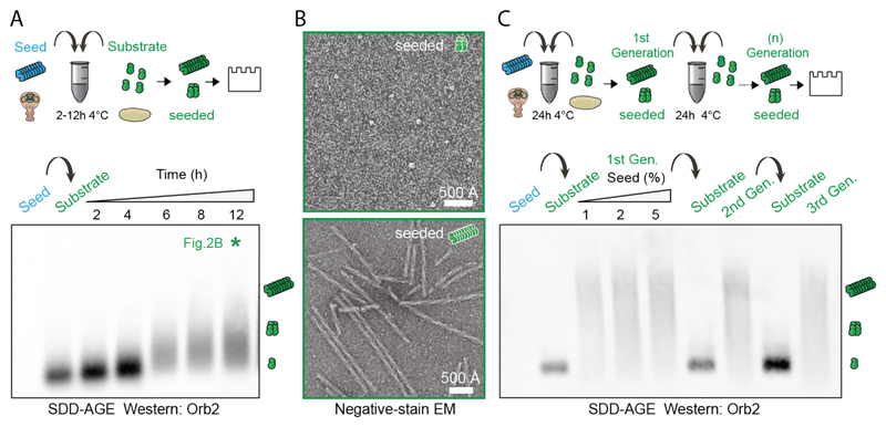
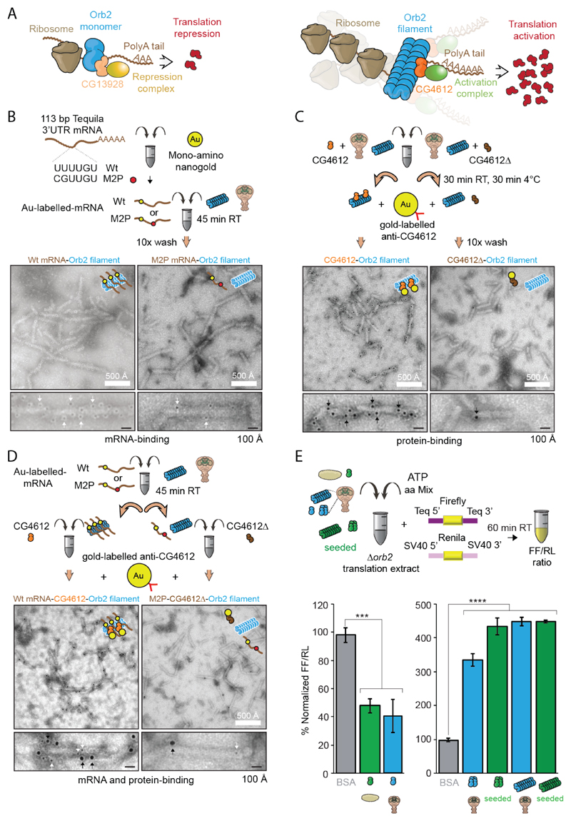
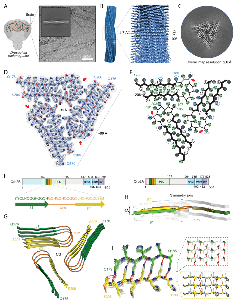
How long-lived memories withstand molecular turnover is a fundamental question. Aggregates of a prion-like RNA-binding protein, cytoplasmic polyadenylation element-binding (CPEB) protein, is a putative substrate of long-lasting memories. We isolated aggregated Drosophila CPEB, Orb2, from adult heads and determined its activity and atomic structure, at 2.6-angstrom resolution, using cryo-electron microscopy. Orb2 formed ~75-nanometer-long threefold-symmetric amyloid filaments. Filament formation transformed Orb2 from a translation repressor to an activator and "seed" for further translationally active aggregation. The 31-amino acid protofilament core adopted a cross-β unit with a single hydrophilic hairpin stabilized through interdigitated glutamine packing. Unlike the hydrophobic core of pathogenic amyloids, the hydrophilic core of Orb2 filaments suggests how some neuronal amyloids could be a stable yet regulatable substrate of memory.


Conflict of interest statement

Competing interests: The authors declare no competing financial interests.;

Figures

Fig. 1
Fig. 1. Purification of different Orb2 species from adult fly head.
(A) Schematic representation of Orb2 purification method from fly head and embryo. (B) Silver stain analysis after each purification step (left) and western blotting after the final purification step (right) by SDS-PAGE and Semi-Denaturing Detergent-Agarose Gel Electrophoresis (SDD-AGE). The % indicates relative Orb2 purity determined by mass spectrometry. The position of monomer, oligomer and filaments are indicated schematically. (C) Western blotting of purified head Orb2 following size-exclusion chromatography. The colored asterisk fractions were visualized under negative-stain EM in F. (D) Silver staining and western blotting of Orb2 protein purified from 0-2h embryo. (E) Western blotting of purified embryonic Orb2 following size-exclusion chromatography. The colored asterisk fraction was visualized under negative-stain EM in F. (F) Negative-stain electron micrographs of purified Orb2 embryonic monomer (green box), head monomer (red box), head oligomer (magenta box) and head filament (blue box).
Fig. 2
Fig. 2. Endogenous Orb2 filament can seed further filament formation.
(A) Time course of monomeric Orb2 aggregation seeded by filaments obtained from adult fly head. The seed (~1 ng) is undetectable in western. Within 6 hours most monomers are aggregated. (B) Negative-stain electron micrographs of seeded oligomers and filaments. (C) Propagation of seeded aggregation through successive rounds. In first round, different seed (1 ng, 2 ng, 5 ng) to monomer (100 ng) ratio was used. In subsequent rounds only 1 ng of seed was used.
Fig. 3
Fig. 3. Biochemical activity of Orb2 monomer, oligomer and filament isolated from adult head.
(A) Schematic representation of the biochemical activity of Orb2. CG13928 binds to Orb2 monomer and recruit translation repression complex (indicated in yellow). CG4612 binds to aggregated Orb2 and recruit translation promoting complex (indicated in green). (B) Negative-stain EM of nanogold-labelled 3' UTR of Tequila mRNA bound to Orb2 filaments. Black dots indicated by white arrows are nanogold particle (~ 2nm) attached to target mRNA. (C) Immuno-EM of CG4612 bound to Orb2 filaments. Black dots indicated by black arrows are gold particles (~ 6nm) attached to CG4612 protein. (D) EM of Orb2 filaments bound to nanogold-labelled 3' UTR of Tequila mRNA and CG4612 protein. The larger gold particle (~ 6nm, black arrows) represents CG4612 protein and smaller gold particle (~2nm, white arrows) represents the mRNA. (E) Translation of Orb2-target mRNA in presence of different Orb2 species, obtained from embryo, adult fly head or seeding reaction. Purified Orb2 monomer represses translation while Orb2 oligomer and filament enhances translation. BSA was used as a control. ***P = 0.0003, ****P < 0.0001; Student’s t-test; two tailed. Data are expressed as mean ± s.e.m.
Fig. 4
Fig. 4. Atomic structure of Orb2 filaments isolated from head.
(A) Cryo-electron micrograph obtained at a defocus of -1.8 µm showing individual Orb2 filaments. Inset: representative reference-free 2D class average showing an entire helical crossover. (B) Side view of the three-dimensional cryo-EM reconstruction illustrating the clear separation of the β-strands. (C) Cryo-EM reconstruction of neuronal Orb2 filament at 2.6 Å. (D) Sharpened, high-resolution cryo-EM maps with the corresponding atomic model overlaid. Unsharpened, 5 Å low-pass-filtered maps are shown as grey outlines; The weaker densities that border sidechains of H182, H186 and H189, correspond to the alternative conformation that those residues adopt. The non-proteinaceous density that runs contiguously across the rungs is highlighted by red arrows. Density maps are shown at a contour level of 2.2 σ (blue) and 1.4 σ (grey). (E) Schematic view of the filament core showing the complementary glutamine packing within the protofilament and the hydrophilic interfaces between protofilament. (F) Primary structure of Orb2A and Orb2B isoforms in fly brain. The prion-like domains are labeled PLD (residues 162-315 for Orb2B and residues 9-162 for Orb2A). The RNA-recognition motifs are labeled RRM and the zinc-finger motifs are labeled ZnF. The N-terminal amino acids preceding the prion-like domain of Orb2A (9 residues) and Orb2B (162 residues) are represented in light red and blue, respectively. Schematic depicting the sequence of the Orb2 filament core, with the observed two β-strands colored in green and yellow as well as the connecting turn in orange. (G) Rendered view of the secondary structure elements in the Orb2 fold, depicted as three successive rungs. (H) Same representation as in G, but in a view perpendicular to the helical axis, revealing the changes in height within a single molecule. (I) Close-up view of the interdigitated glutamine residues. The hydrogen bonds between main chains are shown in blue. The hydrogen bonds involving sidechains are shown in red.

Comment in

References

    1. Josselyn SA, Tonegawa S. Memory engrams: Recalling the past and imagining the future. Science. 2020;367 - PMC - PubMed
    1. Lynch G, Baudry M. The biochemistry of memory: a new and specific hypothesis. Science. 1984;224:1057–1063. - PubMed
    1. Crick F. Memory and molecular turnover. Nature. 1984;312:101. - PubMed
    1. Shorter J, Lindquist S. Prions as adaptive conduits of memory and inheritance. Nat Rev Genet. 2005;6:435–450. - PubMed
    1. Fioriti L, et al. The Persistence of Hippocampal-Based Memory Requires Protein Synthesis Mediated by the Prion-like Protein CPEB3. Neuron. 2015;86:1433–1448. - PubMed

Publication types

MeSH terms