. 2020 Mar 13;367(6483):1240-1246.
doi: 10.1126/science.aaz2924.

Structure of V-ATPase from the mammalian brain

Affiliations

Structure of V-ATPase from the mammalian brain

Yazan M Abbas et al. Science. .

Abstract

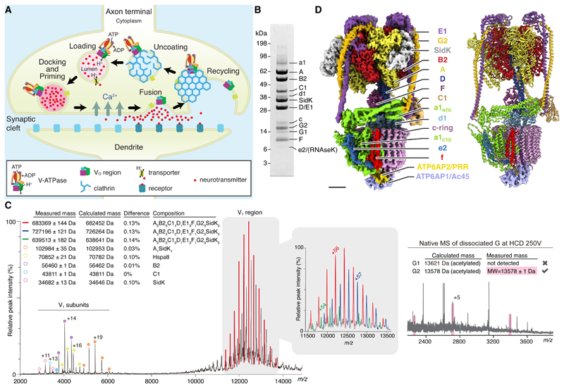
In neurons, the loading of neurotransmitters into synaptic vesicles uses energy from proton-pumping vesicular- or vacuolar-type adenosine triphosphatases (V-ATPases). These membrane protein complexes possess numerous subunit isoforms, which complicates their analysis. We isolated homogeneous rat brain V-ATPase through its interaction with SidK, a Legionella pneumophila effector protein. Cryo-electron microscopy allowed the construction of an atomic model, defining the enzyme's ATP:proton ratio as 3:10 and revealing a homolog of yeast subunit f in the membrane region, which we tentatively identify as RNAseK. The c ring encloses the transmembrane anchors for cleaved ATP6AP1/Ac45 and ATP6AP2/PRR, the latter of which is the (pro)renin receptor that, in other contexts, is involved in both Wnt signaling and the renin-angiotensin system that regulates blood pressure. This structure shows how ATP6AP1/Ac45 and ATP6AP2/PRR enable assembly of the enzyme's catalytic and membrane regions.


Conflict of interest statement

Competing interests: We declare no competing interests.

Figures

Fig. 1
Fig. 1. Overall structure of brain V-ATPase.
(A) Cycle of synaptic vesicle loading, docking and priming, fusion, and recycling. (B) SDS-PAGE of rat brain V-ATPase isolated with 3×FLAG SidK1-278 and gel filtration chromatography. (C) Native mass spectrometry of V1 region (left) and native mass spectrometry of dissociated subunit G (right) at a higher-energy collisional (HCD) voltage of 250 V. The charge state for one peak per subunit is indicated. The table shows the measured mass for each peak (± SD of fit) and the calculated mass depending on subunit composition (Table S5). The difference between calculated and measured masses is indicated. (D) Composite cryoEM map (left) and atomic model (right) of brain V-ATPase in rotational state 1. Scale bar, 25 Å.
Fig. 2
Fig. 2. Structure of the V1 region.
(A) Surface representation. Scale bar, 25 Å. (B) Cross-section through the V1 region viewed from V1 towards VO. ADP is shown in green. (C) Superposition of the catalytic AB pairs (left) and closeup of the conformations of the nucleotide binding sites (right). Scale bar, 5 Å. (D) Interaction of subunits B2, E1, and G2. N and C termini of models are indicated by ‘*’. (E) A continuous β-sheet is formed by E1 and B2 (purple arrowhead).
Fig. 3
Fig. 3. Structure of the VO region.
(A) Surface representation with cryoEM density for subunits f, ATP6AP1/Ac45, and ATP6AP2/PRR. Scale bar, 25 Å. (B) Viewed from V1 with conserved proton-carrying Glu residues as red spheres. The direction of ATP-hydrolysis-driven rotation of the ring is indicated. (C) Cartoon representation viewed parallel to the plane of the lipid bilayer. (D) Proton path through the VO region. (E) Surface representation of a1CTD viewed parallel to the plane of the lipid bilayer. The grey arrow indicates the expected location of an opening between α7 and α8 leading towards the luminal half-channel. (F) Closeup view of the luminal terminus of the luminal half-channel, showing the interaction of subunits f, e2, a1, and the unidentified density. Scale bar, 10 Å.
Fig. 4
Fig. 4. Interaction of subunits within the membrane embedded rotor subcomplex.
(A) Subunits ATP6AP1/Ac45, ATP6AP2/PRR, and c″possess transmembrane α-helices in the centre of the c-ring. (B) ATP6AP2/PRR (top) and ATP6AP1/Ac45 (bottom) possess luminal domains that are absent from the structure. SP, signal peptide; Cyt, cytosolic. (C and D) Subunit d1 interacts with the C termini of ATP6AP1/Ac45 (C) and ATP6AP2/PRR (D). (E) Subunits c″, c(8), and ATP6AP2/PRR interact with the second luminal domain of ATP6AP1/Ac45. Scale bar, 10 Å. (F) Subunits c″, ATP6AP1/Ac45, ATP6AP2/PRR, and d1 create a network of interactions that connect the vesicle lumen and the cytoplasm. Scale bar, 25 Å.

References

    1. Forgac M. Vacuolar ATPases: rotary proton pumps in physiology and pathophysiology. Nat Rev Mol Cell Biol. 2007;8:917–929. - PubMed
    1. Kane PM. The where, when, and how of organelle acidification by the yeast vacuolar H+-ATPase. Microbiol Mol Biol Rev. 2006;70:177–191. - PMC - PubMed
    1. Kane PM. Disassembly and reassembly of the yeast vacuolar H(+)-ATPase in vivo. J Biol Chem. 1995;270:17025–17032. - PubMed
    1. Sumner JP, Dow JA, Earley FG, Klein U, Jager D, Wieczorek H. Regulation of plasma membrane V-ATPase activity by dissociation of peripheral subunits. J Biol Chem. 1995;270:5649–5653. - PubMed
    1. Bodzeta A, Kahms M, Klingauf J. The Presynaptic v-ATPase Reversibly Disassembles and Thereby Modulates Exocytosis but Is Not Part of the Fusion Machinery. Cell Rep. 2017;20:1348–1359. - PubMed

Publication types