. 2020 Mar 12;10(1):4570.
doi: 10.1038/s41598-020-61630-x.

Identification of highly potent and selective inhibitor, TIPTP, of the p22phox-Rubicon axis as a therapeutic agent for rheumatoid arthritis

Affiliations

Identification of highly potent and selective inhibitor, TIPTP, of the p22phox-Rubicon axis as a therapeutic agent for rheumatoid arthritis

Ye-Ram Kim et al. Sci Rep. .

Abstract

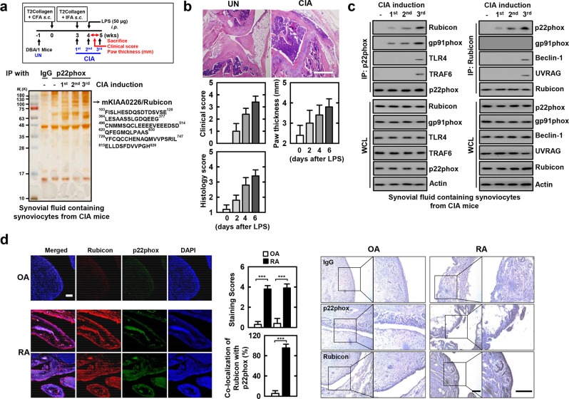
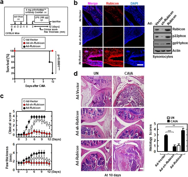
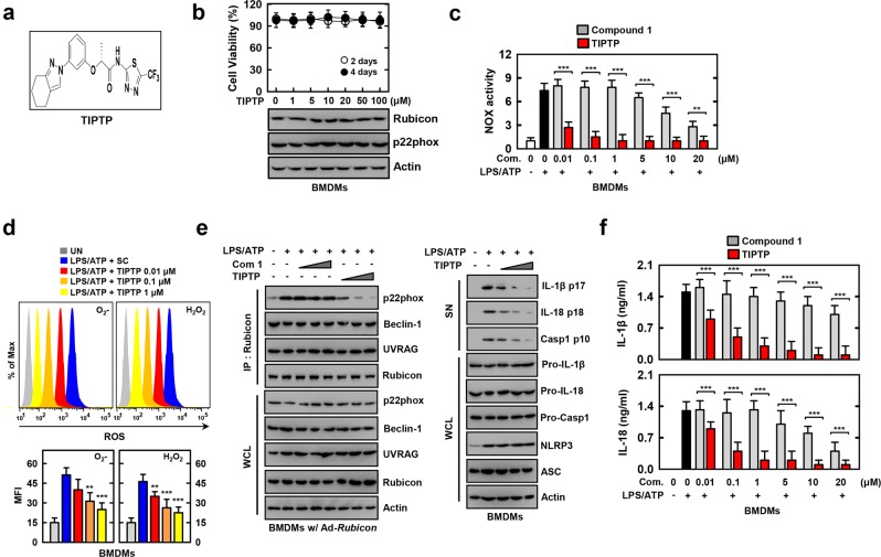
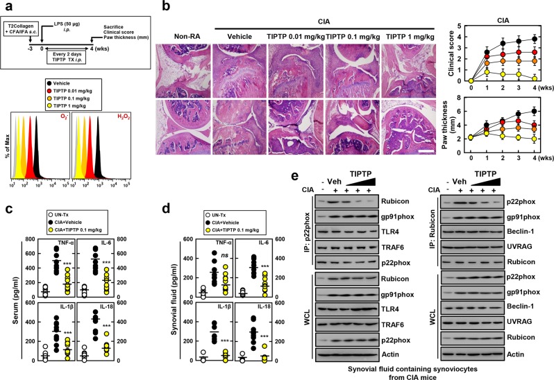
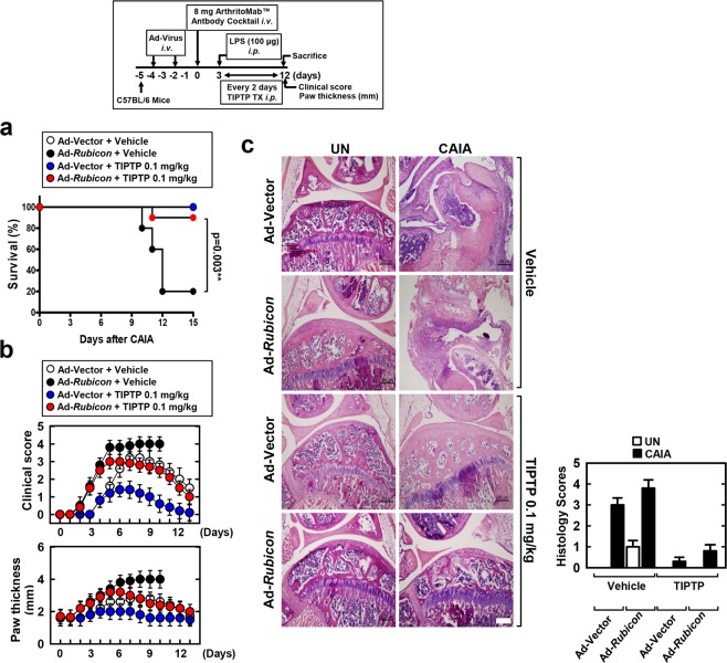
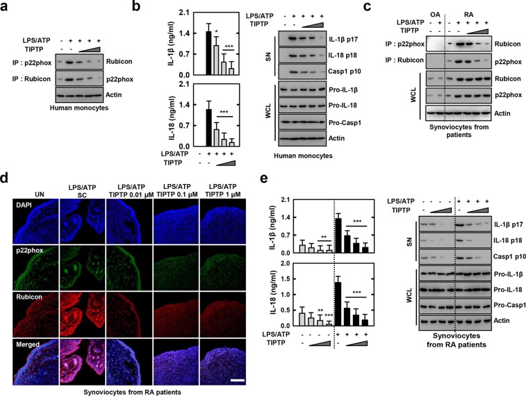
Rheumatoid arthritis (RA) is a chronic inflammatory autoimmune disease linked to oxidative stress, which is associated with significant morbidity. The NADPH oxidase complex (NOX) produces reactive oxygen species (ROS) that are among the key markers for determining RA's pathophysiology. Therefore, understanding ROS-regulated molecular pathways and their interaction is necessary for developing novel therapeutic approaches for RA. Here, by combining mouse genetics and biochemistry with clinical tissue analysis, we reveal that in vivo Rubicon interacts with the p22phox subunit of NOX, which is necessary for increased ROS-mediated RA pathogenesis. Furthermore, we developed a series of new aryl propanamide derivatives consisting of tetrahydroindazole and thiadiazole as p22phox inhibitors and selected 2-(tetrahydroindazolyl)phenoxy-N-(thiadiazolyl)propanamide 2 (TIPTP, M.W. 437.44), which showed considerably improved potency, reaching an IC50 value up to 100-fold lower than an inhibitor that we previously synthesized reported N8 peptide-mimetic small molecule (blocking p22phox-Rubicon interaction). Notably, TIPTP treatment showed significant therapeutic effects a mouse model for RA. Furthermore, TIPTP had anti-inflammatory effects ex vivo in monocytes from healthy individuals and synovial fluid cells from RA patients. These findings may have clinical applications for the development of TIPTP as a small molecule inhibitor of the p22phox-Rubicon axis for the treatment of ROS-driven diseases such as RA.


Conflict of interest statement

The authors declare no competing interests.

Figures

Figure 1
Figure 1
p22phox interaction with Rubicon in human and CIA mice. (a) Schematic of the collagen-induced RA (CIA) model (upper). p22phox complexes purified from synovial fluid containing synoviocytes from CIA mice were subjected to mass spectrometry analysis. Silver stained gel (bottom left) and peptides identified by mass spectrometry analysis (bottom right) The red-colored letters indicate the peptides identified from mass spectrometry analysis. (b) Representative Hematoxylin-Eosin (H&E) staining of the ankle joints of each group (upper); Scale bar, 500 μm. Clinical arthritis, swelling of paws scores (middle), and histopathology scores (bottom). Results are expressed as means ± SD (5 mice per group). (c) Synovial fluid containing synoviocytes from CIA mice for the indicated times, followed by IP with αp22phox (left) or αRubicon (right), followed by IB with αRubicon, αp22phox, and αgp91phox. The anti-p22phox blot was also tested for αTLR4, αTRAF6 binding. The anti-Rubicon blot was also tested for αBeclin-1, or αUVRAG. WCLs were used for IB with αRubicon, αp22phox, αgp91phox, αTLR4, αTRAF6, αBeclin-1, αUVRAG. Actin Western blot was used as a loading control. (d) OA and RA cells were stained with αp22phox (Alexa Fluor 488; green) and αRubicon (Cy3; red). Nuclei were counterstained with DAPI. Cells were visualized by confocal microscopy (left). The middle panel shows the quantitative data of staining intensity of p22phox and Rubicon (upper) and the colocalization index (%) between p22phox and Rubicon (bottom). Immunohistochemical analysis to examine p22phox-DAB (3,3′-diaminobenzidine) and Rubicon-AEC (3-amino-9-ethylcarbazole) expression (right). Representative images from six independent OA and RA patients are shown. Insets, enlargement of outlined areas. Scale bars: 200 μm. The data are representative of five independent experiments with similar results (a, c and d). Statistical analysis was done using the Student’s t-test with Bonferroni adjustment. Data are considered different at p < 0.05. ***p < 0.001. Experimental procedures were described in Supplementary Information.
Figure 2
Figure 2
Alteration of Rubicon gene expression affects CAIA mice mortality. (a) Schematic of the collagen antibody-induced arthritis (CAIA) model (upper). At 48 hr post-injection with Ad-GFP, Ad-shRubicon, or Ad-Rubicon (1 × 10¹³ pfu/kg), twice intravenously via the tail vein, CAIA mice model was established. The survival of CAIA mice was monitored for 12 days and mortality was measured for n = 10 mice per group (lower). Statistical differences, as compared to the Ad-GFP-injected mice, are indicated (log-rank test). (b) Immune-stained with αRubicon or DAPI for Rubicon gene expression (left). IB with αRubicon, αp22phox, or αActin (right). Scale bars: 100 μm. (c) Clinical arthritis score and swelling of paws. Data shown are the means ± SD of three experiments. (d) Representative H&E staining of the ankle joints of each group determined at 9 days of CAIA (left). Scale bars: 200 μm. Histopathology scores (right) from ten mice per group. Statistical significance was determined by two-way analysis of variance (ANOVA) with Tukey’s posttest; ***P < 0.001 compared with Ad-Vector (a). Data shown are the means ± SD of three independent experiments (c). UN, untreated.
Figure 3
Figure 3
TIPTP robustly suppresses LPS/ATP-mediated Rubicon-p22phox interaction and ROS-mediated inflammation. (a) Structure of the TIPTP. (b) MTT assay for cell viability. BMDMs were incubated with TIPTP for the indicated times at the indicated TIPTP concentrations (upper). WCLs were used for IB with α22phox, αRubicon, or αActin (lower). (cf) LPS (100 ng/ml)-primed BMDMs were treated with compounds for 1 h, and then activated with ATP (1 mM) for 30 min. (c) NADPH oxidase activity. (d) FACS analysis for superoxide and hydrogen peroxide (upper). Quantitative analysis of mean fluorescence intensities of ROS (lower). (e) BMDMs were incubated with increasing concentrations of compound 1 and TIPTP (com 2) (***). IP with αRubicon, followed by IB with αp22phox, αBeclin-1, αUVRAG, αRubicon. WCLs were used for IB with αp22phox, αBeclin-1, αUVRAG, αRubicon, or αActin (left). IB analysis of IL-1β p17, IL-18 p18, or caspase-1 p10 in supernatants (SN), and pro-IL-1β, pro-IL-18, or pro-caspase-1 in WCL, with αActin as a loading control (right). (f) Culture supernatants were harvested and analyzed for cytokines by ELISA. The data are representative of three independent experiments with similar results (b and e). Data shown are the means ± SD of three experiments (bd and f). Statistical analysis was done using the Student’s t-test with Bonferroni adjustment (**P < 0.01; ***P < 0.001) compared with LPS/ATP alone (c,d and f). UN, untreated.
Figure 4
Figure 4
TIPTP protects mice from CIA mice. (a) Schematic of the CIA model treated with TIPTP (upper). FACS analysis for superoxide and hydrogen peroxide from synovial fluid containing synoviocytes from CIA (lower). (b) Representative H&E staining of the ankle joints of each group were determined at 4 wks of CIA (left). Clinical arthritis score and swelling of paws (right) from ten mice per group. Scale bars: 500 μm. Serum (c) and Synovial fluid (d) were harvested at 4 wks of CIA and analyzed for cytokines by ELISA. (e) Synovial fluid containing synoviocytes were used for IP with αRubicon or αp22phox, followed by IB with αRubicon, αp22phox, αgp91phox. αRubicon IPs were also blotted for αBeclin-1, or αUVRAG. αp22phox IPs were also blotted for αTLR4, and αTRAF6. WCLs were used for IB with αRubicon, αp22phox, αgp91phox, αTLR4, αTRAF6, αBeclin-1, αUVRAG, or αActin. The data are representative of three independent experiments with similar results (a,b and e). Data shown are the means ± SD of three experiments (bd). Statistical analysis was done using the Student’s t-test with Bonferroni adjustment (***P < 0.001) compared with RA + Vehicle (c and d).
Figure 5
Figure 5
TIPTP protects Rubicon-expressed CAIA mice mortality. Schematic of the CAIA model treated with TIPTP (upper). (a) The survival of CAIA mice was monitored for 15 days and mortality was measured for n = 10 mice per group. Statistical differences, as compared to the Ad-Rubicon + Vehicle, are indicated (log-rank test). (b) Clinical arthritis score and swelling of paws, or (c) representative H&E staining of the ankle joints of each group were determined at 9 days of CAIA (left). Histopathology scores (right) from ten mice per group. Scale bars: 200 μm. The data are representative of three independent experiments with similar results. Statistical significance was determined by two-way analysis of variance (ANOVA) with Tukey’s posttest; **P < 0.01 compared with Ad-Vector+Vehicle (a). Data shown are the means ± SD of three experiments (b and c). UN, untreated.
Figure 6
Figure 6
TIPTP is active for cells from healthy humans or patients with RA. (a and b) LPS-primed human monocytes were treated with TIPTP (0.01, 0.1, 1 μM) for 30 min, and then activated with ATP for the 30 min. (a) IP with αp22phox or αRubicon, followed by IB with αRubicon or αp22phox. Loading control was used for IB with αActin. (b) Culture supernatants were harvested and analyzed for cytokines by ELISA (left). IB analysis of IL-1β p17, IL-18 p18, or caspase-1 p10 in SN, and pro-IL-1β, pro-IL-18, or pro-caspase-1 in WCL, with αActin as a loading control (right). (c) LPS-primed synoviocytes from patients with OA or RA were treated with TIPTP, and then activated with ATP. IP with αp22phox or αRubicon, followed by IB with αRubicon or αp22phox. Loading control was used for IB with αActin. (d) Synoviocytes form RA patients were stained with αp22phox (Alexa Fluor 488; green) and αRubicon (Cy3; red). Nuclei were counterstained with DAPI. Cells were visualized by confocal microscopy. Scale bars: 100 μm. (e) Culture supernatants were harvested and analyzed for cytokines by ELISA (left). IB analysis of IL-1β p17, IL-18 p18, or caspase-1 p10 in SN, and pro-IL-1β, pro-IL-18, or pro-caspase-1 in WCL, with αActin as a loading control (right). The data are representative of three independent experiments with similar results (ae). Data from one of sixteen RA or ten OA patients are shown (c). Data shown are the means ± SD of three experiments and Statistical analysis was done using the Student’s t-test with Bonferroni adjustment (**P < 0.01; ***P < 0.001) compared with LPS/ATP (b and e). SN: supernatant.

References

    1. McInnes IB, Schett G. The pathogenesis of rheumatoid arthritis. N Engl J Med. 2011;365:2205–2219. doi: 10.1056/NEJMra1004965. - DOI - PubMed
    1. Chang KH, et al. Prolong Exposure of NSAID in Patients With RA Will Decrease the Risk of Dementia: A Nationwide Population-Based Cohort Study. Medicine (Baltimore) 2016;95:e3056. doi: 10.1097/MD.0000000000003056. - DOI - PMC - PubMed
    1. Smolen JS, Aletaha D, McInnes IB. Rheumatoid arthritis. Lancet. 2016;388:2023–2038. doi: 10.1016/S0140-6736(16)30173-8. - DOI - PubMed
    1. Bongartz T, et al. Anti-TNF antibody therapy in rheumatoid arthritis and the risk of serious infections and malignancies: systematic review and meta-analysis of rare harmful effects in randomized controlled trials. JAMA. 2006;295:2275–2285. doi: 10.1001/jama.295.19.2275. - DOI - PubMed
    1. Siebert S, Tsoukas A, Robertson J, McInnes I. Cytokines as therapeutic targets in rheumatoid arthritis and other inflammatory diseases. Pharmacol Rev. 2015;67:280–309. doi: 10.1124/pr.114.009639. - DOI - PubMed

Publication types

MeSH terms