

Transcriptional profiling of lung cell populations in idiopathic pulmonary arterial hypertension
- PMID: 32166015
- PMCID: PMC7052475
- DOI: 10.1177/2045894020908782
Transcriptional profiling of lung cell populations in idiopathic pulmonary arterial hypertension
Abstract
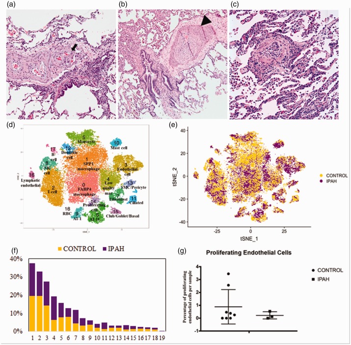
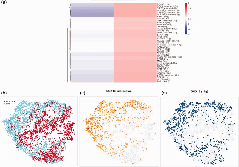
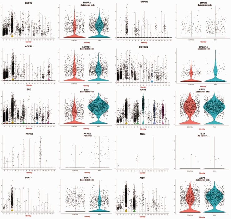
Despite recent improvements in management of idiopathic pulmonary arterial hypertension, mortality remains high. Understanding the alterations in the transcriptome-phenotype of the key lung cells involved could provide insight into the drivers of pathogenesis. In this study, we examined differential gene expression of cell types implicated in idiopathic pulmonary arterial hypertension from lung explants of patients with idiopathic pulmonary arterial hypertension compared to control lungs. After tissue digestion, we analyzed all cells from three idiopathic pulmonary arterial hypertension and six control lungs using droplet-based single cell RNA-sequencing. After dimensional reduction by t-stochastic neighbor embedding, we compared the transcriptomes of endothelial cells, pericyte/smooth muscle cells, fibroblasts, and macrophage clusters, examining differential gene expression and pathways implicated by analysis of Gene Ontology Enrichment. We found that endothelial cells and pericyte/smooth muscle cells had the most differentially expressed gene profile compared to other cell types. Top differentially upregulated genes in endothelial cells included novel genes: ROBO4, APCDD1, NDST1, MMRN2, NOTCH4, and DOCK6, as well as previously reported genes: ENG, ORAI2, TFDP1, KDR, AMOTL2, PDGFB, FGFR1, EDN1, and NOTCH1. Several transcription factors were also found to be upregulated in idiopathic pulmonary arterial hypertension endothelial cells including SOX18, STRA13, LYL1, and ELK, which have known roles in regulating endothelial cell phenotype. In particular, SOX18 was implicated through bioinformatics analyses in regulating the idiopathic pulmonary arterial hypertension endothelial cell transcriptome. Furthermore, idiopathic pulmonary arterial hypertension endothelial cells upregulated expression of FAM60A and HDAC7, potentially affecting epigenetic changes in idiopathic pulmonary arterial hypertension endothelial cells. Pericyte/smooth muscle cells expressed genes implicated in regulation of cellular apoptosis and extracellular matrix organization, and several ligands for genes showing increased expression in endothelial cells. In conclusion, our study represents the first detailed look at the transcriptomic landscape across idiopathic pulmonary arterial hypertension lung cells and provides robust insight into alterations that occur in vivo in idiopathic pulmonary arterial hypertension lungs.
Keywords: endothelial cells; pericytes; pulmonary arterial hypertension; single cell RNA-sequencing.
© The Author(s) 2020.
Figures
References
-
- Farber HW, Miller DP, Poms AD, et al. Five-year outcomes of patients enrolled in the REVEAL registry. Chest 2015; 148: 1043–1054. - PubMed
-
- Tuder RM, Archer SL, Dorfmuller P, et al. Relevant issues in the pathology and pathobiology of pulmonary hypertension. Turk Kardiyol Dern Ars 2014; 42: 5–16. - PubMed
Grants and funding
LinkOut - more resources
Full Text Sources
Molecular Biology Databases
Miscellaneous
