

miRNA-765 mediates multidrug resistance via targeting BATF2 in gastric cancer cells
- PMID: 32166887
- PMCID: PMC7262883
- DOI: 10.1002/2211-5463.12838
miRNA-765 mediates multidrug resistance via targeting BATF2 in gastric cancer cells
Abstract
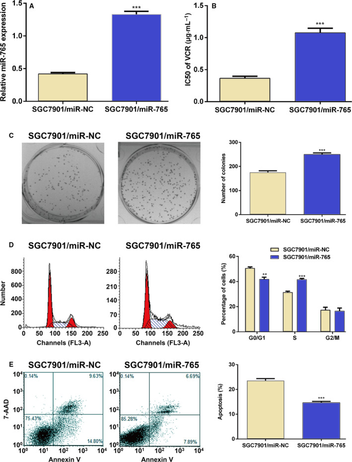
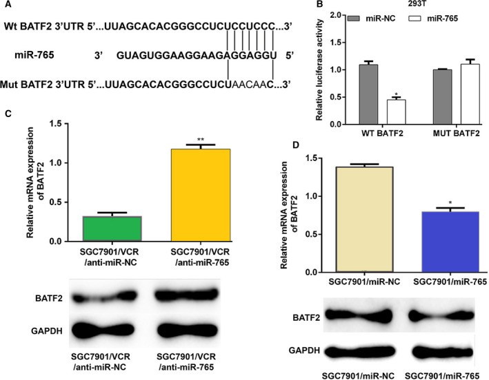
Elucidation of the mechanisms underlying multidrug resistance (MDR) is required to ensure the efficacy of chemotherapy against gastric cancer (GC). To investigate this issue, here we identified that microRNA-765 (miRNA-765) is up-regulated both in MDR GC cell lines and in specimens from patients who are not responding to chemotherapy. In addition, down-regulation of miRNA-765 increased the sensitivity of GC cells to anticancer drugs, whereas its overexpression had the opposite effect. Moreover, miRNA-765 suppressed drug-induced apoptosis and positively regulated the expression of MDR-related genes. Finally, we showed that the basic leucine zipper ATF-like transcription factor 2, a tumor suppressor gene, is the functional target of miRNA-765. In summary, these results suggest that miRNA-765 may promote MDR via basic leucine zipper ATF-like transcription factor 2 in GC cells.
Keywords: basic leucine zipper ATF-like transcription factor 2; gastric cancer; miRNA-765; multidrug resistance.
© 2020 The Authors. Published by FEBS Press and John Wiley & Sons Ltd.
Conflict of interest statement
The authors declare no conflict of interest.
Figures


Similar articles
-
BATF2 reverses multidrug resistance of human gastric cancer cells by suppressing Wnt/β-catenin signaling.In Vitro Cell Dev Biol Anim. 2019 Jun;55(6):445-452. doi: 10.1007/s11626-019-00360-5. Epub 2019 May 28. In Vitro Cell Dev Biol Anim. 2019. PMID: 31140101
-
BATF2 inhibits chemotherapy resistance by suppressing AP-1 in vincristine-resistant gastric cancer cells.Cancer Chemother Pharmacol. 2019 Dec;84(6):1279-1288. doi: 10.1007/s00280-019-03958-4. Epub 2019 Sep 23. Cancer Chemother Pharmacol. 2019. PMID: 31549215
-
Upregulation of miR-34c after silencing E2F transcription factor 1 inhibits paclitaxel combined with cisplatin resistance in gastric cancer cells.World J Gastroenterol. 2020 Feb 7;26(5):499-513. doi: 10.3748/wjg.v26.i5.499. World J Gastroenterol. 2020. PMID: 32089626 Free PMC article.
-
The Roles of microRNAs in Multidrug-Resistance Mechanisms in Gastric Cancer.Curr Mol Med. 2020;20(9):667-674. doi: 10.2174/1566524020666200226124336. Curr Mol Med. 2020. PMID: 32209033 Review.
-
Portrayal of the complex molecular landscape of multidrug resistance in gastric cancer: Unveiling the potential targets.Exp Cell Res. 2025 Jun 1;449(1):114580. doi: 10.1016/j.yexcr.2025.114580. Epub 2025 Apr 29. Exp Cell Res. 2025. PMID: 40306607 Review.
Cited by
-
Gastric cancer: An epigenetic view.World J Gastrointest Oncol. 2022 Jan 15;14(1):90-109. doi: 10.4251/wjgo.v14.i1.90. World J Gastrointest Oncol. 2022. PMID: 35116105 Free PMC article. Review.
-
CD36-BATF2\MYB Axis Predicts Anti-PD-1 Immunotherapy Response in Gastric Cancer.Int J Biol Sci. 2023 Aug 21;19(14):4476-4492. doi: 10.7150/ijbs.87635. eCollection 2023. Int J Biol Sci. 2023. PMID: 37781029 Free PMC article.
-
MicroRNA-122 regulates docetaxel resistance of prostate cancer cells by regulating PKM2.Exp Ther Med. 2020 Dec;20(6):247. doi: 10.3892/etm.2020.9377. Epub 2020 Oct 22. Exp Ther Med. 2020. PMID: 33178345 Free PMC article.
-
Current perspectives on the dysregulated microRNAs in gastric cancer.Mol Biol Rep. 2020 Sep;47(9):7253-7264. doi: 10.1007/s11033-020-05720-z. Epub 2020 Aug 9. Mol Biol Rep. 2020. PMID: 32776162 Review.
-
miR-1273h-5p suppresses CXCL12 expression and inhibits gastric cancer cell invasion and metastasis.Open Med (Wars). 2022 May 16;17(1):930-946. doi: 10.1515/med-2022-0486. eCollection 2022. Open Med (Wars). 2022. PMID: 35647303 Free PMC article.
References
-
- Chen W, Zheng R, Baade PD, Zhang S, Zeng H, Bray F, Jemal A, Yu XQ and He J (2016) Cancer statistics in China, 2015. CA Cancer J Clin 66, 115–132. - PubMed
-
- Cervantes A, Roda D, Tarazona N, Rosello S and Perez‐Fidalgo JA (2013) Current questions for the treatment of advanced gastric cancer. Cancer Treat Rev 39, 60–67. - PubMed
Publication types
MeSH terms
Substances
LinkOut - more resources
Full Text Sources
Medical
Miscellaneous
