. 2020 Apr 17;134(7):695-710.
doi: 10.1042/CS20200016.

Melanocortin therapy ameliorates podocytopathy and proteinuria in experimental focal segmental glomerulosclerosis involving a podocyte specific non-MC1R-mediated melanocortinergic signaling

Affiliations

Melanocortin therapy ameliorates podocytopathy and proteinuria in experimental focal segmental glomerulosclerosis involving a podocyte specific non-MC1R-mediated melanocortinergic signaling

Yingjin Qiao et al. Clin Sci (Lond). .

Abstract

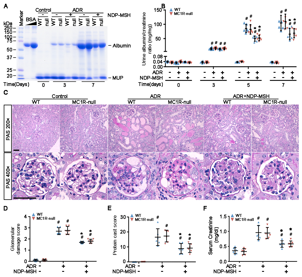
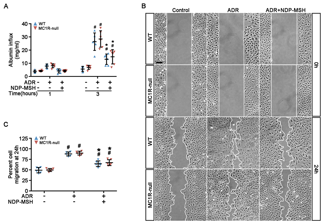
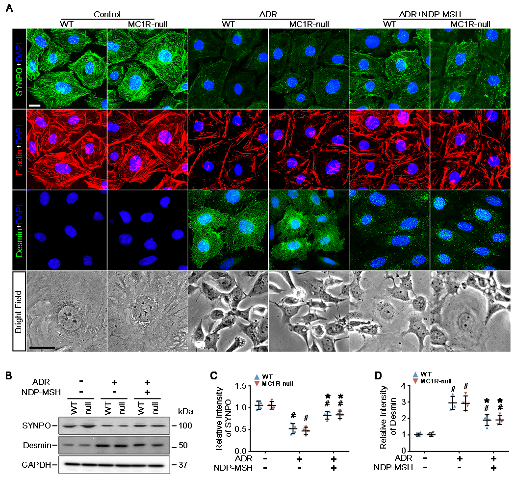
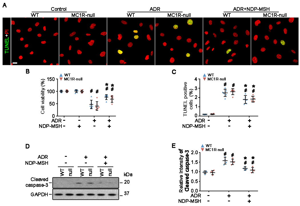
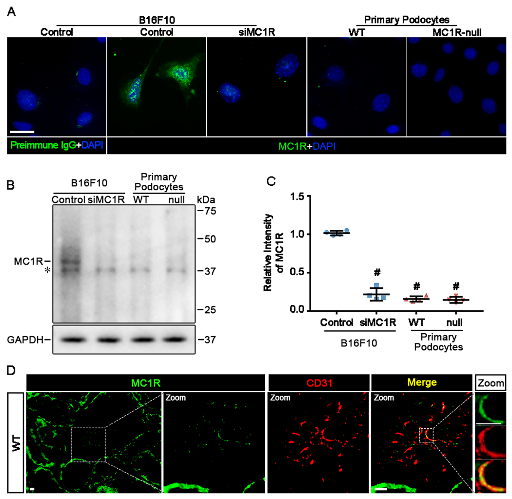
The clinical effectiveness of adrenocorticotropin in inducing remission of steroid-resistant nephrotic syndrome points to a steroidogenic-independent anti-proteinuric activity of melanocortins. However, which melanocortin receptors (MCR) convey this beneficial effect and if systemic or podocyte-specific mechanisms are involved remain uncertain. In vivo, wild-type (WT) mice developed heavy proteinuria and kidney dysfunction following Adriamycin insult, concomitant with focal segmental glomerulosclerosis (FSGS) and podocytopathy, marked by loss of podocin and synaptopodin, podocytopenia and extensive foot process effacement on electron microscopy. All these pathologic findings were prominently attenuated by NDP-MSH, a potent non-steroidogenic pan-MCR agonist. Surprisingly, MC1R deficiency in MC1R-null mice barely affected the severity of Adriamycin-elicited injury. Moreover, the beneficial effect of NDP-MSH was completely preserved in MC1R-null mice, suggesting that MC1R is likely non-essential for the protective action. A direct podocyte effect seems to contribute to the beneficial effect of NDP-MSH, because Adriamycin-inflicted cytopathic signs in primary podocytes prepared from WT mice were all mitigated by NDP-MSH, including apoptosis, loss of podocyte markers, de novo expression of the podocyte injury marker desmin, actin cytoskeleton derangement and podocyte hypermotility. Consistent with in vivo findings, the podoprotective activity of NDP-MSH was fully preserved in MC1R-null podocytes. Mechanistically, MC1R expression was predominantly distributed to glomerular endothelial cells in glomeruli but negligibly noted in podocytes in vivo and in vitro, suggesting that MC1R signaling is unlikely involved in direct podocyte protection. Ergo, melanocortin therapy protects against podocyte injury and ameliorates proteinuria and glomerulopathy in experimental FSGS, at least in part, via a podocyte-specific non-MC1R-mediated melanocortinergic signaling.

Keywords: adrenocorticotropic hormone; apoptosis; cytoskeleton; glomerular disease; podocytes.


Figures

Figure 1.
Figure 1.. MC1R is not involved in ADR nephropathy and is non-essential for the protective effect of NDP-MSH.
Male WT and MC1R-null (null) mice were treated with vehicle or Adriamycin (ADR) in the presence or absence of NDP-MSH co-treatment. Spot urine was collected on indicated time points after ADR or vehicle treatment. All animals were killed on Day 7, and kidneys and serum harvested and examined. (A) Urine samples (1.5 μl) were subjected to SDS-PAGE and staining with Coomassie Brilliant Blue. Bovine serum albumin (BSA, 20, 40, μg) served as standard controls; (B) Quantification of urine albumin levels adjusted with urine creatinine concentrations. #P<0.05 versus non-ADR-injured animals with the same MC1R mutation status at each time point; *P<0.05 versus ADR alone-treated animals with the same MC1R mutation status at each time point (n=6); (C) Representative micrographs demonstrate periodic acid–Schiff (PAS) staining (scale bar=50μm) of mouse kidneys. ADR-induced injury is featured by podocytic swelling and vacuolization, glomerular synechiae, glomerular capillary congestion, collapse and/or obliteration accompanied by hyaline material and/or mesangial matrix expansion as well as protein casts in tubulointerstitium; (D) Semi-quantitative morphometric analysis of PAS-stained kidney sections for glomerular damage scores. #P<0.05 versus non-ADR-injured animals with the same MC1R mutation status; *P<0.05 versus ADR alone-treated animals with the same MC1R mutation status (n=6); (E) Semi-quantitative morphometric analysis of PAS-stained kidney sections for protein cast scores. #P<0.05 versus non-ADR-injured animals with the same MC1R mutation status; *P<0.05 versus ADR alone-treated animals with the same MC1R mutation status (n=6); (F) Serum samples were processed for creatinine assay. #P<0.05 versus non-ADR-injured animals with the same MC1R mutation status; *P<0.05 versus ADR alone-treated animals with the same MC1R mutation status (n=6).
Figure 2.
Figure 2.. MC1R is not required for the NDP-MSH-improved podocytopathy in ADR injured mice.
Male WT and ADR mice were treated as elaborated in Figure 1. (A) Kidney specimens were procured from mice on day 7 after ADR or saline treatment and processed for electron microscopy (scale bar=1μm) and fluorescent immunohistochemistry staining (scale bar=20μm) for indicated molecules, including synaptopodin (SYNPO), desmin and WT-1, or in combination with TUNEL staining for apoptotic cells. Black arrowheads highlight injured podocytes with evident foot process effacement. White arrowheads indicate WT-1+ podocytes with positive staining for desmin or TUNEL; (B) Absolute counting of the number of apoptotic podocytes in glomeruli, expressed as the number of cells positive for both TUNEL and WT-1 in each glomerulus. #P<0.05 versus non-ADR-injured animals with the same MC1R mutation status; *P<0.05 versus ADR alone-treated animals with the same MC1R mutation status(n=6); (C) Absolute counting of the number of foot processes per unit length of glomerular basement membrane (GBM) on electron micrographs#P<0.05 versus non-ADR-injured animals with the same MC1R mutation status; *P<0.05 versus ADR alone-treated animals with the same MC1R mutation status(n=6); (D) Glomeruli were isolated from kidneys by the magnetic beads-based approach and processed for immunoblot analysis for indicated molecules. (E-G) Immunoblots were subjected to densitometric analysis. Data were presented as arbitrary units of densitometric ratios of indicated proteins to GAPDH as folds of the non-ADR-injured WT mice. #P<0.05 versus non-ADR-injured animals with the same MC1R mutation status; *P<0.05 versus ADR alone-treated animals with the same MC1R mutation status (n=6).
Figure 3.
Figure 3.. NDP-MSH directly preserves podocyte filtration barrier function and rectifies podocyte hypermotility in an MC1R independent manner
Primary podocytes derived from WT and MC1R-null mice were grown to confluence and treated with ADR or vehicle in the presence or absence of NDP-MSH (10−7M). (A) Cells were grown on the collagen coated transwell filters and after indicated treatments for 24h paracellular permeability assay was carried out to determine the filtration barrier function of podocytes monolayers. Culture media in the top chamber were collected at 1 or 3h during the paracellular permeability assay and subjected to quantification of the albumin influx across podocyte monolayers. Duration of albumin incubation is shown next to the x-axis. #P < 0.05 vs non-ADR-injured podocytes with the same MC1R mutation status (n = 6); *P < 0.05 vs ADR alone-treated podocytes with the same MC1R mutation status. (n = 6). (B) Primary podocytes were treated and subsequently scratch was processed using a 10μL pipette. Phase-contrast micrographs were taken immediately after wounding (0 h) and after migration for 24 h. Scale bar = 100 μm. (C) Quantification by computerized morphometric analysis of the cell migration area following the indicated treatments. #P < 0.05 vs non-ADR-injured podocytes with the same MC1R mutation status (n = 6); *P < 0.05 vs ADR alone-treated podocytes with the same MC1R mutation status. (n = 6).
Figure 4.
Figure 4.. The protective effect of NDP-MSH against the ADR-elicited cell shape changes, cytoskeleton disarrangement and transdifferentiation is fully preserved in MC1R-null podocytes
Primary podocytes derived from WT and MC1R-null mice were treated with ADR or vehicle in the presence or absence of NDP-MSH (10−7M) for 24h. (A) Cells were fixed and subjected to staining for cytoskeletal F-actin with rhodamine phalloidin and to fluorescent immunocytochemistry staining for synaptopodin (SYNPO) and desmin. Cells were counterstained with 4′,6-diamidino-2-phenylindole (DAPI). Representative micrographs of phase contrast microscopy and florescent microscopy are shown (Scale bar=20μm). (B) Cell lysates were subjected to immunoblot analysis for indicated molecules. (C and D) Immunoblots were subjected to densitometric analysis. Data were presented as arbitrary units of densitometric ratios of indicated proteins to GAPDH as folds of the non-ADR-injured WT cells. #P<0.05 versus non-ADR-injured cells with the same MC1R mutation status; *P<0.05 versus ADR alone treated cells with the same MC1R mutation status (n=4).
Figure 5.
Figure 5.. NDP-MSH directly protects podocytes against the ADR-elicited apoptosis and improves cellular viability via an MC1R-independent mechanism.
Primary podocytes derived from WT and MC1R-null mice were treated with ADR or vehicle in the presence or absence of NDP-MSH (10−7M) for 24 h. (A) Cells were fixed and subjected to TUNEL staining for apoptotic cells. Cells were counterstained with propidium iodide (PI) (Scale bar=20μm). (B) Absolute counting of the numbers of TUNEL positive apoptotic podocytes expressed as percentage of the total number of podocyte nuclei per high-power field. #P < 0.05 vs non-ADR-injured podocytes with the same MC1R mutation status (n = 6); *P < 0.05 vs ADR alone-treated podocytes with the same MC1R mutation status. (n = 6). (C) Cellular viability was assessed by the MTT assay. #P < 0.05 vs non-ADR-injured podocytes with the same MC1R mutation status (n = 6); *P < 0.05 vs ADR alone-treated podocytes with the same MC1R mutation status. (n = 6). (D) Cell lysates were subjected to immunoblot analysis for indicated molecules. (E) Immunoblots were subjected to densitometric analysis. Data were presented as arbitrary units of densitometric ratios of cleaved caspase-3 to GAPDH as folds of the non-ADR-injured WT cells. #P<0.05 versus non-ADR-injured cells with the same MC1R mutation status; *P<0.05 versus ADR alone treated cells with the same MC1R mutation status (n=4).
Figure 6.
Figure 6.. MC1R expression is negligibly noted in podocytes in vitro and in vivo
(A) Primary podocytes derived from WT and MC1R-null mice were cultured under physiologic conditions and processed for fluorescent immunocytochemistry staining for MC1R with 4′,6-diamidino-2-phenylindole (DAPI) counterstaining. B16F10 cells served as a positive control and preimmune IgG control for MC1R staining. B16F10 cells with MC1R silencing (siMC1R) served as a negative control. (Scale bar=30μm). (B) Cells lysate were subjected to immunoblot analysis for MC1R. The MC1R specific band is indicated. The asterisk indicates nonspecific bands probed by the antibody. (C) Immunoblots were subjected to densitometric analysis. Data were presented as arbitrary units of densitometric ratios of MC1R to GAPDH as folds of the control group. #P<0.05 versus control group (n=4). (D) Cryosections of WT kidneys were processed for fluorescent immunohistochemistry staining for MC1R (green). Representative micrographs are shown. Enlarged views show colocalization of MC1R staining (green) with CD31 staining (red) in glomeruli (Scale bars=10μm).

Similar articles

Cited by

References

    1. Rosenberg AZ and Kopp JB (2017) Focal Segmental Glomerulosclerosis. Clinical journal of the American Society of Nephrology : CJASN. 12, 502–517 - PMC - PubMed
    1. D’Agati VD, Kaskel FJ and Falk RJ (2011) Focal segmental glomerulosclerosis. The New England journal of medicine. 365, 2398–2411 - PubMed
    1. Reiser J, Nast CC and Alachkar N (2014) Permeability factors in focal and segmental glomerulosclerosis. Advances in chronic kidney disease. 21, 417–421 - PMC - PubMed
    1. Hogg R, Middleton J and Vehaskari VM (2007) Focal segmental glomerulosclerosis--epidemiology aspects in children and adults. Pediatric nephrology (Berlin, Germany). 22, 183–186 - PMC - PubMed
    1. Fogo AB (2015) Causes and pathogenesis of focal segmental glomerulosclerosis. Nature reviews. Nephrology. 11, 76–87 - PMC - PubMed

Publication types

MeSH terms