Review
. 2020 Mar 13;477(5):985-1008.
doi: 10.1042/BCJ20190468.

Adipocyte lipolysis: from molecular mechanisms of regulation to disease and therapeutics

Affiliations
Review

Adipocyte lipolysis: from molecular mechanisms of regulation to disease and therapeutics

Alexander Yang et al. Biochem J. .

Abstract

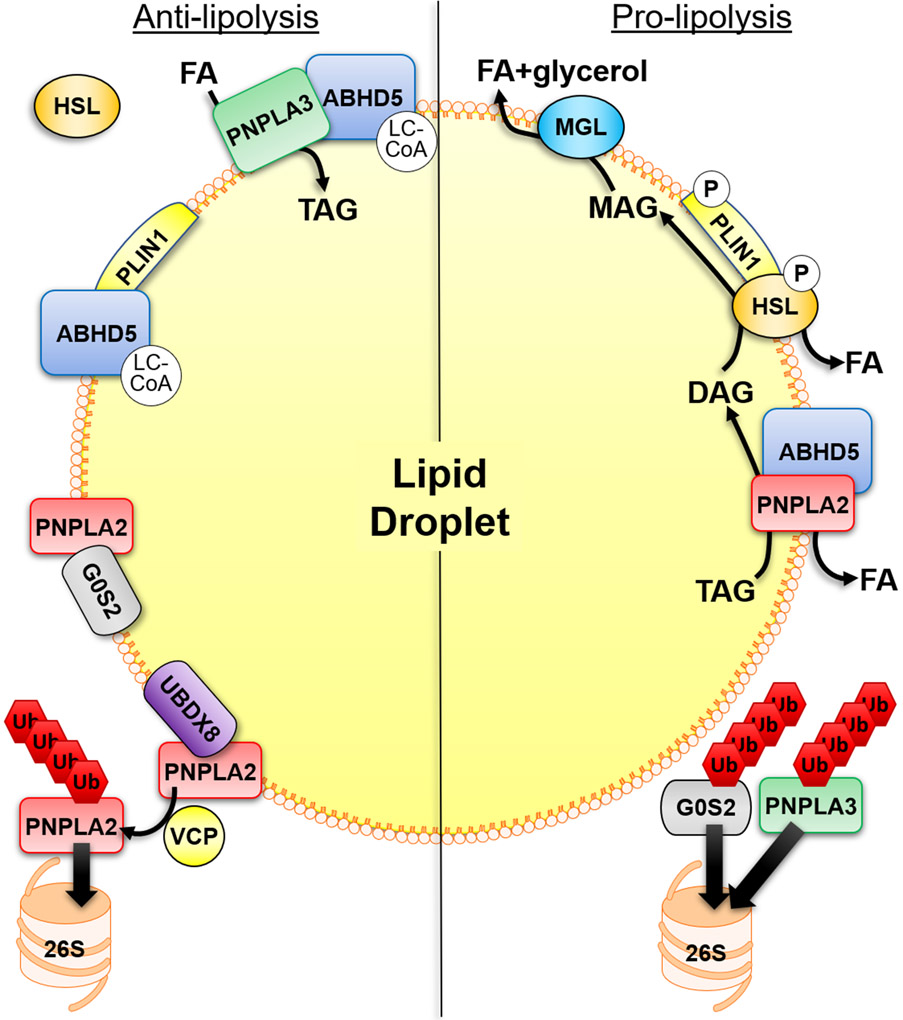
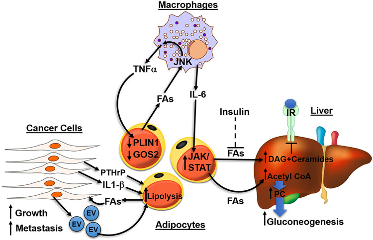
Fatty acids (FAs) are stored safely in the form of triacylglycerol (TAG) in lipid droplet (LD) organelles by professional storage cells called adipocytes. These lipids are mobilized during adipocyte lipolysis, the fundamental process of hydrolyzing TAG to FAs for internal or systemic energy use. Our understanding of adipocyte lipolysis has greatly increased over the past 50 years from a basic enzymatic process to a dynamic regulatory one, involving the assembly and disassembly of protein complexes on the surface of LDs. These dynamic interactions are regulated by hormonal signals such as catecholamines and insulin which have opposing effects on lipolysis. Upon stimulation, patatin-like phospholipase domain containing 2 (PNPLA2)/adipocyte triglyceride lipase (ATGL), the rate limiting enzyme for TAG hydrolysis, is activated by the interaction with its co-activator, alpha/beta hydrolase domain-containing protein 5 (ABHD5), which is normally bound to perilipin 1 (PLIN1). Recently identified negative regulators of lipolysis include G0/G1 switch gene 2 (G0S2) and PNPLA3 which interact with PNPLA2 and ABHD5, respectively. This review focuses on the dynamic protein-protein interactions involved in lipolysis and discusses some of the emerging concepts in the control of lipolysis that include allosteric regulation and protein turnover. Furthermore, recent research demonstrates that many of the proteins involved in adipocyte lipolysis are multifunctional enzymes and that lipolysis can mediate homeostatic metabolic signals at both the cellular and whole-body level to promote inter-organ communication. Finally, adipocyte lipolysis is involved in various diseases such as cancer, type 2 diabetes and fatty liver disease, and targeting adipocyte lipolysis is of therapeutic interest.

Keywords: adipocyte; diabetes; fatty acid; lipolysis; non alcoholic fatty liver disease.


Figures

Figure 1.
Figure 1.. Mechanisms that regulate adipocyte lipolysis on the surface of lipid droplets.
Anti-lipolytic mechanisms of regulation include the binding of ABHD5 to PLIN1 which is regulated allosterically by ABHD5-sensing of long-chain acyl-CoA (LC-CoA). PNPLA2 hydrolytic activity can be inhibited by the interaction with G0S2 which is independent of its interaction with ABHD5. In addition, PNPLA3 can compete with PNPLA2 for binding to ABHD5, thereby sequestering ABHD5 away from PNPLA2. PNPLA3 can also function to sequester fatty acids (FAs) through lysophosphatidic acid acyltransferase or transacylase activity. LC-CoAs also allosterically regulate the interaction between PNPLA3 and ABHD5. UBDX8 can bind PNPLA2 to promote its segregation from the lipid droplet (LD) by VCP, followed by ubiquitination (Ub) and elimination by the proteasome (26S). In the basal state HSL is found mostly in the cytosol. Stimulatory signals lead to the phosphorylation (P) of PLIN1 to release ABHD5 which can bind PNPLA2, and phosphorylation of HSL promotes its trafficking to LDs and interaction with PLIN1. This leads to the sequential hydrolysis of TAG by PNPLA2, hydrolysis of DAG by HSL and hydrolysis of MAG by MGL to three FAs and glycerol. The elimination of G0S2 and PNPLA3 by the ubiquitin-proteasome system (UPS), through currently unknown mechanisms, would be pro-lipolytic.
Figure 2.
Figure 2.. Hormonal control of adipocyte lipolysis.
Activation of the seven transmembrane β-adrenergic receptors (β-AR) on adipocytes by norepinephrine (NE), leads to Gs-dependent activation of adenylyl cyclase (AC) to increase intracellular levels of cyclic-AMP (cAMP). cAMP activates protein kinase A (PKA) to phosphorylate (P) PLIN1, which release ABHD5 to fully activate PNPLA2 TAG hydrolase activity and release a fatty acid (FA). PKA also phosphorylates HSL to promote its translocation to PLIN1 and increase its DAG hydrolase activity to release a FA and produce MAG. MAG is hydrolyzed by MGL to release a final FA and glycerol. Lipolysis can also be stimulated by natriuretic peptide (NP) which signals through the NP receptor-A (NPR-A) to increase guanylyl cyclase (GC)/cyclic-GMP (cGMP) and activation of protein kinase G (PKG). PKG can also phosphorylate HSL and PLIN1. Insulin, the main anti-lipolytic hormone (red lines), signals through the insulin receptor (IR) and insulin receptor substrate 1 and 2 (IRS1/2) to increase activity of phosphoinositide 3-kinase (PI3K) and levels of PIP3. PIP3 activates the kinase AKT, which through currently unknown mechanisms (?), activates phosphodiesterase 3B (PDE3B) to hydrolyze cAMP to AMP. ABHD15, an AKT phosphorylation substrate, is required for the anti-lipolytic action of insulin and can bind to and stabilize PDE3B. Dotted line indicates movement of proteins, while the dashed red line notes currently unclear pathway of regulation.
Figure 3.
Figure 3.. Adipocyte lipolysis produces intracellular signals in the homeostatic regulation of lipid metabolism.
Fatty acids (FA) released by the sequential action of adipocyte lipases and regulators (see text and Figure 2) in response to activation of β-adrenergic receptors (β-ARs) can function as ligands for the nuclear receptor peroxisome proliferator-activated receptors (PPARs). These FAs are thought to traffic to the nucleus by fatty acid binding protein 4 (FABP4) which can interact with ABHD5, sequester FAs, and delivery them to PPARs in the nucleus. In brown adipocytes, the mobilization of FAs can produce ligands for PPARα which traffic to the nucleus within minutes of stimulating lipolysis. This leads to a transcriptional program which upregulates the machinery involved in fatty acid oxidation (FAO) and thermogenesis (UCP1) to match supply with oxidation. In brown adipocytes, FAs are also allosteric regulator of UCP1, the molecular mechanism for heat production. In addition, lipolysis can produce PPARγ ligands which are important for adipocyte differentiation and maintenance of an adipocyte phenotype necessary for sequestration of FAs and preventing lipotoxicity.
Figure 4.
Figure 4.. Adipocyte lipolysis promotes inter-organ crosstalk to regulate whole-body energy homeostasis.
Fatty acids (FAs) released by adipocyte lipolysis can function as PPARα ligands in the liver to upregulate a transcriptional program involved in increasing fatty acid oxidation (FAO) and VLDL secretion. Furthermore, fatty acid ester of hydroxyl fatty acids (FAHFAs) released by adipocytes can act through a Gαi-coupled receptor to suppress hepatic gluconeogenesis. These FAHFAs can also have autocrine effects by upregulating of de novo lipogenesis (DNL) and FA-esterification in adipocytes. FAs released by white adipocytes can activate GRP40 on β-cells in the pancreas to elicit insulin secretion which acts on brown adipose tissue (BAT) to promote greater FA uptake required for FAO. 12,13-diHOME, secreted from brown adipocytes can function in an autocrine manner to similarly promote further uptake and utilization of FAs. FAs released by white adipose tissue can be sensed by afferent nerves which feedback to the brain to increase sympathetic outflow to BAT through the sympathetic nervous system (SNS). Adipocytes can also secrete extracellular vesicles (EVs) in response to lipolysis that can further mediate tissue-tissue communication.
Figure 5.
Figure 5.. Adipocyte lipolysis is implicated in disease such as diabetes, fatty liver disease and cancer.
In diseased states, excessive fatty acids (FAs) activate signaling in surrounding cells to further increase lipolysis in adipocytes, forming a vicious, positive feedback loop. In obesity, adipocytes lose their ability to safely buffer FAs as TAGs, leading to the spillover of FAs which act upon macrophages through JNK signaling to release inflammatory cytokines TNFα and IL-6. Both cytokines function on adipocytes to further increase lipolysis. TNFα decreases PLIN1 and G0S2, inhibitors of PNPLA2, and IL-6 acts through JAK/STAT pathway to increase lipolysis. Under normal physiology, insulin functions to suppress lipolysis, however, in type 2 diabetes (T2D), the failure of insulin to suppress FA release from adipocytes (dashed line) results in excessive FA flux to the liver. FAs in the liver can form DAG and ceramides which act on PKC to decrease insulin sensitivity. Hepatic acetyl-CoA, generated from adipocyte FAs, allosterically upregulates pyruvate carboxylase (PC) to increase gluconeogenesis in the liver. Lastly, cancer cells secrete EVs and cytokine IL-1β increasing lipolysis of adipocytes in the tumor environment. Tumor-derived parathyroid-hormone-related protein (PTHrP) can promote the upregulation of lipolytic enzymes to promote cancer associated cachexia. Cancer cells use FAs generated from lipolysis as fuel to increase growth and invasiveness.

References

    1. Unger RH, Scherer PE. Gluttony, sloth and the metabolic syndrome: a roadmap to lipotoxicity. Trends in Endocrinology & Metabolism. 2010;21(6):345–52. - PMC - PubMed
    1. Edens NK, Leibel RL, Hirsch J. Mechanism of free fatty acid re-esterification in human adipocytes in vitro. J Lipid Res. 1990;31(8):1423–31. - PubMed
    1. Mottillo EP, Bloch AE, Leff T, Granneman JG. Lipolytic Products Activate Peroxisome Proliferator-activated Receptor (PPAR) and in Brown Adipocytes to Match Fatty Acid Oxidation with Supply. Journal of Biological Chemistry. 2012;287(30):25038–48. - PMC - PubMed
    1. Ong KT, Mashek MT, Bu SY, Greenberg AS, Mashek DG. Adipose triglyceride lipase is a major hepatic lipase that regulates triacylglycerol turnover and fatty acid signaling and partitioning. Hepatology. 2011;53(1):116–26. - PMC - PubMed
    1. Zechner R, Zimmermann R, Eichmann Thomas O, Kohlwein Sepp D, Haemmerle G, Lass A, et al. FAT SIGNALS - Lipases and Lipolysis in Lipid Metabolism and Signaling. Cell Metabolism. 2012;15(3):279–91. - PMC - PubMed

Publication types

MeSH terms

Substances