. 2020 Dec;72(6):2090-2108.
doi: 10.1002/hep.31228.

Intestinal Microbiome-Macrophage Crosstalk Contributes to Cholestatic Liver Disease by Promoting Intestinal Permeability in Mice

Affiliations

Intestinal Microbiome-Macrophage Crosstalk Contributes to Cholestatic Liver Disease by Promoting Intestinal Permeability in Mice

Anna Isaacs-Ten et al. Hepatology. 2020 Dec.

Abstract

Background and aims: Mounting evidence supports an association between cholestatic liver disease and changes in the composition of the microbiome. Still, the role of the microbiome in the pathogenesis of this condition remains largely undefined.

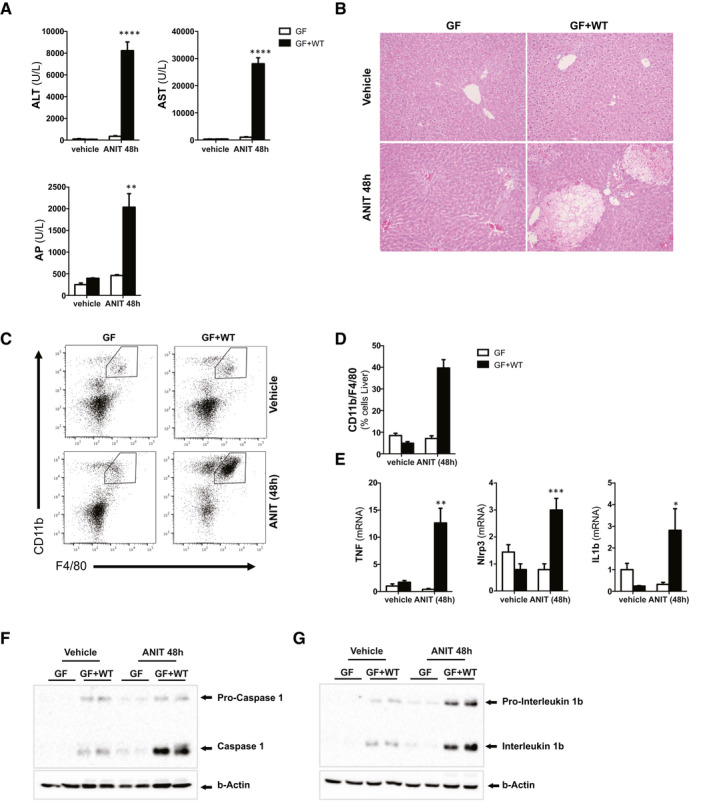
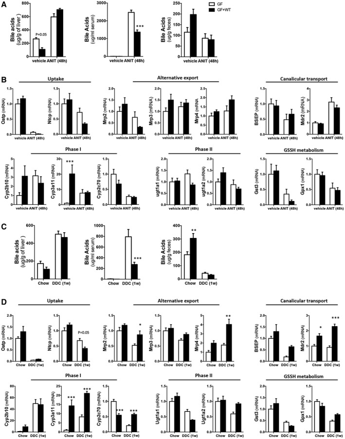
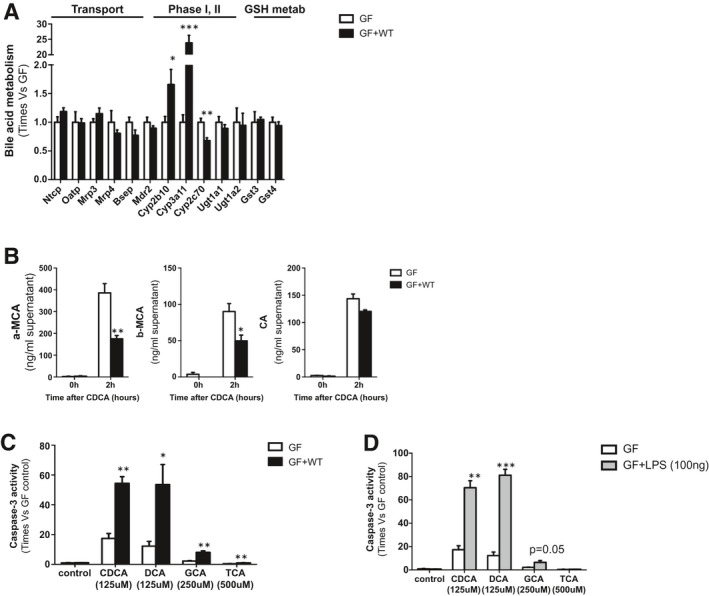
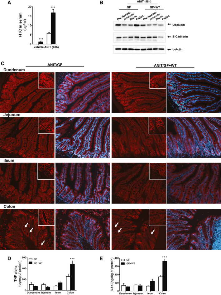
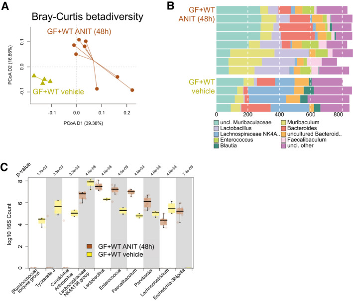
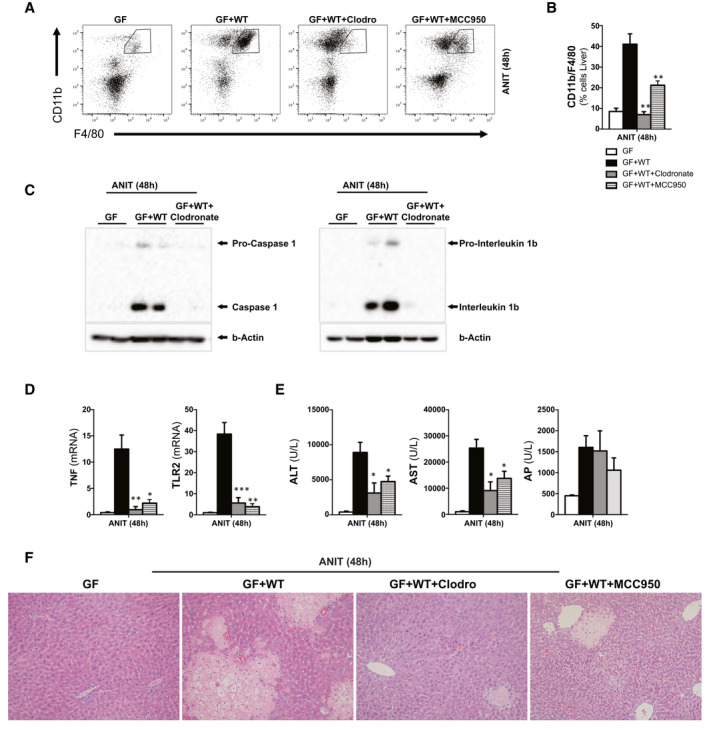
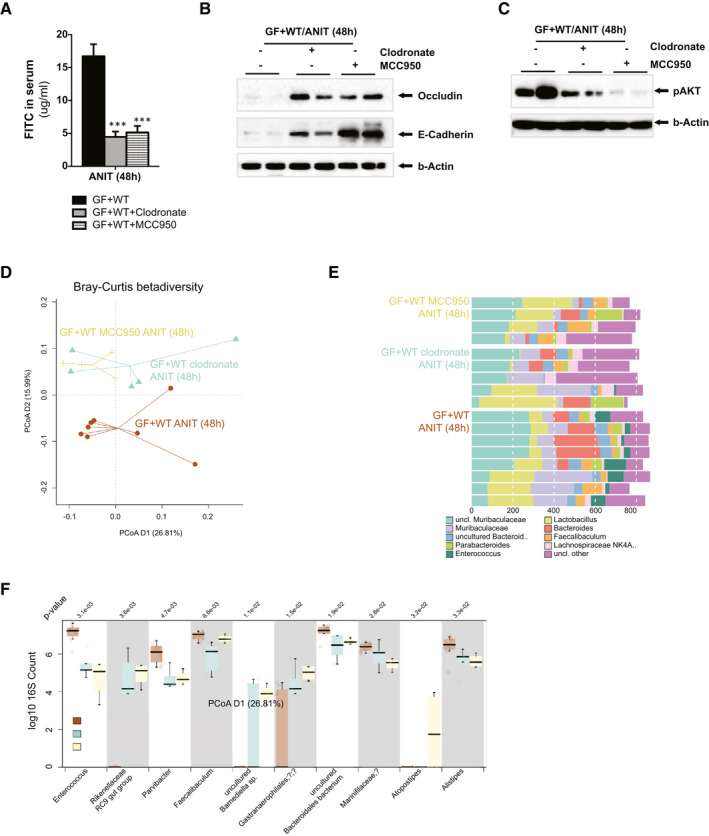
Approach and results: To address this, we have used two experimental models, administering alpha-naphtylisocyanate or feeding a 0.1% 3,5-diethoxycarbonyl-1,4-dihydrocollidine diet, to induce cholestatic liver disease in germ-free mice and germ-free mice conventionalized with the microbiome from wild-type, specific pathogen-free animals. Next, we have inhibited macrophage activation by depleting these cells using clodronate liposomes and inhibiting the inflammasome with a specific inhibitor of NOD-, LRR-, and pyrin domain-containing protein 3. Our results demonstrate that cholestasis, the accumulation of bile acids in the liver, fails to promote liver injury in the absence of the microbiome in vivo. Additional in vitro studies supported that endotoxin sensitizes hepatocytes to bile-acid-induced cell death. We also demonstrate that during cholestasis, macrophages contribute to promoting intestinal permeability and to altered microbiome composition through activation of the inflammasome, overall leading to increased endotoxin flux into the cholestatic liver.

Conclusions: We demonstrate that the intestinal microbiome contributes to cholestasis-mediated cell death and inflammation through mechanisms involving activation of the inflammasome in macrophages.


Figures

Fig. 1
Fig. 1
Absence of a microbiome protects mice from cholestatic‐induced liver injury and inflammation. (A) Levels of blood liver injury and cholestasis markers and (B) H&E staining of liver sections from GF and GF mice conventionalized with WT microbiome (GF + WT) treated with vehicle or ANIT (100 mg/kg) for 48 hours. (C) FC analysis on liver‐isolated immune cells, (D) further quantification, and (E) qPCR analysis on liver extracts showing increased presence of macrophages and inflammation in ANIT/GF + WT mice. (F, G) Western blotting analysis of whole‐liver lysates showing activation of the inflammasome. Images are representative of n ≥ 5 animals per treatment group. Values are mean ± SEM. *P < 0.05; **P < 0.01; ***P < 0.001 (GF vs. GF+WT). Abbreviations: ALT, alanine aminotransferase; AST, aspartate aminotransferase; FC, flow cytometry; H&E, hematoxylin and eosin.
Fig. 2
Fig. 2
Absence of a microbiome protects mice from 0.1% DDC diet–induced liver injury and inflammation. (A) Levels of blood liver injury and cholestasis markers. (B) H&E staining of liver sections from GF and GF mice conventionalized with WT microbiome (GF + WT) fed with chow or 0.1% DDC diet for 1 week. (C) IHC using an anti‐CK19 Ab in paraffin‐embedded liver sections showing milder ductular reaction in GF mice compared to GF + WTs. (D) FC analysis on liver‐isolated immune cells, (E) further quantification, and (F) qPCR analysis on liver extracts showing increased presence of macrophages and inflammation in 0.1% DDC/GF + WT mice. (G) Western blotting analysis of whole‐liver lysates showing activation of the inflammasome. (H) Liver fibrosis was assessed by Sirius Red staining on liver sections. Images are representative of n ≥ 5 animals per treatment group. Values are mean ± SEM. *P < 0.05; **P < 0.01; ***P < 0.001 (GF vs. GF + WT). Abbreviations: Ab, antibody; ALT, alanine aminotransferase; AST, aspartate aminotransferase; FC, flow cytometry; H&E, hematoxylin and eosin; IHC, immunohistochemistry.
Fig. 3
Fig. 3
ANIT and 0.1% DDC promote similar cholestasis in GF and GF + WT mice while showing differences in bile acid metabolism. (A) Quantification of bile acid pool size in livers, serum, and fecal samples from WT and GF + WT mice by MS‐HPLC at 48 hours after ANIT. (B) Gene expression determined by qPCR of xenobiotic and bile acids transporters, phase I and II detoxification, as well as glutathione metabolism in liver samples from GF and GF + WT mice after ANIT. (C) Quantification of bile acid pool size in livers, serum, and fecal samples from WT and GF + WT mice by MS‐HPLC after feeding with 0.1% DDC for 1 week. (D) qPCR of xenobiotic and bile acids metabolism in liver samples from GF and GF + WT mice after 0.1% DDC. Graphs show results from n ≥ 5 animals per treatment group. Values are mean ± SEM. *P < 0.05; **P < 0.01; ***P < 0.001 (GF vs. GF + WT). Abbreviations: GSSH, oxidized glutathione; Mrp3, multidrug resistance‐associated protein 3.
Fig. 4
Fig. 4
Endotoxin sensitizes GF hepatocytes to bile‐acid–induced cell death. (A) qPCR to determine the expression of xenobiotic and bile acid transporters, phase I and phase II detoxification, as well as glutathione metabolism at basal conditions in cultured hepatocytes isolated from GF and GF + WT mice. (B) Quantification of α‐MCA, β‐MCA, and CA in the supernatants of cultured hepatocytes 2 hours after CDCA (125 μM) and DCA (125 μM) stimulation. (C) Caspase 3 activity was determined in isolated hepatocytes from GF and GF + WT mice 2 hours after CDCA (125 μM), DCA (125 μM), GCA (250 μM), and TCA (500 μM) and in hepatocytes (D) pretreated for 4 hours with LPS (100 ng/mL). Values are mean ± SEM. In vitro experiments were done twice in triplicate. *P < 0.05; **P < 0.01; ***P < 0.001 (GF vs. GF + WT). Abbreviations: GSH, glutathione; Mrp3, multidrug resistance‐associated protein 3.
Fig. 5
Fig. 5
The intestinal microbiome exacerbates intestinal permeability during ANIT‐induced cholestasis. (A) Quantification of circulating FITC in serum samples from GF and GF + WT mice after vehicle and ANIT (48 hours). (B) Western blotting analysis on intestinal protein extracts from duodenum, jejunum, ileum, and colon showing reduced expression of tight junctions in GF + WT mice particularly pronounced in the colon. (C) Immunofluorescence staining on intestinal sections supporting reduced apical occludin expression in ANIT/GF + WT mice. (D) TNFα and (E) IL1β protein expression determined by ELISA on protein extracts isolated from duodenum, jejunum, ileum, and colon showing more pronounced inflammation in colons from ANIT/GF + WT mice. Values are mean ± SEM. *P < 0.05; **P < 0.01; ***P < 0.001 (vehicle/GF + WT vs. ANIT/GF + WT). Abbreviation: ELISA, enzyme‐linked immunosorbent assay.
Fig. 6
Fig. 6
Intestinal microbiome composition changes during ANIT‐induced cholestasis. (A) Genus level PCoA (Bray‐Curtis distance) after 16s rRNA sequencing of intestinal microbiome composition. (B) Composition plot of the 10 most abundant taxonomic groups at genus level. Note that because of the often‐incomplete taxonomy, in some cases either the next known taxonomic level was chosen to represent the OTUs, whereas “Lachnospiraceae NK4A 136 group” does refer to a taxonomic group within family Lachnospiraceae. (C) Box plot of genera most significantly different between vehicle/GF + WT (n = 4) and ANIT/GF + WT (n = 8) mice. P value is shown above the plot; after multiple testing, all shown taxa were significantly different. *P < 0.05; **P < 0.01; ***P < 0.001 (vehicle/GF+WT vs. ANIT/GF + WT). Abbreviations: OTUs, operational taxonomic units; PCoA, principal coordinates analysis; uncl., unclassified.
Fig. 7
Fig. 7
Depletion of macrophages and specific inhibition of Nrlp3 protects the liver from cholestasis‐induced liver injury. (A) FC analysis on liver‐isolated immune cells from GF, GF + WT, clodronate/GF+WT, and MCC950/GF + WT mice all treated with ANIT for 48 hours to induce cholestasis and further (B) quantification showing reduced macrophage infiltration in clodronate‐ and MCC950‐treated mice. (C) Western blotting analysis of whole‐liver lysates showing whole and cleaved caspase 1 and whole and cleaved IL1B. (D) qPCR analysis on liver extracts showing decreased inflammation, (E) reduced transaminase levels, and AP and (F) improved liver parenchyma status in H&E staining. n = 4‐8 animals per treatment group were analyzed. Values are mean ± SEM. *P < 0.05; **P < 0.01; ***P < 0.001 (ANIT/GF + WT vs. clodronate/GF + WT and MCC950//GF + WT). Abbreviations: ALT, alanine aminotransferase; AST, aspartate aminotransferase; FC, flow cytometry; H&E, hematoxylin and eosin; TLR2, Toll‐like receptor 2.
Fig. 8
Fig. 8
Macrophage depletion and inflammasome inhibition promote changes in the intestinal microbiome composition and reduce intestinal permeability during ANIT‐induced cholestasis. (A) Quantification of circulating FITC in serum samples from GF + WT, clodronate/GF + WT, and MCC950/GF + WT mice after ANIT (48 hours). (B) Western blotting analysis on colon protein extracts showing increased expression of TJs associated with (C) reduced phosphorylation of AKT in clodronate/GF + WT and MCC950/GF + WT mice. (D) Genus level PCoA (Bray‐Curtis distance) after 16s rRNA sequencing of intestinal microbiome composition. (E) Composition plot of the 10 most abundant genera. (F) Box plot of genera most significantly different between ANIT/GF + WT (n = 8) and clodronate/GF+WT (n = 5) and MCC950/GF+WT (n = 4) mice. P value is shown above the plot, after multiple testing; all shown taxa were significantly different. n = 4‐8 animals per treatment group were analyzed. Values are mean ± SEM. ***P < 0.001 (ANIT/GF + WT vs. clodronate/GF + WT and MCC950//GF + WT). Abbreviations: PCoA, principal coordinates analysis; uncl., unclassified.

References

    1. Hooper LV, Littman DR, Macpherson AJ. Interactions between the microbiota and the immune system. Science 2012;336:1268‐1273. - PMC - PubMed
    1. Konig J, Wells J, Cani PD, Garcia‐Rodenas CL, MacDonald T, Mercenier A, et al. Human intestinal barrier function in health and disease. Clin Transl Gastroenterol 2016;7:e196. - PMC - PubMed
    1. Capaldo CT, Nusrat A. Cytokine regulation of tight junctions. Biochim Biophys Acta 2009;1788:864‐871. - PMC - PubMed
    1. Thevaranjan N, Puchta A, Schulz C, Naidoo A, Szamosi JC, Verschoor CP, et al. Age‐associated microbial dysbiosis promotes intestinal permeability, systemic inflammation, and macrophage dysfunction. Cell Host Microbe 2018;23:570. - PMC - PubMed
    1. Zhang H, Luo XM. Control of commensal microbiota by the adaptive immune system. Gut Microbes 2015;6:156‐160. - PMC - PubMed

Publication types

MeSH terms