Review
. 2020 Apr:31:101490.
doi: 10.1016/j.redox.2020.101490. Epub 2020 Mar 5.

Uncovering sex-specific mechanisms of action of testosterone and redox balance

Affiliations
Review

Uncovering sex-specific mechanisms of action of testosterone and redox balance

Diana Cruz-Topete et al. Redox Biol. 2020 Apr.

Abstract

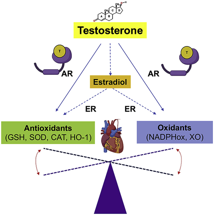
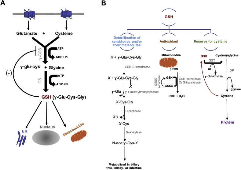
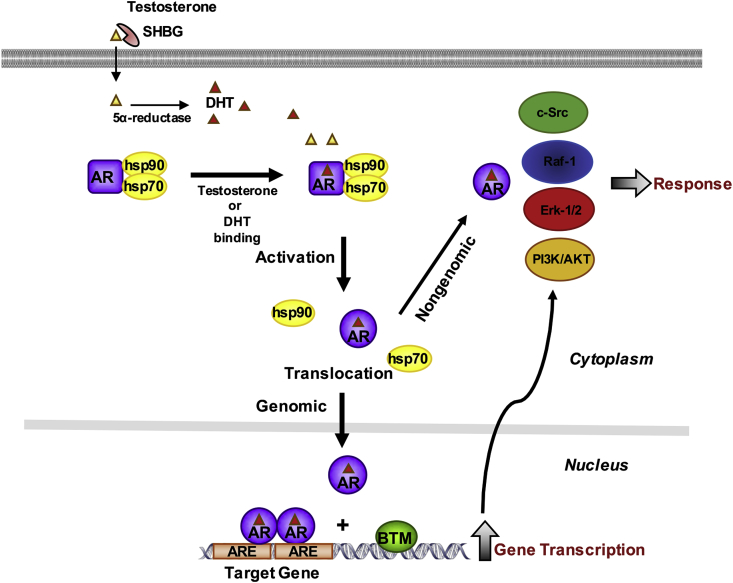
The molecular and pharmacological manipulation of the endogenous redox system is a promising therapy to limit myocardial damage after a heart attack; however, antioxidant therapies have failed to fully establish their cardioprotective effects, suggesting that additional factors, including antioxidant system interactions with other molecular pathways, may alter the pharmacological effects of antioxidants. Since gender differences in cardiovascular disease (CVD) are prevalent, and sex is an essential determinant of the response to oxidative stress, it is of particular interest to understand the effects of sex hormone signaling on the activity and expression of cellular antioxidants and the pharmacological actions of antioxidant therapies. In the present review, we briefly summarize the current understanding of testosterone effects on the modulation of the endogenous antioxidant systems in the CV system, cardiomyocytes, and the heart. We also review the latest research on redox balance and sexual dimorphism, with particular emphasis on the role of the natural antioxidant system glutathione (GSH) in the context of myocardial infarction, and the pro- and antioxidant effects of testosterone signaling via the androgen receptor (AR) on the heart. Finally, we discuss future perspectives regarding the potential of using combing antioxidant and testosterone replacement therapies to protect the aging myocardium.

Keywords: Androgen signaling; Cardiomyocytes; Glutathione; Heart; Oxidative stress; Testosterone.


Conflict of interest statement

Declaration of competing interest The author(s) declare(s) that there is no conflict of interest regarding the publication of this article.

Figures

Image 1
Graphical abstract
Fig. 1
Fig. 1
Glutathione (GSH) Synthesis and role in maintaining systemic redox balance. (A) GSH is synthesized from glutamine, cysteine, and glycine in the cytosol by an ATP-dependent two-step process: 1) This step conjugates cysteine with glutamate, generating γ-glutamyl cysteine (γ-glu-cys) by the action of γ-glutamyl cysteine synthase (γ-GCS, also known as glutamate–cysteine ligase (GCL)) and 2) glutathione synthetase then catalyzes the addition of glycine to γ-glutamylcysteine to form γ-glutamylcysteinylglycine (γ-Glu-Cys-Gly) or GSH. Increased levels of GSH activates a negative feedback inhibition. GHS is then distributed to different areas in the cell, such as the endoplasmic reticulum (ER), the nucleus, and the mitochondria. (B) GSH functions as a detoxification system, antioxidant, and it is the major reserve for cysteine. GSH conjugates with electrophile compounds spontaneously or via enzymatically in reactions catalyzed by GSH-S-transferase. These conjugates are cleaved by γ-glutamyltranspeptidase leaving a cysteinyl-glycine conjugate (X-Cys-Gly). The cysteinyl-glycine bond is then cleaved by dipeptidase. The remaining cysteinyl conjugate (X-Cys) is acetylated by N-acetylase leading to the formation of a mercapturic acid (N-acetyl-Cys-X). This conjugate is then metabolized in the biliary tree, intestine, or kidney. As an antioxidant, when reactive oxygen species (ROS) are produced (mainly by mitochondria), GSH peroxidase or GSH-S-transferase catalyzes the conjugation of GSH to ROS. Oxidized GSH (GSSG) and water are then formed. GSSG can be reduced back to GSH by GSSG reductase. GSH also serves as a source for cysteine. Gamma-Glutamyl Transferase (GGT) catalyze the transfer of the γ-glutamyl moiety of GSH to an amino acid (aa) forming γ-glutamyl amino acid and cysteinylglycine, which is broken down by dipeptidase (DP) to generate cysteine and glycine, which are then transported back into the cell and use for protein synthesis or GSH regeneration.
Fig. 2
Fig. 2
Classical mechanisms of androgen signaling in a target cell. In circulation, testosterone is bound to the serum sex hormone-binding globulin (SHBG). Testosterone dissociates from its binding carrier protein and diffuses freely through the cell membrane. Once in the cytoplasm, testosterone can be converted to its active metabolite 5α-dihydrotestosterone (DHT). Testosterone or DHT can directly bind to the androgen receptor (AR) leading to the dissociation of heat shock proteins (hsp) from the inactive receptor. The ligand-bound receptor then exerts its effects by rapid nongenomic effects by modulating the activity of the Src/Raf-1/Erk-2 pathway or the phosphoinositide 3-kinase (PI3K)/AKT pathway, or by genomic mechanisms involving the activated AR translocation into the nucleus. In the nucleus the androgen receptor binds as homodimer to specific DNA elements present as enhancers in upstream promoter sequences of androgen target genes. Upon AR binding, coactivators are recruited and the basal transcription machinery (BTM) (e.g. RNA-polymerase II [RNA-Pol II], TATA box binding protein [TBP], TBP associating factors [TAF's], general transcription factors [GTF's]) is activated. The interactions between AR, coactivators and the BTM results in gene transcription.
Fig. 3
Fig. 3
Testosterone's effects as a pro- and antioxidant in cardiomyocytes. Testosterone has been reported to increase reactive oxygen species (ROS) production and oxidative stress in cardiomyocytes by signaling via the androgen receptor (AR) through genomic and nongenomic mechanisms (right panel). Activated AR translocates into the nucleus and increases the gene expression of pro-oxidant enzymes (e.g. NAD(P)H oxidases, Xanthine oxidase, and cyclooxygenase 2 (COX-2)) and by increasing the transcription of genes involved in the c-Src and PI3K/Akt pathways, which in turn further exacerbate the activation of pro-oxidant enzymes and increase ROS generation by altering mitochondria function. AR also upregulates the activity of the Proto-oncogene tyrosine kinase Src (c-Src) and phosphoinositide-3-kinase/protein kinase B (PI3K/Akt) pathways by direct protein-protein interactions in the cytosol (nongenomic mechanisms). Testosterone may also increase ROS, via its nongenomic action, through the G protein-coupled receptor, family C, group 6, member A, GPRC6A. Increased ROS levels lead to cardiomyocyte injury, inflammation, lipid accumulation, and eventually cell death, causing heart failure. Recent data suggest that testosterone exerts antioxidant effects, in particular in aging, by a mechanism independent of AR. Although, it is still controversial the proposed mechanism relies on the conversion of testosterone to estradiol, which in turn increases the levels of antioxidant enzymes and reduces oxidative stress in cardiomyocytes by genomic and nongenomic mechanisms (protein-protein interactions with ERKs, MAP and PI3 kinases in the cytoplasm or by binding to the G protein-coupled receptor, family C, group 30, GPR30) (left panel).

References

    1. Benjamin E.J., Muntner P., Alonso A., Bittencourt M.S., Callaway C.W., Carson A.P., Chamberlain A.M., Chang A.R., Cheng S., Das S.R., Delling F.N., Djousse L., Elkind M.S.V., Ferguson J.F., Fornage M., Jordan L.C., Khan S.S., Kissela B.M., Knutson K.L., Kwan T.W., Lackland D.T., Lewis T.T., Lichtman J.H., Longenecker C.T., Loop M.S., Lutsey P.L., Martin S.S., Matsushita K., Moran A.E., Mussolino M.E., O'Flaherty M., Pandey A., Perak A.M., Rosamond W.D., Roth G.A., Sampson U.K.A., Satou G.M., Schroeder E.B., Shah S.H., Spartano N.L., Stokes A., Tirschwell D.L., Tsao C.W., Turakhia M.P., VanWagner L.B., Wilkins J.T., Wong S.S., Virani S.S. American heart association council on E, prevention statistics C and stroke statistics S. Heart disease and stroke statistics-2019 update: a report from the American heart association. Circulation. 2019;139:e56–e528. - PubMed
    1. Yang H.Y., Huang J.H., Hsu C.Y., Chen Y.J. Gender differences and the trend in the acute myocardial infarction: a 10-year nationwide population-based analysis. Sci. World J. 2012;2012 184075. - PMC - PubMed
    1. Anderson R.D., Pepine C.J. Gender differences in the treatment for acute myocardial infarction: bias or biology? Circulation. 2007;115:823–826. - PubMed
    1. Diercks D.B., Owen K.P., Kontos M.C., Blomkalns A., Chen A.Y., Miller C., Wiviott S., Peterson E.D. Gender differences in time to presentation for myocardial infarction before and after a national women's cardiovascular awareness campaign: a temporal analysis from the can rapid risk stratification of unstable Angina patients suppress ADverse outcomes with early implementation (CRUSADE) and the national cardiovascular data registry acute coronary treatment and intervention outcomes network-get with the guidelines (NCDR ACTION registry-GWTG) Am. Heart J. 2010;160:80–87 e3. - PubMed
    1. Granger D.N., Kvietys P.R. Reperfusion injury and reactive oxygen species: the evolution of a concept. Redox Biol. 2015;6:524–551. - PMC - PubMed

Publication types