. 2020 May;80(5):554-562.
doi: 10.1016/j.jinf.2020.02.026. Epub 2020 Mar 10.

COVID-19 spike-host cell receptor GRP78 binding site prediction

Affiliations

COVID-19 spike-host cell receptor GRP78 binding site prediction

Ibrahim M Ibrahim et al. J Infect. 2020 May.

Abstract

Objectives: Understanding the novel coronavirus (COVID-19) mode of host cell recognition may help to fight the disease and save lives. The spike protein of coronaviruses is the main driving force for host cell recognition.

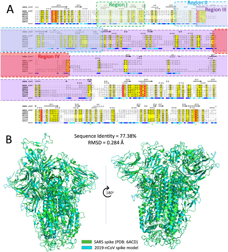
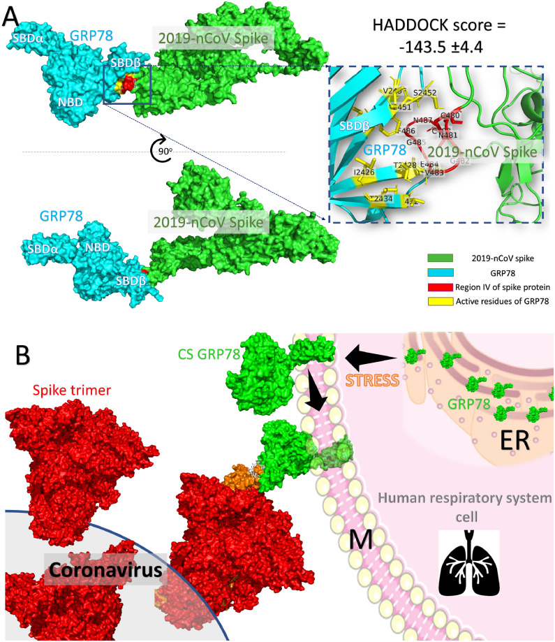
Methods: In this study, the COVID-19 spike binding site to the cell-surface receptor (Glucose Regulated Protein 78 (GRP78)) is predicted using combined molecular modeling docking and structural bioinformatics. The COVID-19 spike protein is modeled using its counterpart, the SARS spike.

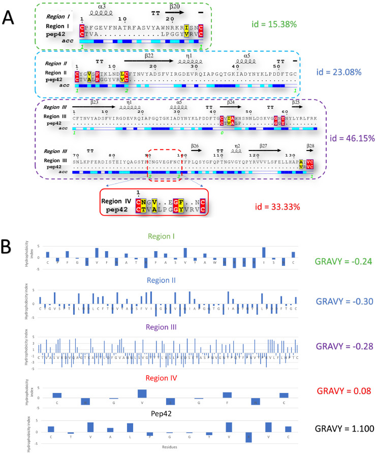
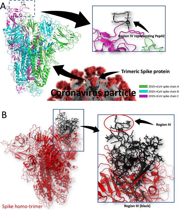
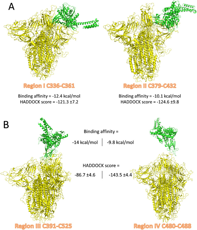
Results: Sequence and structural alignments show that four regions, in addition to its cyclic nature have sequence and physicochemical similarities to the cyclic Pep42. Protein-protein docking was performed to test the four regions of the spike that fit tightly in the GRP78 Substrate Binding Domain β (SBDβ). The docking pose revealed the involvement of the SBDβ of GRP78 and the receptor-binding domain of the coronavirus spike protein in recognition of the host cell receptor.

Conclusions: We reveal that the binding is more favorable between regions III (C391-C525) and IV (C480-C488) of the spike protein model and GRP78. Region IV is the main driving force for GRP78 binding with the predicted binding affinity of -9.8 kcal/mol. These nine residues can be used to develop therapeutics specific against COVID-19.

Keywords: BiP; COVID-19 spike; GRP78; Pep42; Protein-protein docking; Structural bioinformatics.


Conflict of interest statement

Declaration of Competing Interests All of the authors declare that there is no competing interest in this work.

Figures

Fig 1
Fig. 1
(A) Part of the multiple sequence alignment for the spike protein of all of the currently reported human coronaviruses strains (COVID-19, SARS, MERS, NL63, 229E, OC43, and HKU1). The alignment is made using the Clustal Omega web server and is displayed by ESpript 3 software. The red highlighted residues are identical, while yellow highlighted residues are conserved among the seven HCoVs. Secondary structures are represented at the top of the MSA for the COVID-19 spike, while the surface accessibility is shown at the bottom (blue, surface accessible, cyan, partially accessible, and white for buried residues). The four regions of the spike protein are shaded with green, blue, magenta, and red for regions I, II, III, and IV, respectively. (B) Structural superposition of SARS spike structure (green cartoon) and COVID-19 spike model (cyan cartoon). (For interpretation of the references to color in this figure legend, the reader is referred to the web version of this article.)
Fig 2
Fig. 2
(A) Pairwise sequence alignment between Pep42 and four regions of the COVID-19 spike protein model. Secondary structures and surface accessibility are shown at the top and the bottom of the alignment, respectively. The percentage of identity arrows shows beta-sheets on the right side of each alignment. Identical and similar residues are highlighted in red and yellow, respectively. (B) The hydrophobicity index (Kyte & Doolittle) for the four regions of the spike protein model for COVID-19 and the Pep42 peptide. Grand average hydrophobicity (GRAVY) is shown on the right side for each peptide. (For interpretation of the references to color in this figure legend, the reader is referred to the web version of this article.)
Fig 3
Fig. 3
(A) The structure of the spike protein model of COVID-19 in its homotrimer state (colored cartoon). Two chains, A (green) and B (cyan) are in the closed conformation, while chain C (magenta) is the open configuration that makes it able to recognize the host cell receptor. Region IV of the spike (C480-C488), which we suggest is the recognition site for cell-surface GRP78, is shown in the black cartoon in the enlarged panel. (B) The structure of the region III (black carton). (For interpretation of the references to color in this figure legend, the reader is referred to the web version of this article.)
Fig 4
Fig. 4
The structure of the docking complexes of GRP78 (green cartoon) and COVID-19 spike (yellow cartoon) regions I and II (A) and regions III and IV (B). (For interpretation of the references to color in this figure legend, the reader is referred to the web version of this article.)
Fig 5
Fig. 5
(A) The proposed binding mode of the host cell GRP78 (cyan surface) and the COVID-19 spike model (green surface) through region IV (C480-C488) (red surface). The amino acids from the GRP78 SBDβ that interact with the spike protein region IV (red cartoon) are labeled and represented in yellow sticks in the enlarged panel. (B) The proposed recognition mode of the COVID-19 spike (red surface) and cell-surface GRP78 (green surface) through the spike protein region C480-C488. (For interpretation of the references to color in this figure legend, the reader is referred to the web version of this article.)

Comment in

References

    1. Hui D.S., I Azhar E., Madani T.A., Ntoumi F., Kock R., Dar O. Int J Infect Dis. 2020;91:264–266. - PMC - PubMed
    1. Bogoch I.I., Watts A., Thomas-Bachli A., Huber C., Kraemer M.U.G., Khan K. Pneumonia of unknown etiology in Wuhan, China: potential for international spread via commercial air travel. J Travel Med. 2020 - PMC - PubMed
    1. Organization WH. World Health Organization; 2020. Surveillance case definitions for human infection with novel coronavirus ( nCoV): interim guidance v1, January 2020.
    1. Organization WH . World Health Organization; 2020. Laboratory testing of human suspected cases of novel coronavirus ( nCoV) infection: interim guidance, 10 January 2020.
    1. Organization WH . World Health Organization; 2020. Infection prevention and control during health care when novel coronavirus ( nCoV) infection is suspected: interim guidance, January 2020.

MeSH terms

Substances