. 2020 Aug;73(2):349-360.
doi: 10.1016/j.jhep.2020.02.031. Epub 2020 Mar 11.

Alternatively activated macrophages promote resolution of necrosis following acute liver injury

Affiliations

Alternatively activated macrophages promote resolution of necrosis following acute liver injury

Philip Starkey Lewis et al. J Hepatol. 2020 Aug.

Abstract

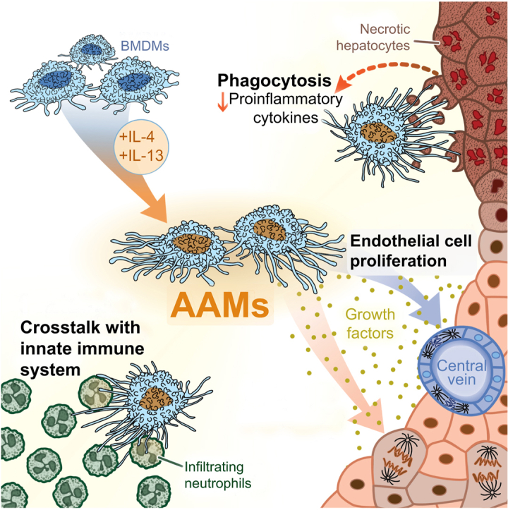
Background & aim: Following acetaminophen (APAP) overdose, acute liver injury (ALI) can occur in patients that present too late for N-acetylcysteine treatment, potentially leading to acute liver failure, systemic inflammation, and death. Macrophages influence the progression and resolution of ALI due to their innate immunological function and paracrine activity. Syngeneic primary bone marrow-derived macrophages (BMDMs) were tested as a cell-based therapy in a mouse model of APAP-induced ALI (APAP-ALI).

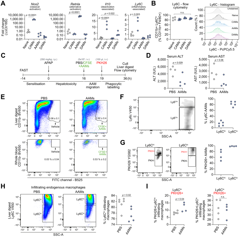
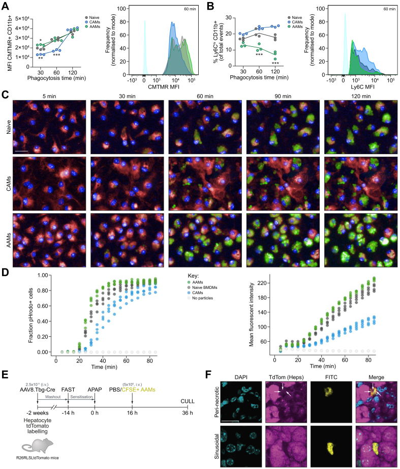
Methods: Several phenotypically distinct BMDM populations were delivered intravenously to APAP-ALI mice when hepatic necrosis was established, and then evaluated based on their effects on injury, inflammation, immunity, and regeneration. In vivo phagocytosis assays were used to interrogate the phenotype and function of alternatively activated BMDMs (AAMs) post-injection. Finally, primary human AAMs sourced from healthy volunteers were evaluated in immunocompetent APAP-ALI mice.

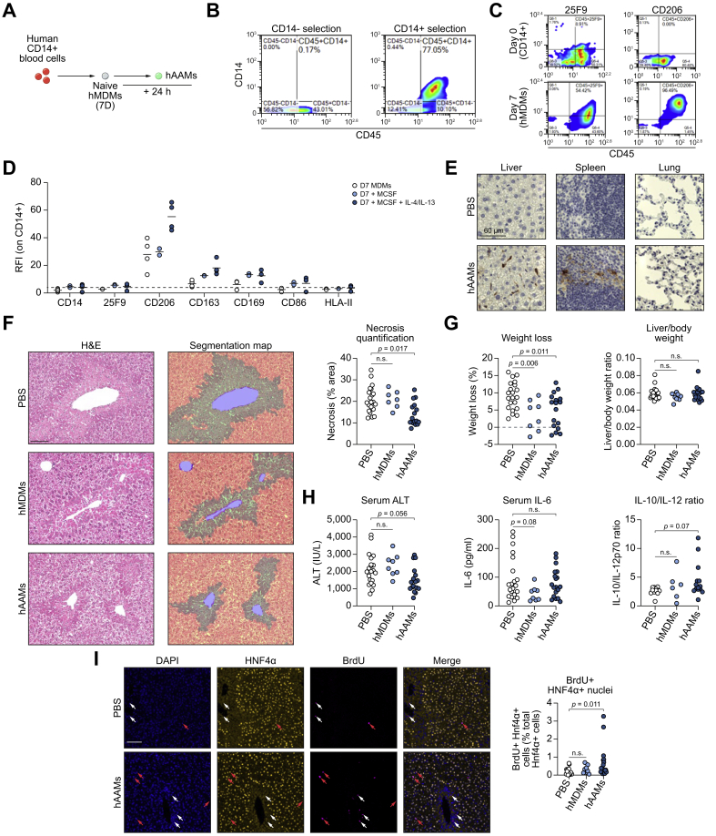
Results: BMDMs rapidly localised to the liver and spleen within 4 h of administration. Injection of AAMs specifically reduced hepatocellular necrosis, HMGB1 translocation, and infiltrating neutrophils following APAP-ALI. AAM delivery also stimulated proliferation in hepatocytes and endothelium, and reduced levels of several circulating proinflammatory cytokines within 24 h. AAMs displayed a high phagocytic activity both in vitro and in injured liver tissue post-injection. Crosstalk with the host innate immune system was demonstrated by reduced infiltrating host Ly6Chi macrophages in AAM-treated mice. Importantly, therapeutic efficacy was partially recapitulated using clinical-grade primary human AAMs in immunocompetent APAP-ALI mice, underscoring the translational potential of these findings.

Conclusion: We identify that AAMs have value as a cell-based therapy in an experimental model of APAP-ALI. Human AAMs warrant further evaluation as a potential cell-based therapy for APAP overdose patients with established liver injury.

Lay summary: After an overdose of acetaminophen (paracetamol), some patients present to hospital too late for the current antidote (N-acetylcysteine) to be effective. We tested whether macrophages, an injury-responsive leukocyte that can scavenge dead/dying cells, could serve as a cell-based therapy in an experimental model of acetaminophen overdose. Injection of alternatively activated macrophages rapidly reduced liver injury and reduced several mediators of inflammation. Macrophages show promise to serve as a potential cell-based therapy for acute liver injury.

Keywords: Acetaminophen; Liver regeneration; Macrophages; Necrosis; Phagocytosis.


Conflict of interest statement

Conflict of interest P.S.L., L.F., and S.J.F. have patents pending, entitled ‘Macrophage-based therapy’ in national territories of USA, Europe, Japan, China and Australia. These patents have been derived from PCT/GB2017/052769 filed 18/09/2017 and claim priority from UK application 1615923.8 filed 19/09/2016. Both of the original patents have now been abandoned because the original UK patent and PCT patent are no longer live and have now been replaced by the national patents. Please refer to the accompanying ICMJE disclosure forms for further details.

Figures

None
Graphical abstract
Fig. 1
Fig. 1
Injection of AAMs reduces necrosis and stimulates liver regeneration following APAP-ALI. (A) Four macrophage populations, derived from mouse BM, were generated for testing: 1. unstimulated BMDMs (naïve), 2. CAMs, 3. AAMs, and 4. DAMs. (B) Study design: injection of macrophages (1 × 106, i.v.) or PBS alone into APAP-ALI mice at 16 h, before cull at 36 h. (C) Serum ALT activity (left panel) and AST activity (right panel) in APAP-ALI mice receiving indicated treatments (D) Representative liver histological stains from APAP-ALI mice receiving indicated treatments, necrosis quantification in right panel (E) Representative Ly6G immunohistochemical stains in liver from APAP-ALI mice with indicated treatments. Black arrows indicate Ly6G-positive cells, quantification in right panel. (F) Representative images of HMGB1 immunohistochemical stains of liver tissue from APAP-ALI mice with indicated treatments. Black arrows indicate HMGB1-negative nuclei, quantification in right panel. (G) Representative immunofluorescence stains of BrdU incorporation (yellow nuclei, indicated by white arrows) in liver with DAPI counterstain (cyan) from APAP-ALI mice with indicated treatments. Quantification in right panel. (H/I) Dual immunofluorescence stains of BrdU (yellow), and either HNF4α (H) or ERG (I) (magenta), with DAPI counterstain (cyan) in AAM-treated liver tissue. White arrows indicate dual-positive cells, quantification in right panel. All data shown are n = 6–12 mice per group (white circles — individual vehicle controls; grey circles — cell-transfer reference group; blue circles — polarised-BMDM-treated mice). Scale bars — 100 μm unless otherwise indicated. p values indicated in panels, Kruskal-Wallis test for (C/D/E/F/G/H). n.s., not significant. One-way ANOVA for I. AAMs, alternatively activated macrophages; ALI, acute liver injury; ALT, alanine aminotransferase; APAP, acetaminophen; APAP-ALI, APAP-induced ALI; AST, aspartate aminotransferase; BM, bone marrow; BMDMs, bone marrow-derived macrophages; CAMs, classically activated macrophages; DAMs, deactivated macrophages.
Fig. 2
Fig. 2
Injection of AAMs reduces several serum proinflammatory cytokines following APAP-ALI. (A) Serum concentrations of proinflammatory cytokines measured in APAP-ALI mice receiving indicated treatments (n = 10–12 per group; some sera had undetectable IL-12p70). (B) IL-6 levels in liver homogenates from APAP-ALI mice receiving indicated treatments (n = 5–11 per group) (C) Relative expression of indicated genes (using 2−ΔΔCT method; standardised to PBS-treated controls, after GAPDH normalisation) in liver tissue of APAP-ALI mice receiving indicated treatments (n = 6–10 per group). In A–C, white circles — individual vehicle controls; grey circles — cell-transfer reference group; blue circles — AAM-treated mice. (D) Study design: Plasma biomarkers were measured daily in APAP-ALI mice following PBS/AAM-treatment. (E/F/G) Plasma biomarkers (left panels) and change in plasma biomarkers from point-of-treatment (right panels) for plasma ALT activity (E), AST activity (F), and miR-122 levels (G). Shaded area represents treatment phase. Grouped values represents mean ± SD for AAM-treatment (blue) and PBS-treatment (white). Plasma miR-122 levels are presented as relative quantitation (using 2-ΔΔCT method; standardised to pre-APAP-ALI levels (−96 h), after let-7d normalisation). p values provided in panels; n.s., not significant, ∗p <0.05, ∗∗p <0.01. Kruskal-Wallis tests for A/B/C (Csf1/Ccl5), one-way ANOVA for C (Tgfb, Il6, Cxcl1), mixed-effects model for (E/F/G). AAMs, alternatively activated macrophages; ALI, acute liver injury; ALT, alanine aminotransferase; APAP, acetaminophen; APAP-ALI, APAP-induced ALI.
Fig. 3
Fig. 3
Injected AAMs are primarily Ly6Clo and highly phagocytic in situ. (A) Relative gene expression in BMDM populations (determined by 2−ΔΔCT method; standardised to naïve BMDMs, after 18S normalisation): Nos2 — CAM-associated gene, Retnla — AAM-associated gene, Il10 — DAM-associated gene, Ly6C — proinflammatory-associated gene (n = 3/4 biological replicates per group). (B) Ly6C status in BMDM populations (flow cytometry quantification, left panel; cytometry histograms, right panel) (C) Study design: CFSE-labelled AAMs injected (5 × 106, i.v.) at 16 h in APAP-ALI mice, 3 h before PKH26 (fluorescent phagocytic tracer). Cull at 36 h. (D) Serum ALT activity (left) and AST (right) in APAP-ALI mice treated with PBS or AAMs (n = 4 per group). (E) Gating shows CFSE+ AAMs in liver digests (top panels) and whole blood (bottom panels) in AAM-treated APAP-ALI mice. (F). Representative flow plot of Ly6C status in retrieved CFSE+ AAMs (gating: left panel; quantification: right panel, circles represent digests from individual mice). (G) Representative flow plots showing PKH uptake in Ly6Chi (left panel) and Ly6Clo (middle panel) AAMs, quantification in right panel. (H) Representative flow plots show Ly6C status in infiltrating endogenous macrophages in liver digests from APAP-ALI mice treated with PBS (left panel) or AAMs (middle panel), quantification in right panel. (I) Quantification of PKH uptake in Ly6Chi (left panel) and Ly6Clo (right panel) infiltrating endogenous macrophages. p values provided in panels, n.s. not significant. One-way ANOVA (A), unpaired t test (D, I), or Mann-Whitney U test (H) were performed. AAMs, alternatively activated macrophages; ALI, acute liver injury; ALT, alanine aminotransferase; APAP, acetaminophen; APAP-ALI, APAP-induced ALI; AST, aspartate aminotransferase; BM, bone marrow; BMDMs, bone marrow-derived macrophages; CAMs, classically activated macrophages; DAMs, deactivated macrophages.
Fig. 4
Fig. 4
Murine AAMs are highly phagocytic. (A) Phagocytosis quantification in naïve BMDMs (dark grey), AAMs (green), or CAMs (blue) during incubation with CMTMR-labelled apoptotic thymocytes. MFI of CMTMR-positive macrophage populations (left), and representative histograms (right). (B) Ly6C status in BMDM populations during incubation with CMTMR-labelled apoptotic thymocytes at indicated times (left panel); representative histograms (right panel). Coloured circles represent individual preparations connected by lines (mean value, n = 3). (C) Representative images of real-time phagocytosis at indicated times. Naïve BMDMs (top row), CAMs (middle row) and AAMs (bottom row) are shown (Deep Red CellMask, red; NucBlue, blue). Phagocytosis indicated by intracellular fluorescence (green). (D) Phagocytosis quantification: pHrodo-positive cell fraction (left) and total cell MFI (right). (E) Study design: Hepatocytes were Tdtomato-labelled by delivery of hepatotropic AAV8 virus delivering Cre-recombinase to R26RLSLtdTomato mice. Tdtomato-positive APAP-ALI mice received CFSE-labelled AAMs (5 × 106, i.v.) at 16 h, before cull (36 h). (F) Panels show representative confocal immunofluorescence of liver tissue (max intensity projection from 7 slices; 2.4 μm) in each channel: DAPI (cyan), TdTom (TdTomato+ hepatocytes, magenta), FITC (CFSE+ AAMs, yellow), and merged images. Faint punctate TdTomato+ debris were visible inside vesicles in peri-necrotic macrophages (top row, white arrow heads). ∗p <0.05, ∗∗p <0.01, ∗∗∗p <0.001, scale bars 20 μm. Two-way ANOVA (A/B). AAMs, alternatively activated macrophages; AAV, adeno-associated virus; ALI, acute liver injury; APAP, acetaminophen; APAP-ALI, APAP-induced ALI; AST, aspartate aminotransferase; BM, bone marrow; BMDMs, bone marrow-derived macrophages; CAMs, classically activated macrophages; DAMs, deactivated macrophages; MFI, mean fluorescence intensity.
Fig. 5
Fig. 5
Injection of hAAMs reduces necrosis in APAP-ALI immunocompetent mice. (A) hMDMs were differentiated from CD14+ cells isolated from healthy volunteer buffy coats, before incubation with hCSF-1 for 7 days. hAAMs were generated by stimulating hMDMs overnight with hCSF1, hIL-4, and hIL-13. (B) Representative flow cytometry plots demonstrating CD14-enrichment using CliniMACS® beads (C) Representative flow cytometry plots showing macrophage maturity markers (25F9, left panels; CD206, right panels) in CD14+ cells (top) and hMDMs (bottom) (D) Flow cytometry quantification of hMDMs (white), hMDMs stimulated with CSF-1 alone (light blue), and fully-stimulated hAAMs (dark blue), spots represent individual donors. (E) Panels show representative anti-FITC immunohistochemical stains in liver (left), spleen (centre), and lung (right) from APAP-ALI mice treated with PBS (top row) or hAAMs (bottom row). (F) Representative histological staining (left) and necrosis segmentation map (centre) in APAP-ALI mice with indicated treatments. Necrosis quantification in right panel (n ≥7 mice per group). (G) Percentage weight loss (left panel) and liver/body weight ratio (right panel) of APAP-ALI mice with indicated treatments (n ≥8 per group) (H). Serum injury/inflammatory markers in APAP-ALI mice receiving indicated treatments (n ≥6 per group; serum ALT activity, left panel; serum IL-6, centre panel; serum IL-10/-12p70 ratio, right panel, some sera has undetectable IL-12p70 levels). (I) Representative dual immunofluorescence images of HNF4α (yellow) and BrdU (magenta) against DAPI (counterstain, blue) in liver tissue from APAP-ALI mice treated with PBS (top row) or hAAMs (bottom row). White arrowheads indicate BrdU-positive cells, red arrowheads indicate dual-positive BrdU-positive HNF4α-positive cells (quantification right panel; n≥ 8). Scale bars 100 μm, unless otherwise indicated. p values indicated in panels, n.s. not significant. One-way ANOVA test in F, G (weight loss %), H (ALT), or Kruskal-Wallis test in G (liver/body ratio), H (IL-6, IL-10/-12 ratio), and I. ALI, acute liver injury; APAP, acetaminophen; APAP-ALI, APAP-induced ALI; hAAMs, alternatively activated macrophages; hMDMs, human monocyte-derived macrophages.

Similar articles

Cited by

References

    1. Larson A.M., Polson J., Fontana R.J., Davern T.J., Lalani E., Hynan L.S. Acetaminophen-induced acute liver failure: results of a United States multicenter, prospective study. Hepatology. 2005;42:1364. - PubMed
    1. Lee W.M. Acetaminophen and the U.S. acute liver failure study group: lowering the risks of hepatic failure. Hepatology. 2004;40:6. - PubMed
    1. Lee W.M. Acetaminophen-related acute liver failure in the United States. Hepatol Res. 2008;38:S3. - PubMed
    1. Prescott L.F., Illingworth R.N., Critchley J.A.J.H. Intravenous N-acetylcysteine: the treatment of choice for paracetamol poisoning. Br Med J. 1979;2:1097. - PMC - PubMed
    1. Rolando N., Wade J., Davalos M., Wendon J., Philpott-Howard J., Williams R. The systemic inflammatory response syndrome in acute liver failure. Hepatology. 2000;32:734–739. - PubMed

Publication types

MeSH terms