. 2020 May:206:110997.
doi: 10.1016/j.jinorgbio.2020.110997. Epub 2020 Jan 11.

Hydrolyzed Ce(IV) salts limit sucrose-dependent biofilm formation by Streptococcus mutans

Affiliations

Hydrolyzed Ce(IV) salts limit sucrose-dependent biofilm formation by Streptococcus mutans

Lopa Bhatt et al. J Inorg Biochem. 2020 May.

Abstract

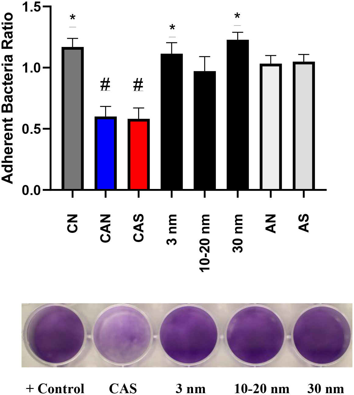
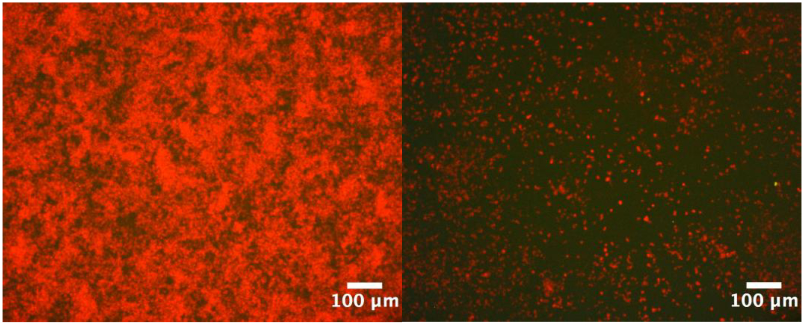
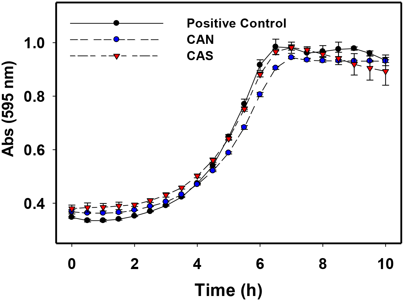
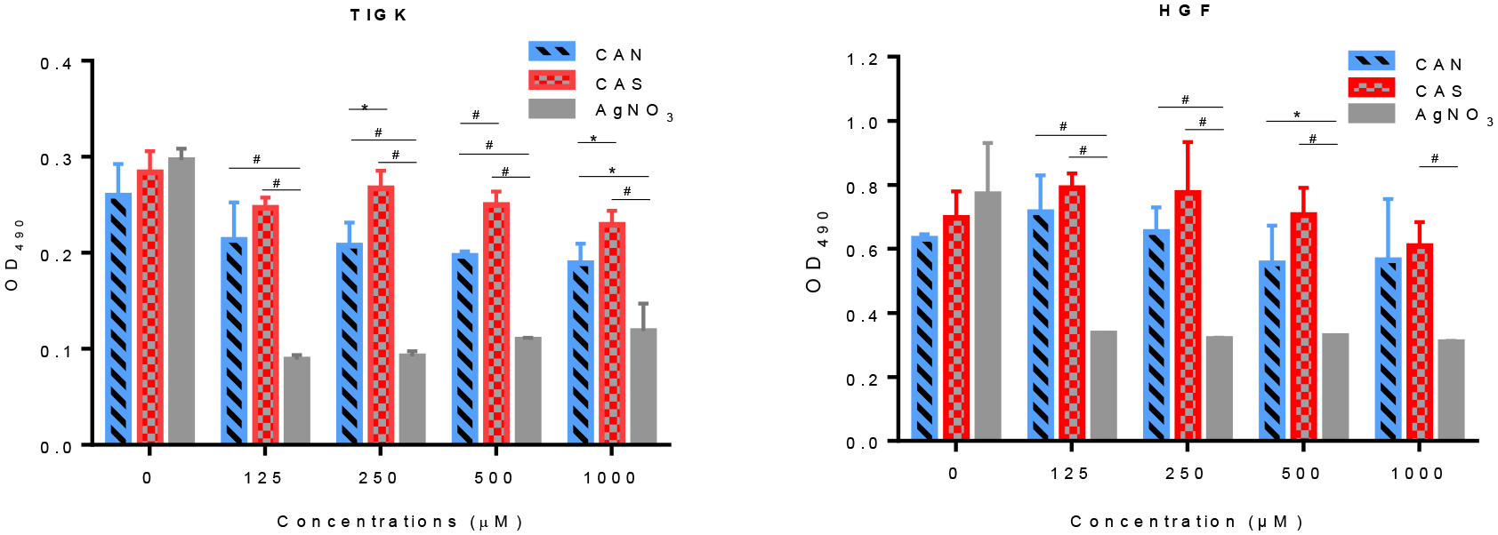
Several studies have focused on the antimicrobial effects of cerium oxide nanoparticles (CeO2-NP) but few have focused on their effects on bacteria under initial biofilm formation conditions. Streptococcus mutans is a prolific biofilm former contributing to dental caries in the presence of fermentable carbohydrates and is a recognized target for therapeutic intervention. CeO2-NP derived solely from Ce(IV) salt hydrolysis were found to reduce adherent bacteria by approximately 40% while commercial dispersions of "bare" CeO2-NP (e.g., 3 nm, 10-20 nm, 30 nm diameter) and Ce(NO3)3·6H2O were either inactive or observed to slightly increase biofilm formation under similar in vitro conditions. Planktonic growth and dispersal assays support a non-bactericidal mode of biofilm inhibition active in the initial phases of S. mutans biofilm production. Human cell proliferation assays suggest only minor effects of hydrolyzed Ce(IV) salts on cellular metabolism at concentrations up to 1 mM Ce, with less observed toxicity compared to equimolar concentrations of AgNO3. The results presented herein have implications in clinical dentistry.

Keywords: Biofilm; Ce(IV); Dental; Inhibition; Streptococcus mutans.


Conflict of interest statement

Declaration of competing interest We report no conflicts of interest in relation to this study or the materials utilized.

Figures

Figure 1.
Figure 1.
The ratio of adherent bacteria (compared to positive control) from the treatment of S. mutans UA159 with Ce-containing agents (125 μM Ce) [CN = Ce(NO3)3•6H2O] and ammonium salts (AN, AS) (1 mM) BHI, 1% sucrose, 37°C, 5% CO2 for 20 h. *p<0.05, #p<0.0001, student t test.
Figure 2.
Figure 2.
Microscopic images (10 x) of adherent S. mutans UA159 grown for 20 h at 37 °C, 5% CO2 in BHI. The untreated wells (left) displayed significantly more biofilm growth than 250 μM CAS treated wells (right).
Figure 3.
Figure 3.
Planktonic growth curves of S. mutans UA159 with 500 μM CAN and CAS treated cells in BHI at 37°C and 5% CO2 for 10 h. Measurements were run in triplicate. Error bars represent standard deviation of the triplicate measurements.
Figure 4.
Figure 4.
MTS cell proliferation assays utilizing (left) TIGK and (right) HGF in the presence of hydrolyzed CAN (red), CAS (blue) and AgNO3 (gray) for 24 h at 37°C, 5% CO2. *p<0.05, #p<0.01, t test.
Figure 5.
Figure 5.
Distribution of hydrolyzed [Ce(NO3)6]2- (pH 1.8) and CeO2-NP (3 nm, Strem) (pH 4.5) in 30 mM NO3.
Figure 6.
Figure 6.
Distribution of (left) 5 mM [Ce(NO3)6]2- (pH 1.8) and (right) 5 mM CeO2-NP (3 nm, Strem) (pH 4.5) in 30 mM NO3 and with 40 mM KCl.
Figure 7.
Figure 7.
Distribution of hydrolyzed [Ce(NO3)6]2- (500 μM Ce) in 50 mM KH2PO4 (pH 7.4)
Figure 8.
Figure 8.
Low-magnification high-angle annular dark field (HAADF) of (a) hydrolyzed [Ce(NO3)6]2- and (b) CeO2-NP (3nm, Strem) in graphene liquid cells. Inserts show high-resolution images of the nano-particles revealing lattice fringes. Electron energy-loss spectroscopy (EELS) of the Ce M-edges for (c) hydrolyzed [Ce(NO3)6]2- and (d) CeO2-NP (3nm, Strem) in graphene liquid cells, showing the M4/M5 ratio expected for a Ce(IV) valence state.

References

    1. Dewhirst FE, Chen T, Izard J, Paster BJ, Tanner AC, Yu WH, Lakshmanan A, Wade WG, J Bacteriol, vol. 192, 2010, pp. 5002–5017. - PMC - PubMed
    1. Klein MI, Hwang G, Santos PH, Campanella OH, Koo H, Front Cell Infect Microbiol, vol. 5, 2015, pp. 10. - PMC - PubMed
    1. Lemos JA, Palmer SR, Zeng L, Wen ZT, Kajfasz JK, Freires IA, Abranches J, Brady LJ, Microbiol Spectr, vol. 7, 2019. - PMC - PubMed
    1. Davis IJ, Richards H, Mullany P, Oral Microbiol Immunol, vol. 20, 2005, pp. 191–194. - PubMed
    1. Mijnendonckx K, Leys N, Mahillon J, Silver S, Van Houdt R, Biometals, vol. 26, 2013, pp. 609–621. - PubMed

Publication types