. 2020 Apr 17;295(16):5350-5361.
doi: 10.1074/jbc.RA120.012724. Epub 2020 Mar 13.

The transcriptional regulator IscR integrates host-derived nitrosative stress and iron starvation in activation of the vvhBA operon in Vibrio vulnificus

Affiliations

The transcriptional regulator IscR integrates host-derived nitrosative stress and iron starvation in activation of the vvhBA operon in Vibrio vulnificus

Garam Choi et al. J Biol Chem. .

Abstract

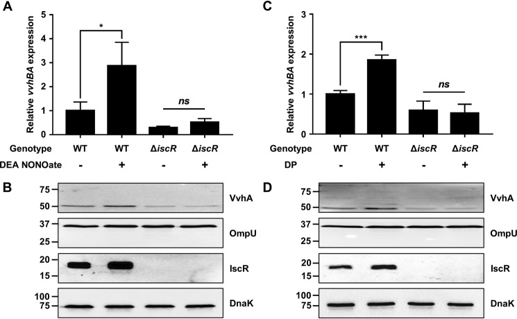
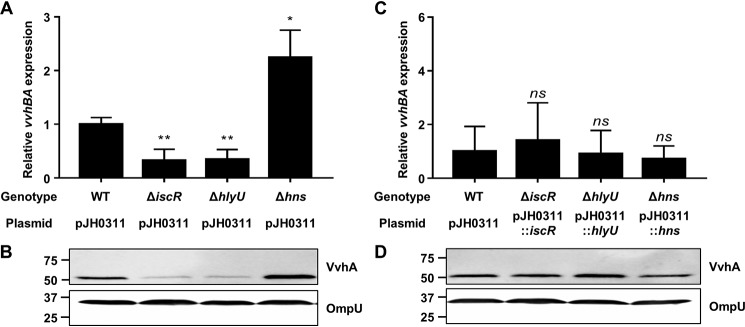
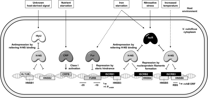
For successful infection of their hosts, pathogenic bacteria recognize host-derived signals that induce the expression of virulence factors in a spatiotemporal manner. The fulminating food-borne pathogen Vibrio vulnificus produces a cytolysin/hemolysin protein encoded by the vvhBA operon, which is a virulence factor preferentially expressed upon exposure to murine blood and macrophages. The Fe-S cluster containing transcriptional regulator IscR activates the vvhBA operon in response to nitrosative stress and iron starvation, during which the cellular IscR protein level increases. Here, electrophoretic mobility shift and DNase I protection assays revealed that IscR directly binds downstream of the vvhBA promoter P vvhBA , which is unusual for a positive regulator. We found that in addition to IscR, the transcriptional regulator HlyU activates vvhBA transcription by directly binding upstream of P vvhBA , whereas the histone-like nucleoid-structuring protein (H-NS) represses vvhBA by extensively binding to both downstream and upstream regions of its promoter. Of note, the binding sites of IscR and HlyU overlapped with those of H-NS. We further substantiated that IscR and HlyU outcompete H-NS for binding to the P vvhBA regulatory region, resulting in the release of H-NS repression and vvhBA induction. We conclude that concurrent antirepression by IscR and HlyU at regions both downstream and upstream of P vvhBA provides V. vulnificus with the means of integrating host-derived signal(s) such as nitrosative stress and iron starvation for precise regulation of vvhBA transcription, thereby enabling successful host infection.

Keywords: H-NS; HlyU; Host-derived signal; IscR; Vibrio vulnificus; bacterial toxin; bacterial transcription; gene regulation; gene transcription; host-pathogen interaction; transcription factor.


Conflict of interest statement

The authors declare that they have no conflicts of interest with the contents of this article

Figures

Figure 1.
Figure 1.
Expression of vvhBA upon exposure to murine blood and RAW 264.7 cells. A, the genes induced in V. vulnificus upon exposure to murine blood were identified by RNA-seq analysis. vvhBA was selected as the most highly induced extracellular toxin-encoding gene, and its induction was confirmed by qRT-PCR. Each column represents the vvhBA transcript level in V. vulnificus exposed to murine blood relative to M9G (negative control). Error bars represent the S.E. calculated using DeSeq2 for RNA-seq and the S.D. for qRT-PCR. B, V. vulnificus was exposed to DMEM (negative control) or RAW 264.7 cells in the presence or absence of l-NMMA. The vvhBA transcript levels were determined by qRT-PCR, and the vvhBA transcript level in the cells exposed to DMEM without l-NMMA was set to 1. Error bars represent the S.D. *, p < 0.05; ***, p < 0.0005; ns, not significant.
Figure 2.
Figure 2.
The effect of iscR mutation on vvhBA expression. Total RNA and proteins were isolated from the V. vulnificus strains grown aerobically to an A600 of 0.3. A and C, the vvhBA transcript levels were determined by qRT-PCR, and the vvhBA transcript levels in the WT were set to 1. Error bars represent the S.D. **, p < 0.005; ***, p < 0.0005; ns, not significant. B and D, the secreted VvhA and OmpU (internal control), and cellular IscR or IscR3CA and DnaK (internal control) protein levels were determined by Western blot analysis. Molecular size markers (Bio-Rad) are shown in kDa. WT (pJH0311) and WT, a WT; ΔiscR (pJH0311) and ΔiscR, an iscR-deletion mutant; ΔiscR (pJH0311::iscR), an iscR-complemented strain with pKK1531; iscR3CA, a strain expressing apo-locked IscR.
Figure 3.
Figure 3.
The effect of nitrosative stress and iron starvation on vvhBA and IscR expression. Total RNA and proteins were isolated from the V. vulnificus strains grown aerobically to an A600 of 0.3 and then exposed to 25 μm DEA NONOate for 20 min (A and B) or 50 μm DP for 10 min (C and D). A and C, the vvhBA transcript levels were determined by qRT-PCR, and the vvhBA transcript levels in the WT unexposed to DEA NONOate (A) or DP (C) were set to 1. Error bars represent the S.D. *, p < 0.05; ***, p < 0.0005; ns, not significant. B and D, the secreted VvhA and OmpU (internal control), and cellular IscR and DnaK (internal control) protein levels were determined by Western blot analysis. Molecular size markers (Bio-Rad) are shown in kDa. WT, a WT; ΔiscR, an iscR-deletion mutant.
Figure 4.
Figure 4.
Specific binding of IscR to PvvhBA and sequences of the PvvhBA regulatory region. A, a 501-bp DNA of the PvvhBA regulatory region (5 nm) was radiolabeled and then incubated with increasing amounts of IscR as indicated. For competition analysis, various amounts of the unlabeled DNA fragment were used as a self-competitor and added to a reaction mixture containing 5 nm radiolabeled DNA and 30 nm IscR. B1, a DNA-IscR complex; F, free DNA. B, the same DNA of the PvvhBA regulatory region (32.3 nm) was labeled with 6-FAM, incubated with increasing amounts of IscR as indicated, and then digested with DNase I. The regions protected by IscR are indicated by black boxes (ISCRB1, ISCRB2, ISCRB3). Nucleotide numbers shown are relative to the transcription start site of vvhBA, which was determined previously (17). C, sequence analysis of the PvvhBA regulatory region. The transcription start site of vvhBA and the putative translational initiation codon of VvhB are indicated by solid and dashed bent arrows, respectively. The putative −10 and −35 regions are underlined and the putative ribosome-binding site (AGGA) is boldface. The binding sequences of IscR are shown with the black boxes as described above. The binding sequences of HlyU (HLYUB; a white box) and H-NS (HNSB1, HNSB2, HNSB3, HNSB4, HNSB5, HNSB6; gray boxes) were determined later in this study (Fig. 6, C and D). The consensus sequences of the IscR-binding Type 2 DNA motif are indicated above the V. vulnificus DNA sequences. W, A or T; Y, C or T; R, A or G; x, any nucleotide.
Figure 5.
Figure 5.
IscR and HlyU activate, but H-NS represses the vvhBA transcription. Total RNA and proteins were isolated from the V. vulnificus strains grown aerobically to an A600 of 0.3. A and C, the vvhBA transcript levels were determined by qRT-PCR, and the vvhBA transcript levels in the WT were set to 1. Error bars represent the S.D. *, p < 0.05 relative to the WT; **, p < 0.005; ns, not significant. B and D, the secreted VvhA and OmpU (internal control) protein levels were determined by Western blot analysis. Molecular size markers (Bio-Rad) are shown in kDa. WT (pJH0311), a WT; ΔiscR (pJH0311), an iscR-deletion mutant; ΔhlyU (pJH0311), a hlyU-deletion mutant; Δhns (pJH0311), an hns-deletion mutant; ΔiscR (pJH0311::iscR), an iscR-complemented strain with pKK1531; ΔhlyU (pJH0311::hlyU), a hlyU-complemented strain with pZW1510; Δhns (pJH0311::hns), an hns-complemented strain with pGR1713.
Figure 6.
Figure 6.
Specific binding of HlyU and H-NS to PvvhBA. A and B, a 501-bp DNA of the PvvhBA regulatory region (5 nm) was radiolabeled and then incubated with increasing amounts of HlyU (A) or H-NS (B) as indicated. For competition analysis, various amounts of the unlabeled DNA fragment were used as a self-competitor and added to a reaction mixture containing 5 nm radiolabeled DNA and 30 nm HlyU (A) or 300 nm H-NS (B). B2, a DNA-HlyU complex; B3, a DNA-H-NS complex; F, free DNA. C and D, the same DNA of the PvvhBA regulatory region (32.3 nm) was labeled with 6-FAM, incubated with increasing amounts of HlyU (C) or H-NS (D) as indicated, and then digested with DNase I. The regions protected by HlyU and H-NS are indicated by a white box (HLYUB) and gray boxes (HNSB1, HNSB2, HNSB3, HNSB4, HNSB5, HNSB6), respectively. Nucleotide numbers shown are relative to the transcription start site of vvhBA.
Figure 7.
Figure 7.
Interactions between IscR, HlyU, and H-NS in binding to PvvhBA. A and B, a 501-bp DNA of the PvvhBA regulatory region (5 nm) was radiolabeled and then incubated with a mixture of 200 nm H-NS and increasing amounts of either IscR (A) or HlyU (B) as indicated. C, the same radiolabeled DNA (5 nm) was incubated with a mixture of 30 nm HlyU and increasing amounts of IscR as indicated. B1, a DNA-IscR complex; B2, a DNA-HlyU complex; B3, a DNA-H-NS complex; B4, a DNA-IscR-HlyU complex; F, free DNA.
Figure 8.
Figure 8.
IscR relieves H-NS repression of vvhBA in cooperation with HlyU in vivo. Total RNA and proteins were isolated from the V. vulnificus strains grown aerobically to an A600 of 0.3. A and C, the vvhBA transcript levels were determined by qRT-PCR, and the vvhBA transcript levels in the WT were set to 1. Error bars represent the S.D. *, p < 0.05; **, p < 0.005; ns, not significant. B and D, the secreted VvhA and OmpU (internal control), and cellular IscR or IscR3CA, H-NS, HlyU, and DnaK (internal control) protein levels were determined by Western blot analysis. Molecular size markers (Bio-Rad) are shown in kDa. WT, a WT; Δhns, an hns-deletion mutant; ΔiscRΔhns, an iscR hns double-deletion mutant; iscR3CAΔhns, an hns-deletion mutant expressing apo-locked IscR; ΔiscR, an iscR-deletion mutant; ΔhlyU, a hlyU-deletion mutant; ΔiscRΔhlyU, an iscR hlyU double-deletion mutant; iscR3CAΔhlyU, a hlyU-deletion mutant expressing apo-locked IscR.
Figure 9.
Figure 9.
A proposed model for the regulation of vvhBA by multiple transcriptional regulators during host infection. Upon entering the host, V. vulnificus induces vvhBA expression in response to drastic environmental changes. IscR, along with HlyU, which is preferentially produced in the host (23), activates vvhBA by relieving H-NS repression by sensing nitrosative stress and iron starvation. Additionally, CRP activates vvhBA expression via Class I activation under certain nutrient-depleted conditions (17, 21). Meanwhile, a repressive interaction of H-NS and Fur at PvvhBA would be relieved in response to the increase in temperature and iron starvation in the host, respectively (32, 48). Taken together, the transcriptional regulators integrate diverse host-derived signals to collaboratively regulate vvhBA transcription during infection. Solid lines indicate activation of vvhBA by positive regulators, whereas dashed lines show relieved repression of vvhBA by negative regulators in the host. The transcription start site of vvhBA and the putative translational initiation codon of VvhB are indicated by solid bent arrows. The putative −10 and −35 regions, and ribosome-binding site (RBS) are underlined. ISCRB, an IscR-binding site; HLYUB, a HlyU-binding site; HNSB, an H-NS-binding site; CRPB, a CRP-binding site; FURB, a Fur-binding site.

Similar articles

Cited by

References

    1. Green J., Rolfe M. D., and Smith L. J. (2014) Transcriptional regulation of bacterial virulence gene expression by molecular oxygen and nitric oxide. Virulence 5, 794–809 10.4161/viru.27794 - DOI - PMC - PubMed
    1. Fang F. C., Frawley E. R., Tapscott T., and Vázquez-Torres A. (2016) Bacterial stress responses during host infection. Cell Host Microbe 20, 133–143 10.1016/j.chom.2016.07.009 - DOI - PMC - PubMed
    1. Matson J. S., Withey J. H., and DiRita V. J. (2007) Regulatory networks controlling Vibrio cholerae virulence gene expression. Infect. Immun. 75, 5542–5549 10.1128/IAI.01094-07 - DOI - PMC - PubMed
    1. Py B., Moreau P. L., and Barras F. (2011) Fe-S clusters, fragile sentinels of the cell. Curr. Opin. Microbiol. 14, 218–223 10.1016/j.mib.2011.01.004 - DOI - PubMed
    1. Miller H. K., and Auerbuch V. (2015) Bacterial iron-sulfur cluster sensors in mammalian pathogens. Metallomics 7, 943–956 10.1039/C5MT00012B - DOI - PMC - PubMed

Publication types

LinkOut - more resources